. 2010 Jun 1;498(1):62-73.
doi: 10.1016/j.abb.2010.04.002. Epub 2010 Apr 11.

Protein kinase CK2-mediated phosphorylation of HDAC2 regulates co-repressor formation, deacetylase activity and acetylation of HDAC2 by cigarette smoke and aldehydes

Affiliations

Protein kinase CK2-mediated phosphorylation of HDAC2 regulates co-repressor formation, deacetylase activity and acetylation of HDAC2 by cigarette smoke and aldehydes

David Adenuga et al. Arch Biochem Biophys. .

Abstract

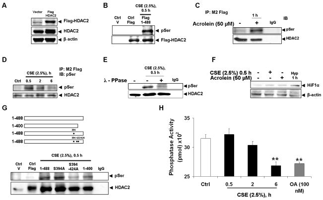
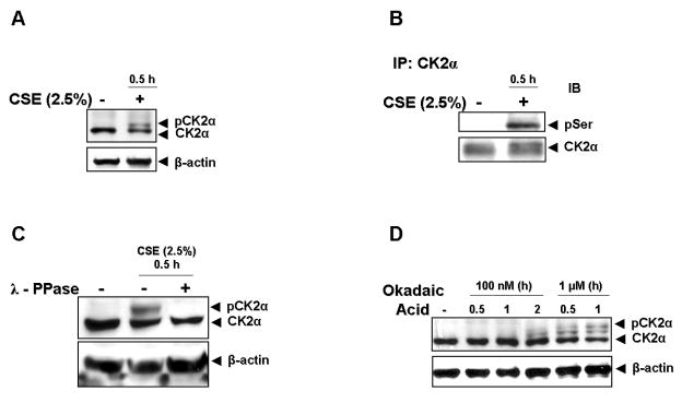
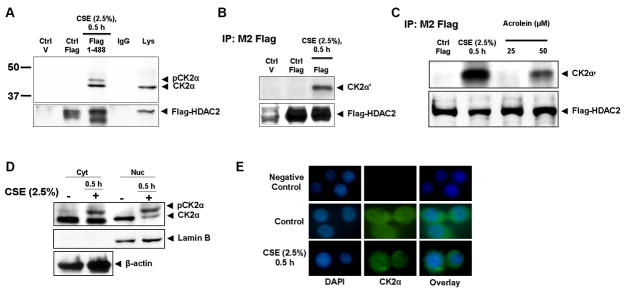
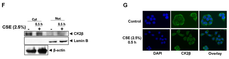
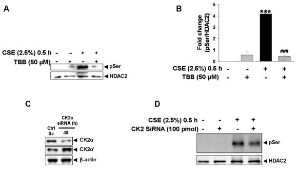
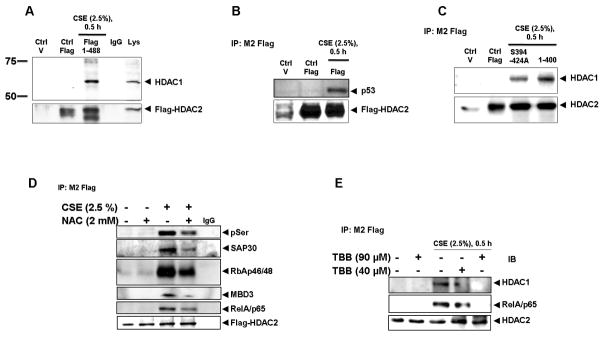
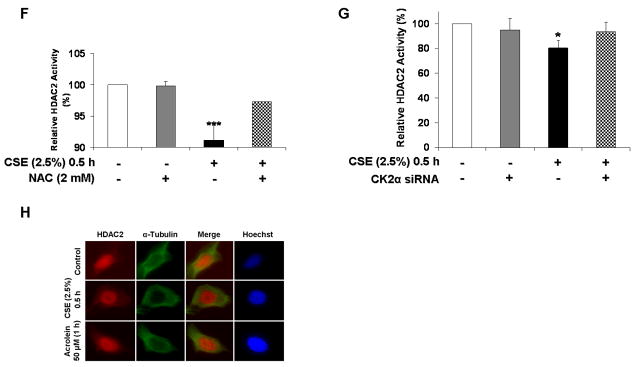
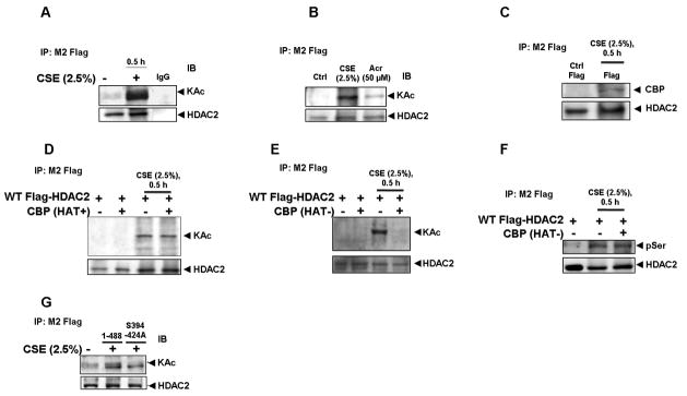
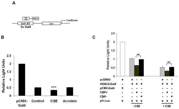
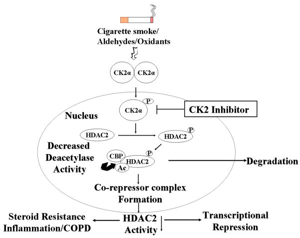
Histone deacetylase 2 (HDAC2) mediates the repression of pro-inflammatory genes by deacetylating core histones, RelA/p65 and the glucocorticoid receptor. Reduced level of HDAC2 is associated with steroid resistant inflammation caused by cigarette smoke (CS)-derived oxidants and aldehydes. However, the molecular mechanisms regulating HDAC2 in response to CS and aldehydes is not known. Here, we report that CS extract, and aldehyde acrolein induced phosphorylation of HDAC2 which was abolished by mutations at serine sites S(394), S(411), S(422) and S(424). HDAC2 phosphorylation required direct interaction with serine-phosphorylated protein kinase CK2alpha and involved reduced HDAC2 deacetylase activity. Furthermore, HDAC2 phosphorylation was required for HDAC2 interaction with transcription factors, co-repressor complex formation, CBP recruitment, acetylation on lysine residues and modulates transrepression activity. Thus, phospho-acetylation of HDAC2 negatively regulates its deacetylase activity which has implications in steroid resistance in chronic inflammatory conditions.


Figures

Figure 1
Figure 1. HDAC2 phosphorylation requires serine sites S394, S422, S424 and is independent of phosphatase inhibition
(A) Bronchial epithelial H292 cells were transfected with plasmid vector or flag-tagged WT HDAC2 (1-488) for 24 h using Lipofectamine 2000. 5 – 10 μg proteins were separated on a 7.5% SDS-PAGE gel and flag proteins detected with M2 monoclonal anti-flag antibodies. (B) H292 cells were transfected with flag-tagged WT HDAC2 for 24 h and then treated with CSE (2.5%) for 0.5 h, (C) acrolein (50 μM) for 1 h or (D) CSE (2.5%) for 0.5, 2 and 6 h. Immunoprecipitated flag proteins were probed with phosphoserine monoclonal antibodies by western blot. (E) Immunoprecipitated beads from H292 cells transfected with flag-tagged HDAC2 and treated with CSE (2.5%) were incubated with lambda PPase buffer or 400 units of lambda PPase for 30 minutes at 30 °C. Washed beads were then eluted with 2x Laemmli sample buffer and separated on a 7.5% SDS-PAGE gel. Blots were probed with phosphoserine antibodies. (F) Lysates from H292 cells treated with CSE (2.5 %), acrolein (50 μM) or exposed to hypoxia for 1 h were separated by SDS-PAGE gel and blots probed for HIF1A. (G) H292 cells were transfected with plasmid vector, flag-tagged WT HDAC2 (1-488) or S394A, S394/411/422/424A and 1-400 aa HDAC2 mutants for 24 h with Lipofectamine 2000. Cells were then treated with CSE (2.5%) for 0.5 h. Serine phosphorylation of immunoprecipitated flag proteins was determined by western blot. (H) H292 cells were treated with or without CSE (2.5%) for 0.5, 2 and 6 h with serine/threonine phosphatase (okadaic acid) used as a positive control. PP2A activity was determined by incubating 5 μg protein with PP2A specific-peptide substrate RRA(pT)VA and PP2A activity determined by reading optical density with microplate reader using a 630 nm filter (n = 4, ** p < 0.01).
Figure 2
Figure 2. Cigarette Smoke Extract (CSE) induces CK2α serine phosphorylation
(A) Bronchial epithelial H292 cells were treated with CSE (2.5 %) for 0.5 h. 10 μg protein was separated by SDS-PAGE and CK2α detected by western blot. (B) CK2α from control or CSE-treated H292 cells was immunoprecipitated and analyzed for serine phosphorylation. (C) Lysates from H292 cells treated with media alone or CSE were incubated with 400 units of lambda phosphatase for 30 minutes at 30 °C before separation by SDS-PAGE. CK2α expression was determined by western blot. (D) H292 cells were treated with indicated concentrations of okadaic acid for indicated time periods. CK2α expression was determined by western blot.
Figure 3
Figure 3. Cigarette Smoke Extract (CSE) and acrolein induce HDAC2 interaction with catalytic subunits of CK2
H292 cells were transfected with plasmid vector or flag-tagged WT HDAC2 (1-488) for 24 h using Lipofectamine 2000 and then treated with or without CSE (2.5 %) for 0.5 h. Flag proteins were immunoprecipitated and binding to (A) CK2α and (B) CK2α′ determined by western blot. (C) H292 cells transfected with flag-tagged WT HDAC2 were treated with CSE (2.5 %) for 0.5 h, and acrolein (25 μM and 50 μM) for 1 h. CK2α′ binding to immunoprecipitated flag-tagged HDAC2 was determined by western blot. (D) H292 cells were treated with or without CSE (2.5%) for 0.5 h. Nuclear and cytoplasmic extracts were probed for CK2α or (F) CK2β expression by western blot. (E) H292 cells were treated with CSE (2.5%) for 0.5 h, washed and fixed with 4% PFA. Slides were incubated with CK2α or (G) CK2β goat polyclonal antibody (1:100) overnight at 4 °C. Slides were mounted with ProLong Gold antifade reagent (Invitrogen) and images analyzed by a Leica confocal microscope.
Figure 4
Figure 4. CK2 is required for CSE-induced HDAC2 phosphorylation
(A) H292 cells transfected with flag-tagged WT HDAC2 were pre-treated with TBB (50 μM) for 2 h prior to treatment with CSE (2.5%) for 0.5 h. Immunoprecipitated flag proteins were analyzed for serine phosphorylation by western blot. (B) Relative density of phospho-serine expression normalized to immunoprecipitated HDAC2. (C) H292 cells were transiently transfected with scrambled or 100 pmol CK2α siRNA for 24 h. Cells were washed and incubated for a further 24 h. Whole cell lysates were separated by SDS-PAGE and CK2α and CK2α′ expression analyzed by western blot. (D) H292 cells transfected with CK2α siRNA for 24 h were transfected for a further 24 h with flag-tagged WT HDAC2. Cells were then exposed to CSE (2.5 %) for 0.5 h and immunoprecipitated flag proteins analyzed for serine phosphorylation by western blot (n = 3, *** ### P<0.001).
Figure 5
Figure 5. Cigarette Smoke Extract (CSE)-induced HDAC2 phosphorylation regulates co-repressor formation, interaction with transcription factors and deacetylase activity but not cellular localization
(A) H292 cells were transfected with plasmid vector or flag-tagged WT HDAC2 (1-488) for 24 h using Lipofectamine 2000 and then treated with or without CSE (2.5 %) for 0.5 h. Flag proteins were immunoprecipitated and binding to HDAC1 and (B) p53 determined by western blot. (C) H292 cells were transfected with plasmid vector, flag-tagged WT HDAC2 (1-488) or S394/411/422/424A and 1-400 aa HDAC2 mutants for 24 h with Lipofectamine 2000. Cells were then treated with CSE (2.5%) for 0.5 h. HDAC1 interaction with immunoprecipitated flag proteins was determined by western blot. (D) Flag-tagged HDAC2-transfected H292 cells were pre-treated with NAC (2 mM) for 2 h prior to 0.5 h treatment with CSE (2.5 %). Immunoprecipitated flag proteins were separated by SDS-PAGE and binding to SAP30, RbAp46/48, MBD3 and RelA/p65 analyzed by western blot. (E) Flag-tagged HDAC2-transfected H292 cells were pre-treated with TBB 40 or 90 μM for 2 h or 0.5 h respectively prior to treatment with CSE (2.5 %) for 0.5 h. Immunoprecipitated flag proteins were analyzed for HDAC1 and p65 binding by western blot. (F) Immunoprecipitated flag proteins from NAC pre-treatment experiment were analyzed for HDAC deacetylase activity using a deacetylase activity kit. (G) H292 cells were transfected with siRNA targeting CK2α for 24 h. Cells were then washed and transfected with flag-tagged WT HDAC2 for a further 24 h prior to treating with CSE (2.5 %) for 0.5 h. Immunoprecipitated flag proteins were analyzed for deacetylase activity. (H) H292 cells were grown on coverslips and treated with CSE (2.5%) or acrolein (50 μM) for 0.5 and 1 h respectively. Cells were fixed with 4 % PFA then incubated with HDAC2 rabbit polyclonal antibody overnight at 4 °C. Images from slides, mounted with ProLong Gold antifade reagent, were then analyzed with an Olympus inverted microscope (n = 3, * P<0.05, ** P<0.01).
Figure 6
Figure 6. HDAC2 acetylation requires phosphorylation-induced CBP recruitment
(A) H292 cells, transfected with flag-tagged WT HDAC2 were treated with CSE (2.5%) for 0.5 h and (B) Acrolein (50 μM) for 1 h. Immunoprecipitated flag proteins were analyzed for global acetylation by western blot. (C) Immunoprecipitated flag proteins from H292 cells treated with CSE (2.5%) for 0.5 h were analyzed for CBP binding by western blot. (D) H292 cells were transiently co-transfected with CBP WT or (E) CBP HAT mutant for 24 h prior to CSE treatment. Immunoprecipitated flag-tagged proteins were analyzed for lysine residue acetylation or (F) serine phosphorylation by western blot. G) H292 cells were transfected with plasmid vector, flag-tagged WT HDAC2 (1-488) or S394/411/422/424A mutant for 24 h with Lipofectamine 2000. Cells were treated with CSE (2.5%) for 0.5 h. Acetylation of immunoprecipitated flag proteins was determined by western blot.
Figure 7
Figure 7. HDAC2 acetylation is critical for HDAC2 functional activity
(A) Schematic for yeast Gal4 2-hybrid assay. HDAC2-Gal4 plasmids were co-transfected with a pFR-luciferase reporter tagged to a promoter with 5 tandem yeast Gal 4 repeats. (B) H292 cells were co-transfected with either pCMX-Gal4 or Gal4-HDAC2 plasmids with a pFR-luciferase reporter plasmid. Cells were treated for 0.5 or 1 h with CSE (2.5%) or acrolein (50 μM) respectively. 20 μg cell lysates were analyzed for luciferase expression (n=3). (C) H292 cells were co-transfected with pcDNA3 vector, CBP wild-type or CBP (HAT-) plasmids with HDAC2-Gal4 and a pFR-luciferase reporter. Cells were exposed to either media alone or CSE (2.5%) for 0.5 h. 20 μg cell lysates were analyzed for luciferase expression (n=3).
Figure 8
Figure 8. Schematic for oxidant/aldehyde regulation of HDAC2
Schematic for cigarette smoke/oxidant/aldehyde-mediated regulation of HDAC2. CK2α-mediated phosphorylation of HDAC2 induces increased co-repressor complex formation and transcription factor recruitment. Phospho-acetylation of HDAC2 leads to decreased in its deacetylase activity and hence steroid resistance.

Similar articles

Cited by

References

    1. de Ruijter AJ, van Gennip AH, Caron HN, Kemp S, van Kuilenburg AB. Biochem J. 2003;370:737–749. - PMC - PubMed
    1. Sengupta N, Seto E. J Cell Biochem. 2004;93:57–67. - PubMed
    1. Villagra A, Sotomayor EM. E. Seto Oncogene. 29:157–173. - PubMed
    1. Kramer OH. Trends Pharmacol Sci. 2009;30:647–655. - PubMed
    1. Hassig CA, Fleischer TC, Billin AN, Schreiber SL, Ayer DE. Cell. 1997;89:341–347. - PubMed

Publication types

MeSH terms