

Distinct patterns of kidney and liver cyst growth in pkd2(WS25/-) mice
- PMID: 20388629
- PMCID: PMC2980992
- DOI: 10.1093/ndt/gfq195
Distinct patterns of kidney and liver cyst growth in pkd2(WS25/-) mice
Abstract
Background: Autosomal dominant polycystic kidney disease (ADPKD) is a common genetic disease that results in the development of cystic kidneys and liver. Pkd2(WS25/-) mice are a key genetic mouse model of human ADPKD that recapitulate the 'molecular recessive' nature of human ADPKD. Providing the foundation for future long-term studies, the present work documents distinct patterns of long-term cyst growth in the kidneys and liver of male and female pkd2(WS25/-) mice.
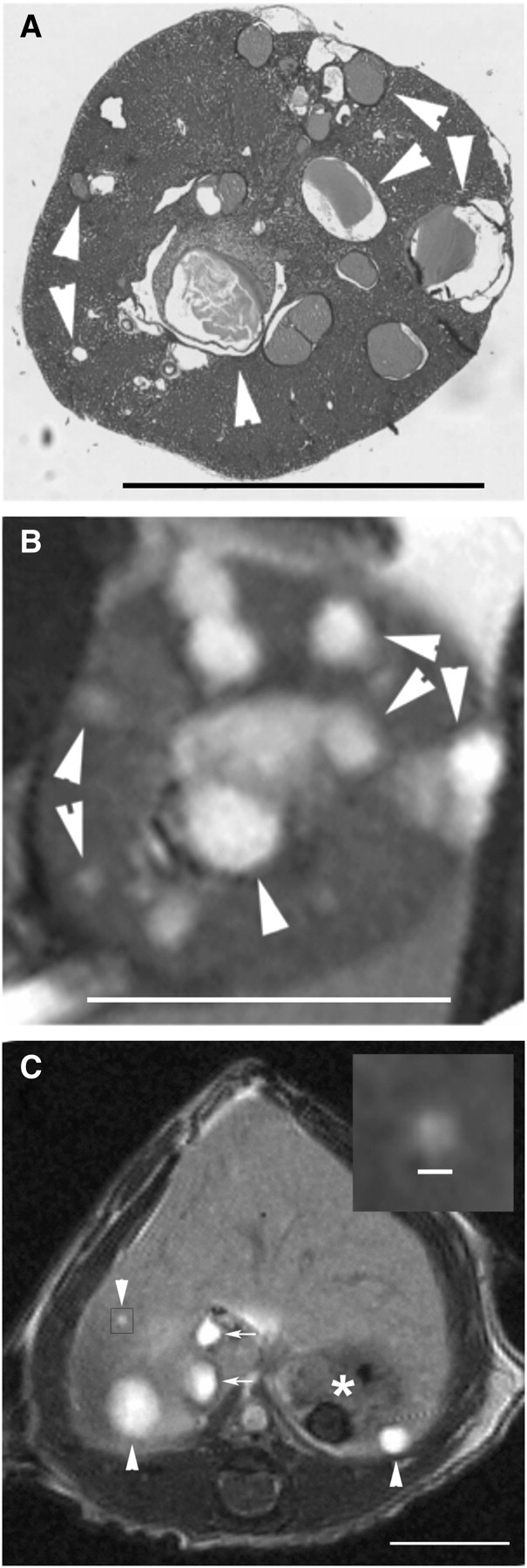
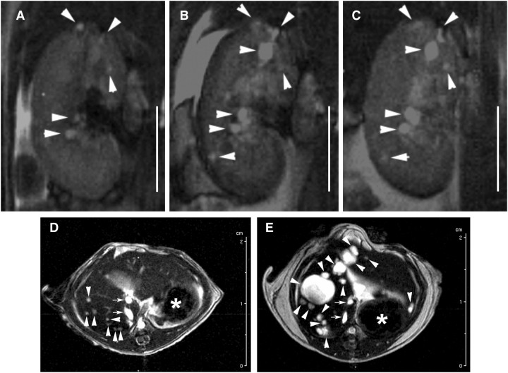
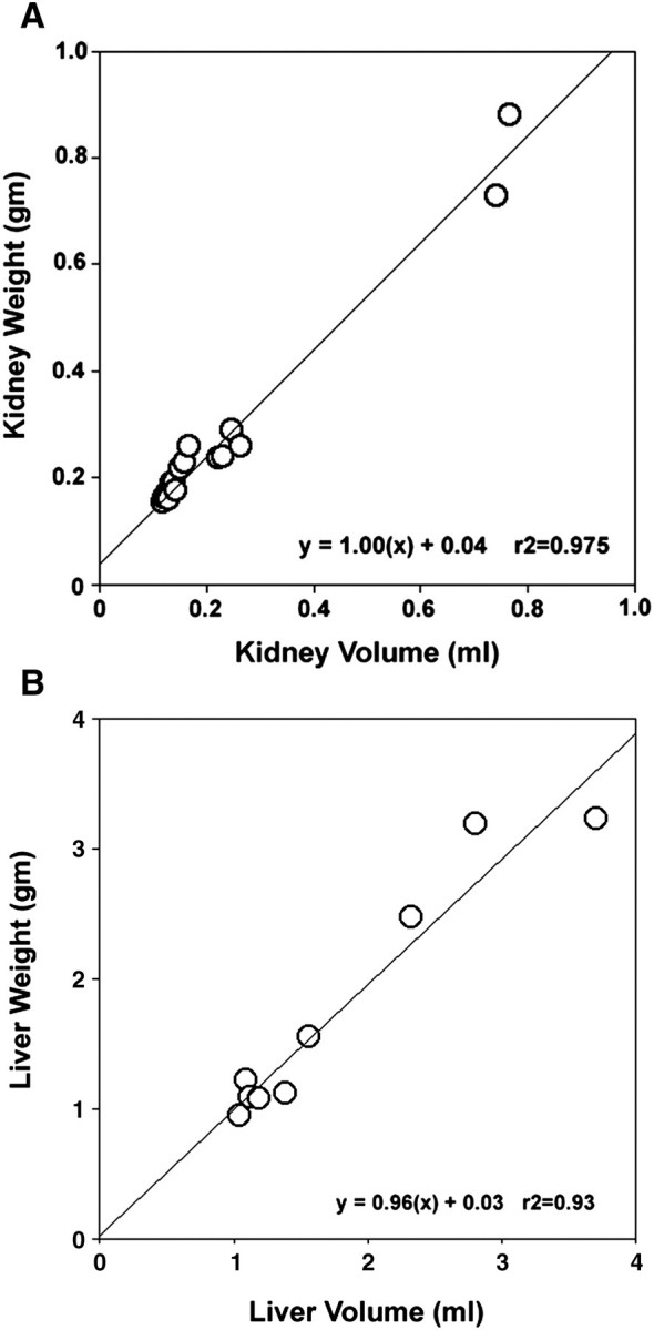
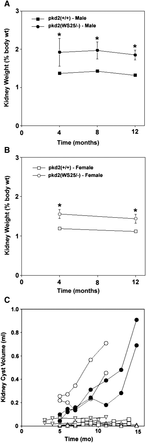
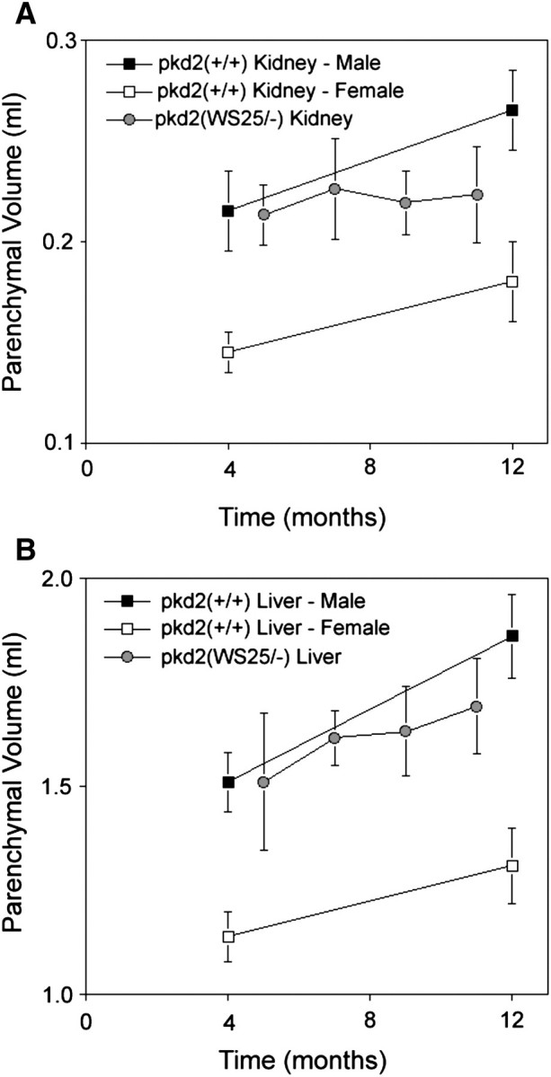
Methods: Gravimetric measurements documented the progression of kidney and liver growth in male and female pkd2(WS25/-) mice over 12 months. A fast imaging with steady-state precision-magnetic resonance imaging (FISP-MRI) technique to measure kidney and liver organ and cyst volumes was optimized and validated. Longitudinal FISP-MRI analyses of changes in cyst volumes were performed in pkd2(WS25/-) mice over 15 months.
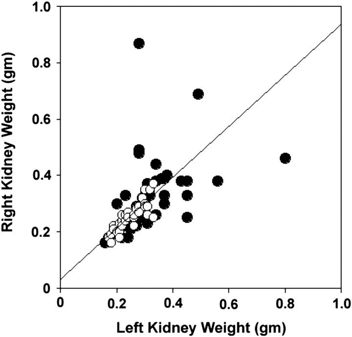
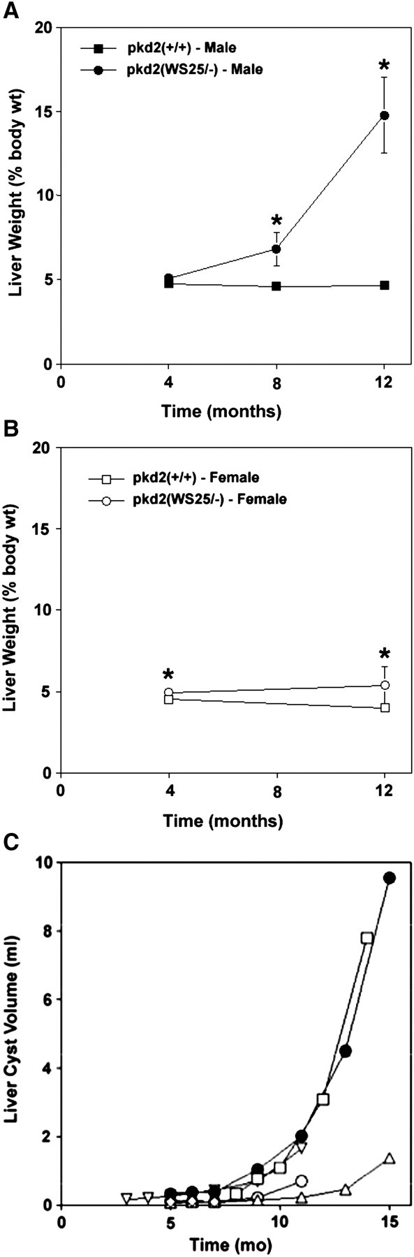
Results: Male and female pkd2(WS25/-) mice had significant increases in kidney weights after 4 months of age. The progression of kidney growth was minimal after 4 months of age. Liver cyst growth in male pkd2(WS25/-) mice was minimal after 4 months of age but showed an accelerated rate of growth after 8 months of age. Female pkd2(WS25/-) mice also showed accelerated growth but this was delayed in time when compared with male pkd2(WS25/-) mice.
Conclusions: Pkd2(WS25/-) mice are a genetic mouse model that recapitulates the early phenotypic characteristics of human ADPKD kidney cystogenesis. Male pkd2(WS25/-) mice consistently display a late progression in liver growth that is seen in clinically impacted livers of human ADPKD patients.
Figures
Similar articles
-
VEGF receptor inhibition blocks liver cyst growth in pkd2(WS25/-) mice.Am J Physiol Cell Physiol. 2007 Jul;293(1):C419-28. doi: 10.1152/ajpcell.00038.2007. Epub 2007 May 2. Am J Physiol Cell Physiol. 2007. PMID: 17475663
-
Inhibition of Cdc25A suppresses hepato-renal cystogenesis in rodent models of polycystic kidney and liver disease.Gastroenterology. 2012 Mar;142(3):622-633.e4. doi: 10.1053/j.gastro.2011.11.036. Epub 2011 Dec 7. Gastroenterology. 2012. PMID: 22155366 Free PMC article.
-
Nonselective Cyclooxygenase Inhibition Retards Cyst Progression in a Murine Model of Autosomal Dominant Polycystic Kidney Disease.Int J Med Sci. 2019 Jan 1;16(1):180-188. doi: 10.7150/ijms.27719. eCollection 2019. Int J Med Sci. 2019. PMID: 30662341 Free PMC article.
-
Molecular genetics and mechanism of autosomal dominant polycystic kidney disease.Mol Genet Metab. 2000 Jan;69(1):1-15. doi: 10.1006/mgme.1999.2943. Mol Genet Metab. 2000. PMID: 10655152 Review.
-
Clinical and genetic characteristics of Korean autosomal dominant polycystic kidney disease patients.Korean J Intern Med. 2021 Jul;36(4):767-779. doi: 10.3904/kjim.2021.176. Epub 2021 Jul 1. Korean J Intern Med. 2021. PMID: 34237823 Free PMC article. Review.
Cited by
-
Automated total kidney volume measurements in pre-clinical magnetic resonance imaging for resourcing imaging data, annotations, and source code.Kidney Int. 2021 Mar;99(3):763-766. doi: 10.1016/j.kint.2020.07.040. Epub 2020 Aug 20. Kidney Int. 2021. PMID: 32828755 Free PMC article.
-
Quantifying Memory CD8 T Cells Reveals Regionalization of Immunosurveillance.Cell. 2015 May 7;161(4):737-49. doi: 10.1016/j.cell.2015.03.031. Cell. 2015. PMID: 25957682 Free PMC article.
-
Hypoxia-inducible factor-1α (HIF-1α) and autophagy in polycystic kidney disease (PKD).Am J Physiol Renal Physiol. 2011 May;300(5):F1235-43. doi: 10.1152/ajprenal.00348.2010. Epub 2011 Jan 26. Am J Physiol Renal Physiol. 2011. PMID: 21270095 Free PMC article.
-
Nanosized contrast agents to noninvasively detect kidney inflammation by magnetic resonance imaging.Adv Chronic Kidney Dis. 2013 Nov;20(6):488-99. doi: 10.1053/j.ackd.2013.06.001. Adv Chronic Kidney Dis. 2013. PMID: 24206601 Free PMC article. Review.
-
Antisense-mediated angiotensinogen inhibition slows polycystic kidney disease in mice with a targeted mutation in Pkd2.Am J Physiol Renal Physiol. 2015 Feb 15;308(4):F349-57. doi: 10.1152/ajprenal.00478.2014. Epub 2014 Dec 23. Am J Physiol Renal Physiol. 2015. PMID: 25537744 Free PMC article.
References
-
- Wall WJ. Liver transplantation for polycystic liver disease. N Engl J Med. 2007;356:1560. - PubMed
-
- King BF, Reed JE, Bergstrahl EJ, Sheedy PF, Torres VE. Quantification and longitudinal trends of kidney, renal cyst and renal parenchymal volumes in ADPKD. J Am Soc Nephrol. 2000;11:1505–1511. - PubMed
-
- Sise C, Kusaka M, Wetzel LH, et al. Volumetric determination of progression in ADPKD by computed tomography. Kidney Int. 2000;58:2492–2501. - PubMed
-
- Chapman AB, Guay-Woodford LM, Grantham JJ, et al. Renal structure in early ADPKD: the consortium for radiologic imaging studies of polycystic kidney disease cohort. Kidney Int. 2003;64:1035–1045. - PubMed
Publication types
MeSH terms
Substances
Grants and funding
LinkOut - more resources
Full Text Sources
Medical
Molecular Biology Databases
Miscellaneous
