

Low CD4+ T-cell levels and B-cell apoptosis in vertically HIV-exposed noninfected children and adolescents
- PMID: 20388660
- PMCID: PMC3107461
- DOI: 10.1093/tropej/fmq024
Low CD4+ T-cell levels and B-cell apoptosis in vertically HIV-exposed noninfected children and adolescents
Abstract
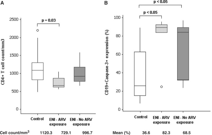
Lymphocyte subsets, activation markers and apoptosis were assessed in 20 HIV-exposed noninfected (ENI) children born to HIV-infected women who were or not exposed to antiretroviral (ARV) drugs during pregnancy and early infancy. ENI children and adolescents were aged 6-18 years and they were compared to 25 age-matched healthy non-HIV-exposed children and adolescents (Control). ENI individuals presented lower CD4(+) T cells/mm(3) than Control group (control: 1120.3 vs. ENI: 876.3; t-test, p = 0.030). ENI individuals had higher B-cell apoptosis than Control group (Control: 36.6%, ARV exposed: 82.3%, ARV nonexposed: 68.5%; Kruskal-Wallis, p < 0.05), but no statistical difference was noticed between those exposed and not exposed to ARV. Immune activation in CD4(+) T, CD8(+) T and in B cells was comparable in ENI and in Control children and adolescents. Subtle long-term immune alterations might persist among ENI individuals, but the clinical consequences if any are unknown, and these children require continued monitoring.
Figures
Similar articles
-
Imbalance of naive and memory T lymphocytes with sustained high cellular activation during the first year of life from uninfected children born to HIV-1-infected mothers on HAART.Braz J Med Biol Res. 2008 Aug;41(8):700-8. doi: 10.1590/s0100-879x2008000800011. Braz J Med Biol Res. 2008. PMID: 18797705
-
Specific patterns of CD4-associated immunosenescence in vertically HIV-infected subjects.Clin Microbiol Infect. 2013 Jun;19(6):558-65. doi: 10.1111/j.1469-0691.2012.03934.x. Epub 2012 Jun 27. Clin Microbiol Infect. 2013. PMID: 22735071
-
HIV-1-infected children on HAART: immunologic features of three different levels of viral suppression.Cytometry B Clin Cytom. 2007 Jan 15;72(1):14-21. doi: 10.1002/cyto.b.20152. Cytometry B Clin Cytom. 2007. PMID: 17041945 Clinical Trial.
-
Characterizing the immune system after long-term undetectable viral load in HIV-1-infected children.J Clin Immunol. 2003 Jul;23(4):279-89. doi: 10.1023/a:1024536816684. J Clin Immunol. 2003. PMID: 12959220
-
The CD4/CD8 ratio as a marker T-cell activation, senescence and activation/exhaustion in treated HIV-infected children and young adults.AIDS. 2013 Jun 1;27(9):1513-6. doi: 10.1097/QAD.0b013e32835faa72. AIDS. 2013. PMID: 23435292
Cited by
-
Delayed acquisition of Plasmodium falciparum antigen-specific CD4(+) T cell responses in HIV-exposed uninfected Malawian children receiving daily cotrimoxazole prophylaxis.Malar J. 2016 May 10;15(1):264. doi: 10.1186/s12936-016-1318-2. Malar J. 2016. PMID: 27165269 Free PMC article.
-
HIV-Exposed Uninfected Infants Have Increased Regulatory T Cells That Correlate With Decreased T Cell Function.Front Immunol. 2019 Mar 26;10:595. doi: 10.3389/fimmu.2019.00595. eCollection 2019. Front Immunol. 2019. PMID: 30972079 Free PMC article.
-
Impaired haemophilus influenzae type b transplacental antibody transmission and declining antibody avidity through the first year of life represent potential vulnerabilities for HIV-exposed but -uninfected infants.Clin Vaccine Immunol. 2014 Dec;21(12):1661-7. doi: 10.1128/CVI.00356-14. Epub 2014 Oct 8. Clin Vaccine Immunol. 2014. PMID: 25298109 Free PMC article.
-
Immune and Metabolic Alterations in Children with Perinatal HIV Exposure.Viruses. 2023 Jan 18;15(2):279. doi: 10.3390/v15020279. Viruses. 2023. PMID: 36851493 Free PMC article. Review.
-
Immune development in HIV-exposed uninfected children born to HIV-infected women.Rev Inst Med Trop Sao Paulo. 2017 Jun 1;59:e30. doi: 10.1590/S1678-9946201759030. Rev Inst Med Trop Sao Paulo. 2017. PMID: 28591258 Free PMC article.
References
-
- European Collaborative Study. Mother-to-child transmission of HIV infection in the era of highly active antiretroviral therapy. Clin Infect Dis. 2005;40:458–65. - PubMed
-
- Gesner M, Papaevangelou V, Kim M, et al. Alteration in the proportion of CD4 T lymphocytes in a subgroup of human immunodeficiency virus-exposed-non-infected children. Pediatrics. 1994;93:624–30. - PubMed
-
- Clerici M, Saresella M, Colombo F, et al. T-lymphocyte maturation abnormalities in uninfected newborns and children with vertical exposure to HIV. Blood. 2000;96:3866–71. - PubMed
Publication types
MeSH terms
Substances
Grants and funding
LinkOut - more resources
Full Text Sources
Medical
Research Materials
