. 2010 Jun;139(6):1057-66.
doi: 10.1530/REP-10-0069. Epub 2010 Apr 13.

Expression of basigin in reproductive tissues of estrogen receptor-{alpha} or -{beta} null mice

Affiliations

Expression of basigin in reproductive tissues of estrogen receptor-{alpha} or -{beta} null mice

Li Chen et al. Reproduction. 2010 Jun.

Abstract

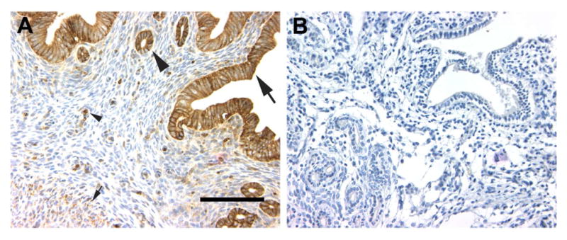
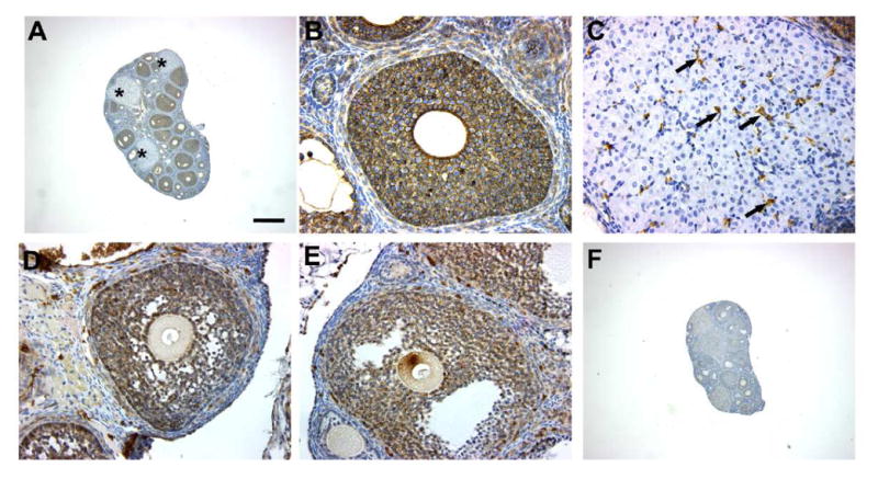
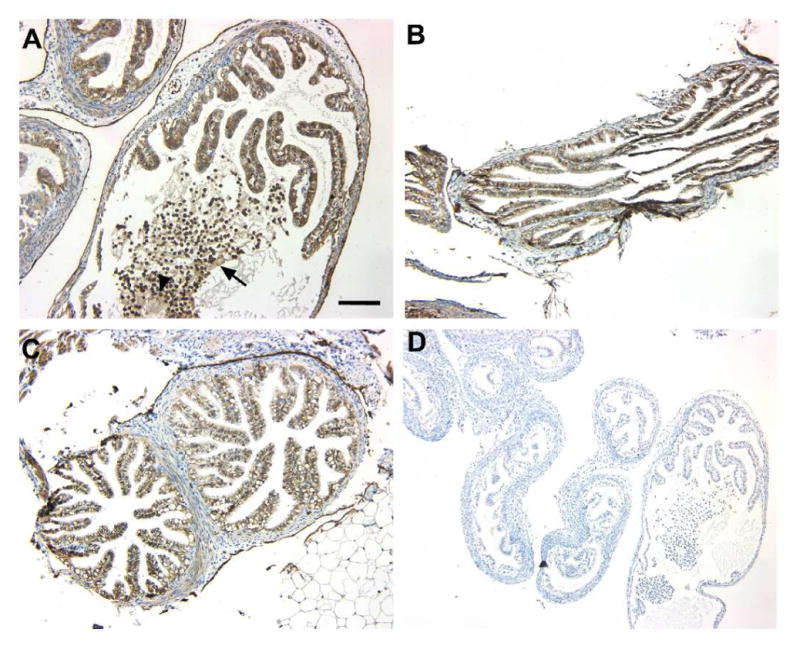
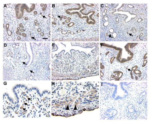
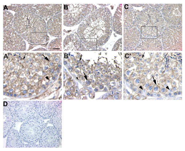
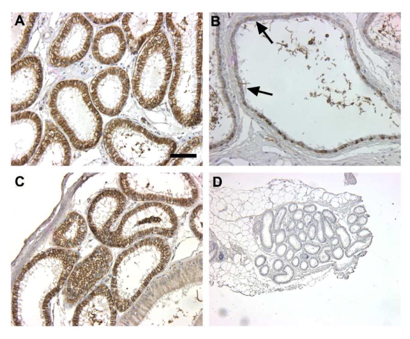
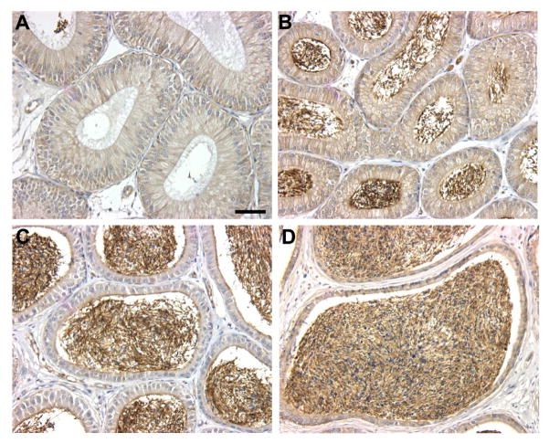
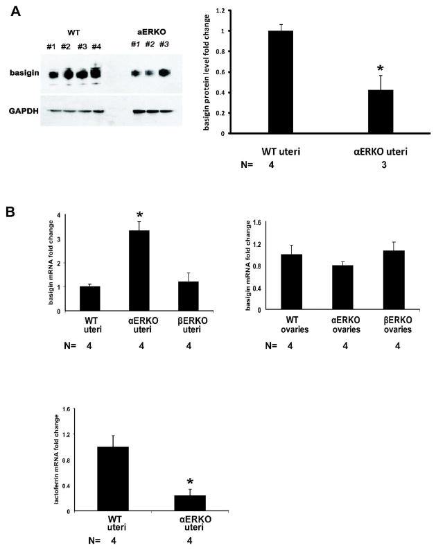
Basigin plays important roles in both male and female reproduction because basigin (Bsg) null male and female mice are infertile. The aim of the present study was to determine whether basigin expression in reproductive organs requires estrogen receptor-alpha (ESR1, ERalpha) or -beta (ESR2, ERbeta). Expression of basigin protein in the testis, ovary, and male and female reproductive tracts was studied in adult wild-type (WT), Esr1-null (alphaERKO), and Esr2-null (betaERKO) mice by immunohistochemistry and immunoblotting. Basigin mRNA levels in ovary and uterus were examined by quantitative RT-PCR. In females, basigin protein expression was observed mainly in granulosa and interstitial cells of the ovary and epithelial cells of the proximal oviduct in all genotypes. Basigin protein was also expressed in the uterine epithelium at proestrus and estrus in WT and betaERKO mice but not in alphaERKO mice. However, a higher level of basigin mRNA was observed in uteri of alphaERKO mice compared with WT and betaERKO mice. In males, basigin was expressed in Leydig cells and all germ cells except spermatogonia in all genotypes. Basigin was present in epithelial cells lining the efferent ductules in WT and betaERKO mice, but expression was greatly reduced in alphaERKO mice. In epididymal ducts, basigin expression was observed in epithelial cells in the caput and cauda in all genotypes. These data suggest that expression of basigin protein requires ESR1, but not ESR2, in the uterus and efferent ductules, but is independent of estrogen receptor in the ovary, oviduct, testis, and epididymis.


Figures

Figure 1
Figure 1
Basigin immunoreactivity in the uteri (estrus) of WT (A) and Bsg-null (B) female mice. Uterine luminal (A, big arrow) and glandular (A, big arrow head) epithelia and erythrocytes (A, small arrow head) in the endometrium are strongly positive for basigin in the WT mouse at estrus. Basigin is also positive in myometrium (A, small arrow), whereas there is no basigin immunoreactivity in the Bsg-null uterus (B). Scale bar = 50μm.
Figure 2
Figure 2
Basigin immunoreactivity in ovaries of WT (A, B and C), αERKO (D) and βERKO (E) mice. Ovarian sections were stained with goat anti-mouse basigin antibody (A–E) or goat non-specific IgG (F). In the ovaries of WT mice basigin is strongly positive in the granulosa cells of follicles, interstitial cells. In the corpora lutea basigin is expressed by erythrocytes (arrows in C) in the blood vessels, not by the corpus luteum cells (C). The similar expression pattern is observed in αERKO (D) and βERKO (E) ovaries. Scale bar = 250μm for A, F, and 25μm for B, C, D and E. Stars in A, corpus luteum; arrows in C, erythrocytes.
Figure 3
Figure 3
Basigin immunoreactivity in oviducts of WT (A), αERKO (B) and βERKO (C) mice. Sections were stained with goat anti-mouse basigin antibody (A–C) or goat non-specific IgG (D). Basigin expression is evident on the surface of ciliated and non-ciliated epithelial cells of WT, αERKO and βERKO mice. Basigin expression is observed on the surface of cumulus oophorus cells (A, arrow) surrounding the ovulated oocyte (A, arrow head) that were retained in the oviduct of WT mice at oestrus. Scale bar = 50μm for A, B, C and 100μm for D.
Figure 4
Figure 4
Basigin immunoreactivity in uteri of WT (A, B, C, and D), αERKO (E, G and H) and βERKO (F) mice. Uteri were collected at prooestrus (A), oestrus (B), metoestrus (C) or dioestrus (D) from WT mice, and at oestrus from βERKO mice (F). Sections were stained with goat anti-mouse basigin antibody (A–H) or goat non-specific IgG (I). Basigin immunoreactivity is intense in luminal and glandular epithelium of WT mice at prooestrus (A), oestrus (B), weak at metoestrus (C), very low or absent at dioestrus (D). Basigin expression is seen on the surface of erythrocytes (arrows in A, B, C and D) and myometrium (data not shown) in the WT uteri at all stages of oestrous cycle. In the αERKO uteri basigin is not observed in the luminal or glandular epithelium, whereas basigin immunoreactivity is detectable in erythrocytes (arrows in G) and myometrium (arrow heads in H). Scale bar = 50μm for A, B, C, D, F, I, 25μm for E and 15μm for G and H.
Figure 5
Figure 5
Basigin immunoreactivity in testes of WT (A, A′), αERKO (B, B′) and βERKO (C, C′) mice. Sections were stained with goat anti-mouse basigin antibody (A–C) or goat non-specific IgG (D). A′, B′, and C′ are the enlarged views of the highlighted areas in A, B, and C, respectively. In WT mice, strong basigin immunoreactivity is observed on the cell surface of Leydig cells, spermatocytes, spermatid and sperm tails, while undetectable in spermatogonia. The same pattern of basigin expression is observed in αERKO (B) and βERKO (C) mice. Big arrow head, Sertoli cells; big arrow, spermatocytes; small arrow, round spermatids; small arrow head, spermatogonia. Scale bar = 50μm for A, B, C and D, and 15μm for A′, B′, and C′.
Figure 6
Figure 6
Basigin immunoreactivity in efferent ductules of WT (A), αERKO (B) and βERKO (C) mice. Sections were stained with goat anti-mouse basigin antibody (A–C) or goat non-specific IgG (D). Intense basigin immunoreactivity is observed in the ciliated and non-ciliated epithelial cells of WT (A) and βERKO (C) mice. Basigin immunoreactivity is greatly diminished or absent in epithelial cells of αERKO efferent ductules (B, arrow). Scale bar= 50μm for A, B, C and 200μm for D.
Figure 7
Figure 7
Basigin immunoreactivity in epididymis of WT mice. Moderate basigin immunoreactivity is observed on the lateral surface of epithelial cells in caput (B) and apical surface in cauda (D), while weak immunoreactivity is detected on the lateral surface of epithelial cells in the initial segment (A) and apical surface in the corpus (C). Scale bar = 50μm.
Figure 8
Figure 8
Basigin protein and mRNA levels in uteri of WT and ERKO mice. Uterine protein or RNA were isolated from whole uteri and subjected to immunoblotting (A) or quantitative RT-PCR analyses (B). For immunoblotting basigin bands were normalized to GAPDH bands. Basigin protein levels of αERKO uteri are 40% of those of WT mice, while basigin mRNA levels of αERKO uteri were 3-fold increased compared with those of WT (B). Basigin mRNA levels in uteri and ovaries of βERKO mice were comparable to those of WT mice (B). The data are presented as fold changes relative to tissues of WT mice and the numbers of samples are indicated. Data are means ± S.E. The Star indicates the statistic difference (P < 0.05) compared with WT tissues.

Similar articles

Cited by

References

    1. Ambros V. The functions of animal microRNAs. Nature. 2004;431:350–355. - PubMed
    1. Arora K, Gwinn WM, Bower MA, Watson A, Okwumabua I, MacDonald HR, Bukrinsky MI, Constant SL. Extracellular cyclophilins contribute to the regulation of inflammatory responses. J Immunol. 2005;175:517–522. - PMC - PubMed
    1. Biswas C. Collagenase stimulation in cocultures of human fibroblasts and human tumor cells. Cancer Lett. 1984;24:201–207. - PubMed
    1. Biswas C, Zhang Y, DeCastro R, Guo H, Nakamura T, Kataoka H, Nabeshima K. The human tumor cell-derived collagenase stimulatory factor (renamed EMMPRIN) is a member of the immunoglobulin superfamily. Cancer Res. 1995;55:434–439. - PubMed
    1. Chang H, Ni H, Ma XH, Xu LB, Kadomatsu K, Muramatsu T, Yang ZM. Basigin expression and regulation in mouse ovary during the sexual maturation and development of corpus luteum. Mol Reprod Dev. 2004;68:135–141. - PubMed

Publication types

MeSH terms

LinkOut - more resources