

Review
doi: 10.1136/thx.2009.128025.
Stem cells as vectors for antitumour therapy
Affiliations
- PMID: 20388765
- PMCID: PMC3401681
- DOI: 10.1136/thx.2009.128025
Item in Clipboard
Review
Stem cells as vectors for antitumour therapy
Thorax.
2010 Apr.
Abstract
Recent research suggests that mesenchymal stem cells (MSCs) are able to migrate specifically to tumours and their metastases throughout the body. This has led to considerable excitement about the possibility of modifying these cells to express anticancer molecules and using them as specific targeted anticancer agents. However, there are concerns that systemically delivered MSCs may have non-desirable effects, and there are also many unanswered questions including the mechanism of tumour homing. This review investigates the different MSC-delivered anticancer agents, addresses the questions and concerns, and tries to place this potential therapy in future cancer management.
Figures
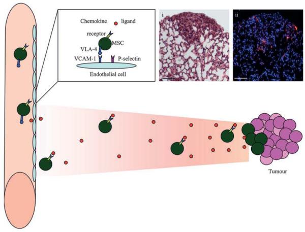
Mesenchymal stem cells (MSCs) home to tumours. MSCs are believed to extravasate similarly to leucocytes with adhesion molecules and integrins. They express very late antigen 4 (VLA-4) which binds to its counterpart adhesion molecule vascular cell adhesion molecule-1 (VCAM-1) on the endothelium. The expression of P-selectin has also been shown to be important for this process. MSCs migrate towards tumours in response to chemokines, which bind to the chemokine receptors on MSCs. The specific DiI-labelled (red) MSC homing is demonstrated in histological sections of lung metastases with (i) H&E and (ii) fluorescent microscopy of contiguous sections with DAPI (blue) nuclear counterstaining. Scale bars represent 20 μm.
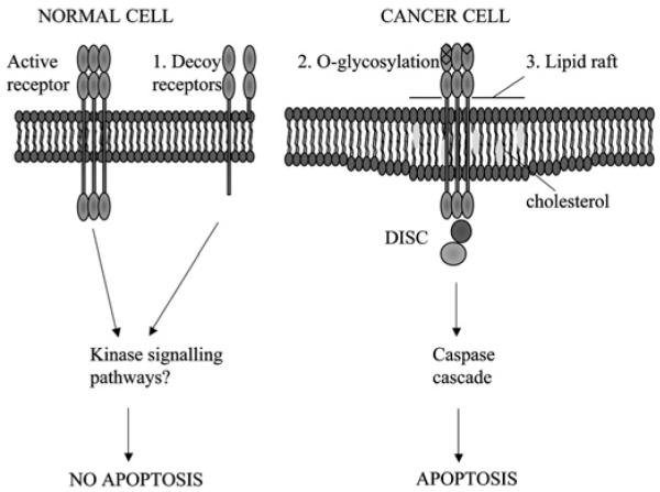
Cancer cells are specifically sensitive to TRAIL-induced apoptosis. Possible mechanisms to explain the selective apoptosis of cancer cell to TRAIL include (1) increase in decoy receptors (which lack the intracellular death domain) in normal cells which may activate anti-apoptotic kinase signalling pathways, (2) O-glycosylation of receptors and (3) their location in lipid rafts enhancing death inducing signalling complex (DISC) formation.
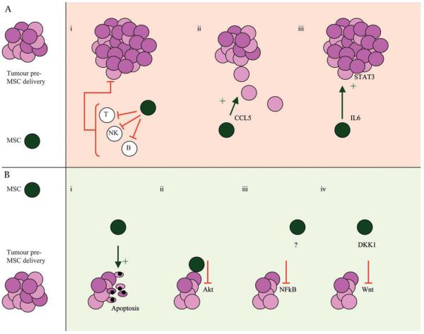
Mesenchymal stem cells (MSCs) may have both growth inhibitory and stimulatory effects on tumours. The literature is divided on the impact that MSCs have on tumour growth with both (A) growth stimulatory and (B) inhibitory effects described. (A) Possible mechanisms for increased tumour proliferation include (i) reduced immunosurveillance secondary to MSC-induced T cell, B cell and natural killer (NK) cell suppression; (ii) promotion of motility, invasion and metastasis of cancer cells by CCL5 release; and (iii) interleukin 6 (IL6) release leading to STAT-3 phosphorylation and tumour cell proliferation. (B) Possible mechanisms for tumour inhibition include (i) promotion of cancer cell cycle arrest and apoptosis; (ii) inhibition of Akt activity; (iii) downregulation of nuclear factor κB (NFκB); and (iv) downregulation of the Wnt pathway by the release of the soluble inhibitor Dickkopf1 (DKK1), both of which reduce cancer cell proliferation.
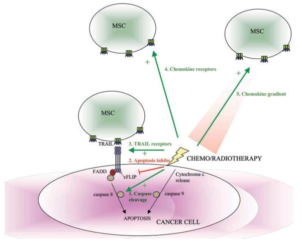
Synergy of chemotherapy and radiotherapy with mesenchymal stem cell (MSC)-delivered anticancer agents. Chemotherapy and radiotherapy cause DNA damage and cancer cell apoptosis by the intrinsic apoptosis pathway, whereas TRAIL leads to cancer cell apoptosis by the extrinsic apoptosis pathway. Chemotherapy and radiotherapy can enhance the TRAIL sensitivity of cancer cells by (1) activating caspase cleavage; (2) inhibiting apoptotic inhibitors (eg, c-Flice-like inhibitory protein (c-FLIP)); and (3) increasing TRAIL receptor number and their location within the lipid raft. Radiotherapy can increase the homing of systemically-delivered MSCs to tumours by increasing (4) MSC chemokine receptor expression and (5) chemokine release by the tumours.
References
-
- Jemal A, Siegel R, Ward E, et al. Cancer statistics, 2007. CA Cancer J Clin. 2007;57:43–66. - PubMed
-
- Bonnet D. Biology of human bone marrow stem cells. Clin Exp Med. 2003;3:140–9. - PubMed
-
- Dvorak HF. Tumors: wounds that do not heal. Similarities between tumor stroma generation and wound healing. N Engl J Med. 1986;315:1650–9. - PubMed
-
- Ishii G, Sangai T, Oda T, et al. Bone-marrow-derived myofibroblasts contribute to the cancer-induced stromal reaction. Biochem Biophys Res Commun. 2003;309:232–40. - PubMed
-
- Direkze NC, Hodivala-Dilke K, Jeffery R, et al. Bone marrow contribution to tumor-associated myofibroblasts and fibroblasts. Cancer Res. 2004;64:8492–5. - PubMed
Publication types
MeSH terms
Grants and funding
LinkOut - more resources
Full Text Sources
Other Literature Sources
Medical