. 2010 May 1;70(9):3594-605.
doi: 10.1158/0008-5472.CAN-09-3565. Epub 2010 Apr 13.

Blueberry phytochemicals inhibit growth and metastatic potential of MDA-MB-231 breast cancer cells through modulation of the phosphatidylinositol 3-kinase pathway

Affiliations

Blueberry phytochemicals inhibit growth and metastatic potential of MDA-MB-231 breast cancer cells through modulation of the phosphatidylinositol 3-kinase pathway

Lynn S Adams et al. Cancer Res. .

Abstract

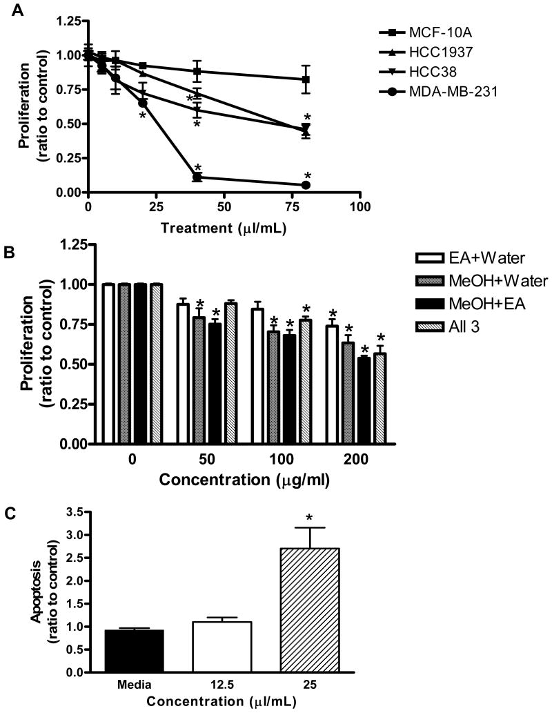
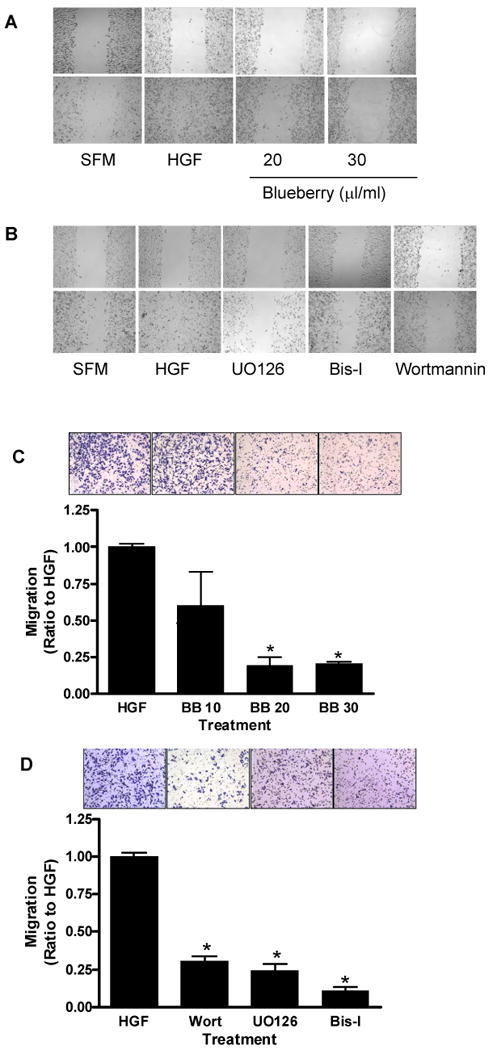
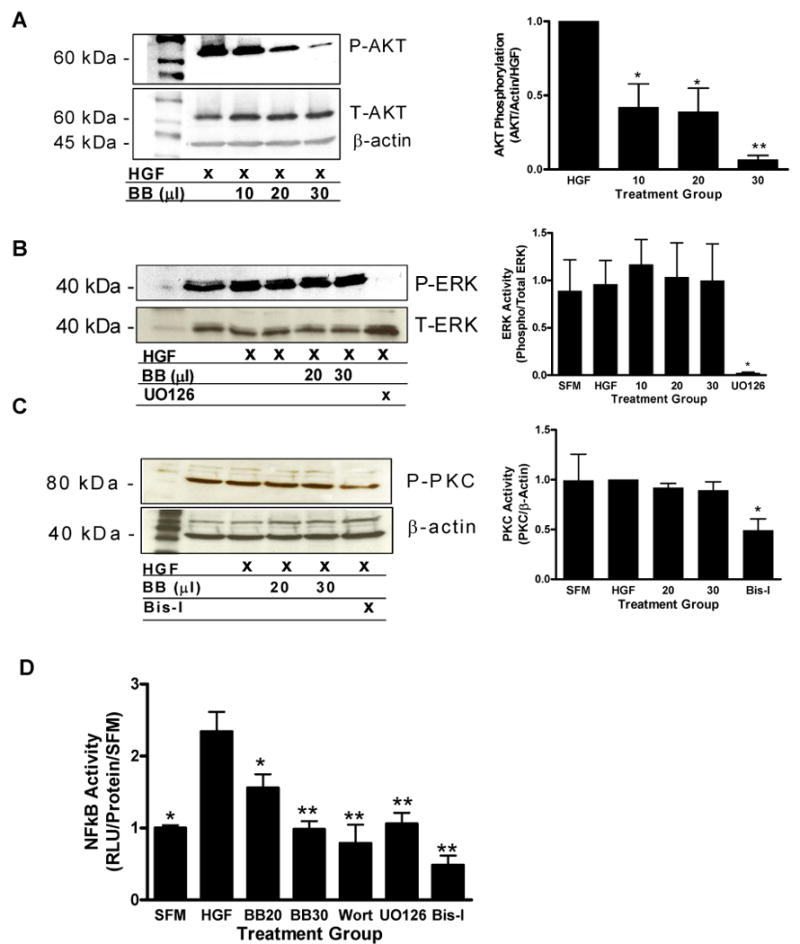
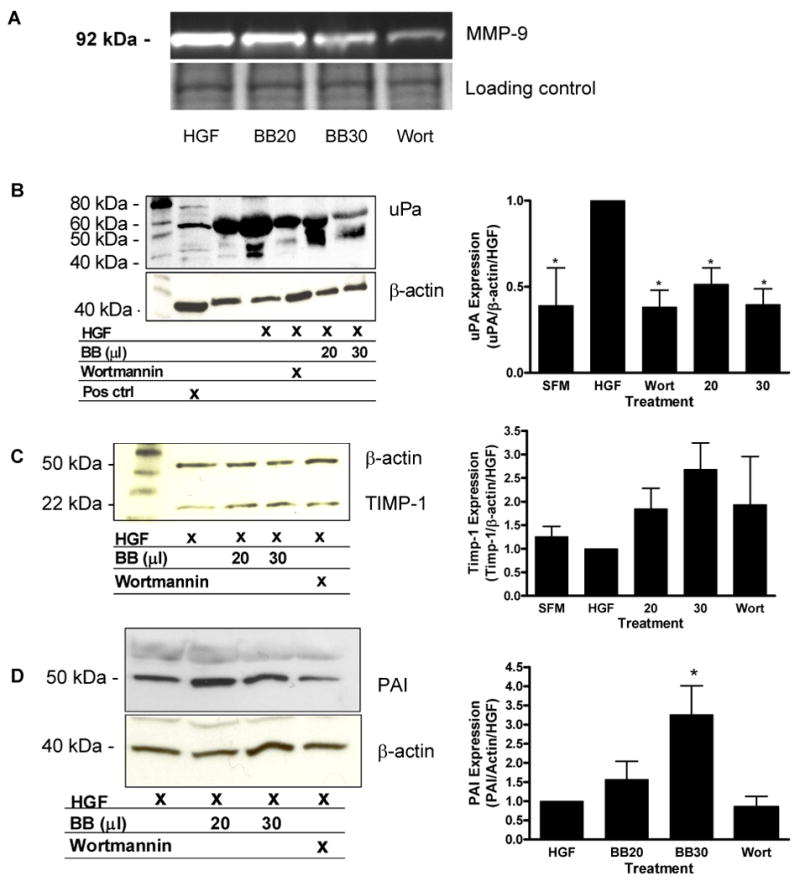
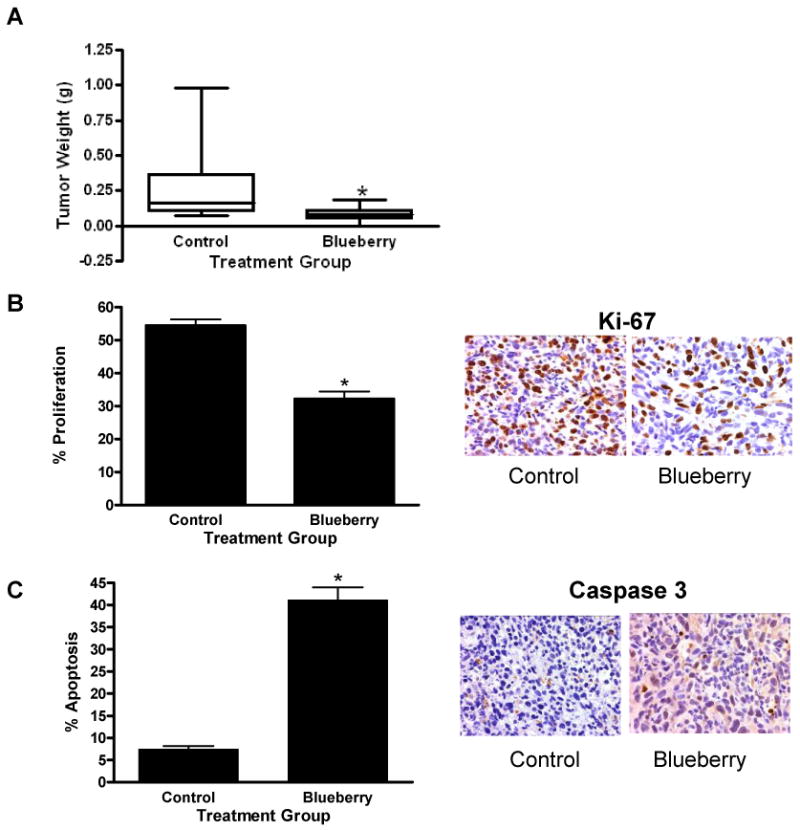
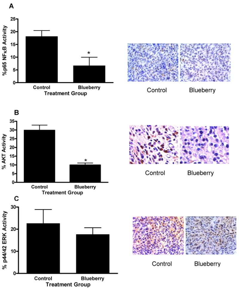
Dietary phytochemicals are known to exhibit a variety of anticarcinogenic properties. This study investigated the chemopreventive activity of blueberry extract in triple-negative breast cancer cell lines in vitro and in vivo. Blueberry decreased cell proliferation in HCC38, HCC1937, and MDA-MB-231 cells with no effect on the nontumorigenic MCF-10A cell line. Decreased metastatic potential of MDA-MB-231 cells by blueberry was shown through inhibition of cell motility using wound-healing assays and migration through a polyethylene terephthalate membrane. Blueberry treatment decreased the activity of matrix metalloproteinase-9 and the secretion of urokinase-type plasminogen activator while increasing tissue inhibitor of metalloproteinase-1 and plasminogen activator inhibitor-1 secretion in MDA-MB-231 conditioned medium as shown by Western blotting. Cell signaling pathways that control the expression/activation of these processes were investigated via Western blotting and reporter gene assay. Treatment with blueberry decreased phosphatidylinositol 3-kinase (PI3K)/AKT and NFkappaB activation in MDA-MB-231 cells, where protein kinase C and extracellular signal-regulated kinase (ERK) were not affected. In vivo, the efficacy of blueberry to inhibit triple-negative breast tumor growth was evaluated using the MDA-MB-231 xenograft model. Tumor weight and proliferation (Ki-67 expression) were decreased in blueberry-treated mice, where apoptosis (caspase-3 expression) was increased compared with controls. Immunohistochemical analysis of tumors from blueberry-fed mice showed decreased activation of AKT and p65 NFkappaB signaling proteins with no effect on the phosphorylation of ERK. These data illustrate the inhibitory effect of blueberry phytochemicals on the growth and metastatic potential of MDA-MB-231 cells through modulation of the PI3K/AKT/NFkappaB pathway.


Figures

Figure 1
Figure 1. MDA-MB-231 cell proliferation and apoptosis
(A) Breast cell lines were treated with increasing concentrations of blueberry extract for 72 hours (B) MDA-MB-231 cells were treated with increasing concentrations of blueberry fractions (EA = ethyl acetate, MeOH = methanol, water fractions) for 72 hours. Proliferation was assessed using the CellTiter-Glo Luminescent Cell Viability Assay. Data are expressed as a ratio of treated samples to untreated controls, mean ± SE. Asterisk indicates a significant difference (n ≥ 9, p ≤ 0.01) compared to untreated controls. (C) Cells were exposed to blueberry extract at indicated concentrations for 72h, harvested and analyzed using the Cell Death Detection ELISAPLUS Assay. Values are means ± SE. Data are expressed as absorbance at 405 nm of each treated sample divided by control. Asterisk indicates a significant difference (n ≥ 12, p ≤ 0.01) compared to untreated controls.
Figure 2
Figure 2. HGF-induced motility and migration in MDA-MB-231 cells
Confluent monolayers were scratched with a plastic pipette tip and incubated in serum free medium (SFM) in the presence of either HGF (40 ng/ml), or HGF plus blueberry (BB) extract (20 and 30 μl/ml) (A) or various cell signaling inhibitors (B) for 24 h. Migration of MDA-MB-231 cells through a PET membrane (0.8 micron Transwell culture inserts) was evaluated in the presence or absence of blueberry (C) or various cell signaling inhibitors (D) after 24 hours of treatment. Quantification of the number of migrating cells from three separate experiments. Data is expressed as a ratio to HGF-treated cells; mean ± SEM. Asterisk indicates significant difference from HGF-treated cells (n ≥ 3, p ≤ 0.01).
Figure 3
Figure 3. ERK, NFκB and AKT activation in MDA-MB-231 tumor cells
The phosphorylation of AKT (A), ERK (B) and PKC (C) in cell lysates from blueberry (BB), wortmannin (Wort), UO126 or Bis-I treated cells was determined via western blotting and chemiluninescence. Bar graphs indicate quantification of at least three separate blots using Quantity One software (Biorad). NFκB activity was evaluated via reporter gene assay (D). Luciferase activity was assayed after 24 hours and normalized to total protein. Data are expressed as relative luciferase units as a ratio of treated samples to serum free (SFM) samples. Values represent mean ± SEM, asterisks indicate significant difference from HGF treated controls (single: p ≤ 0.05, double: p ≤ 0.01), n ≥ 3.
Figure 4
Figure 4. Modulation of metastatic proteins
(A) Inhibition of MMP-9 activity in conditioned medium from MDA-MB-231 cells treated with HGF (40 ng/ml) and blueberry (BB) extract (20 and 30 μl/ml) or wortmannin (Wort) (0.2 μM) for 24h was evaluated via gelatin zymography. Bands were visualized on a Kodak Gel Logic 200 imaging system with the Kodak 1D software. Equal amounts of sample run on 10% acrylamide gels and stained with coomassie blue as loading control. Conditioned medium from cells was evaluated for modulation of uPA (B), TIMP-1 (C) and PAI (D) secretion via western blotting and chemiluminescence. Bar graphs indicate quantification of at least three separate blots using Quantity One software (Biorad). Data represent mean in each group (n ≥ 3 ± SEM). Asterisk indicates statistical significance from HGF-treated cells (p ≤ 0.05).
Figure 5
Figure 5. Inhibition of breast tumor growth
Seven to 8-week-old athymic, nude female mice were gavage fed daily with 100 μl blueberry extract or water. One week after the start of gavage, mice were s.c. injected in the right flank with MDA-MB-231 cells (1×10⁶). After 6 weeks, mice were euthanized; tumors were removed, weighed and sent for histologic staining. Tumor weights are shown (A), Ki-67 antibody staining for cell proliferation (B), and cleaved caspase-3 for apoptosis (C) was evaluated via immunohistochemistry. Bar graphs indicate quantification of six random fields; stained and unstained cells were counted and divided by the total number of cells counted to generate the percentage of positive cells in each group. Data represent mean in each group (n ≥ 6 ± SEM). Asterisk indicates statistical significance from control group, (p ≤ 0.01).
Figure 6
Figure 6. ERK, NFκB and AKT activation in MDA-MB-231 tumor xenografts
Tumors from blueberry or water treated mice were antibody stained for phospho NFκB (A), AKT (B), or ERK (C) and evaluated via immunohistochemistry. Bar graphs indicate quantification of six random fields; stained and unstained cells were counted and divided by the total number of cells counted to generate the percentage of positive cells in each group. Data represent mean in each group (n ≥ 6 ± SEM). Asterisk indicates statistical significance from control group, (p ≤ 0.01).

References

    1. Reis-Filho JS, Tutt AN. Triple negative tumours: a critical review. Histopathology. 2008;52:108–18. - PubMed
    1. Cleator S, Heller W, Coombes RC. Triple-negative breast cancer: therapeutic options. The lancet oncology. 2007;8:235–44. - PubMed
    1. van de Rijn M, Perou CM, Tibshirani R, et al. Expression of cytokeratins 17 and 5 identifies a group of breast carcinomas with poor clinical outcome. The American journal of pathology. 2002;161:1991–6. - PMC - PubMed
    1. Foulkes WD, Brunet JS, Stefansson IM, et al. The prognostic implication of the basal-like (cyclin E high/p27 low/p53+/glomeruloid-microvascular-proliferation+) phenotype of BRCA1-related breast cancer. Cancer research. 2004;64:830–5. - PubMed
    1. Tsuda H, Takarabe T, Hasegawa F, Fukutomi T, Hirohashi S. Large, central acellular zones indicating myoepithelial tumor differentiation in high-grade invasive ductal carcinomas as markers of predisposition to lung and brain metastases. The American journal of surgical pathology. 2000;24:197–202. - PubMed

Publication types

MeSH terms