. 2010 Apr 15;70(8):3177-88.
doi: 10.1158/0008-5472.CAN-09-3379. Epub 2010 Apr 13.

Tumor site-specific silencing of NF-kappaB p65 by targeted hollow gold nanosphere-mediated photothermal transfection

Affiliations

Tumor site-specific silencing of NF-kappaB p65 by targeted hollow gold nanosphere-mediated photothermal transfection

Wei Lu et al. Cancer Res. .

Abstract

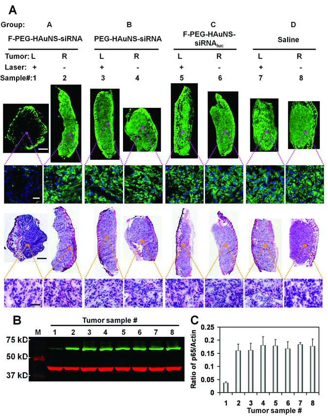
NF-kappaB transcription factor is a critical regulator of the expression of genes involved in tumor formation and progression. Successful RNA interference (RNAi) therapeutics targeting NF-kappaB is challenged by small interfering RNA (siRNA) delivery systems, which can render targeted in vivo delivery, efficient endolysosomal escape, and dynamic control over activation of RNAi. Here, we report near-IR (NIR) light-inducible NF-kappaB downregulation through folate receptor-targeted hollow gold nanospheres carrying siRNA recognizing NF-kappaB p65 subunit. Using micro-positron emission tomography/computed tomography imaging, the targeted nanoconstructs exhibited significantly higher tumor uptake in nude mice bearing HeLa cervical cancer xenografts than nontargeted nanoparticles following i.v. administration. Mediated by hollow gold nanospheres, controllable cytoplasmic delivery of siRNA was obtained on NIR light irradiation through photothermal effect. Efficient downregulation of NF-kappaB p65 was achieved only in tumors irradiated with NIR light but not in nonirradiated tumors grown in the same mice. Liver, spleen, kidney, and lung were not affected by the treatments, in spite of significant uptake of the siRNA nanoparticles in these organs. We term this mode of action "photothermal transfection." Combined treatments with p65 siRNA photothermal transfection and irinotecan caused substantially enhanced tumor apoptosis and significant tumor growth delay compared with other treatment regimens. Therefore, photothermal transfection of NF-kappaB p65 siRNA could effectively sensitize the tumor to chemotherapeutic agents. Because NIR light can penetrate the skin and be delivered with high spatiotemporal control, therapeutic RNAi may benefit from this novel transfection strategy while avoiding unwanted side effect.


Figures

Figure 1
Figure 1
Photothermal-induced siRNA release. (A) Scheme for bioconjugation of HAuNS-siRNA and photothermal-induced siRNA release. (B) Absorption spectra of the HAuNS-siRNA solution before and after NIR laser irradiation (800 nm) at different energy power levels for 60 s (Left) and at 50 mW/cm2 for different time periods (Right). (C) TEM images of uranyl acetate-stained HAuNS and HAuNS-siRNA before and after NIR laser irradiation. Lower panel shows TEM images at a higher magnification. Arrow, the integrated siRNA layer present on the surface of HAuNS. Bar, 50 nm. (D) XPS spectra of HAuNS-siRNA showing the detachment of siRNA from HAuNS after NIR laser irradiation at 50 mW/cm2 for 60s.
Figure 2
Figure 2
Intracellular trafficking of F-PEG-HAuNS-siRNA following NIR laser irradiation. (A) Scheme for the synthesis of F-PEG-HAuNS-siRNA and their proposed intracellular itinerary following NIR light irradiation. (B) Photothermal-induced endo-lysosomal escape of Dy547-labeled siRNA. Green represents LysoTracker green labeled endo-lysosomes; red represents Dy547-labeled siRNA. Bar, 10 µm. (C) TEM images of intracellular distribution of F-PEGHAuNS-siRNA with or without NIR light treatment. Arrows in upper panel indicate the nanoparticles remain attachment to the rim of endocytic vesicles. Arrows in lower panel represent indiscernible membrane boundary of the endocytic vesicles. Higher magnification images (lower panel, right three images) show some parts of the membrane of endocytic vesicles disappeared, resulting in endo-lysosmal escape of the nanoparticles as depicted by the schemes below. (D) Z-stack images showing the dissociation of siRNA from the HAuNS after laser irradiation. Red, Dy547-labeled siRNA; green, scattering signal of HAuNS; blue, cell nuclei counterstained with DAPI. Arrows, siRNA colocalized with HAuNS. Bar, 10 µm.
Figure 3
Figure 3
Photothermal transfection of F-PEG-HAuNS-siRNA and enhanced chemosensitivity to irinotecan in HeLa cells. (A) Western blot of p65 expression in HeLa cells. Left panel, p65 expression at 48 h following different transfection procedures. M, molecular weight marker; Lane 1, cells without any treatment; Lane 2, Lipofectamine 2000 plus siRNA; Lane 3, free siRNA; Lanes 4 and 5, PEG-HAuNS-siRNA and F-PEG-HAuNS-siRNAluc both plus NIR laser irradiation (50 mW/cm2 for 60 s); Lanes 6–8, F-PEG-HAuNS-siRNA plus NIR laser irradiation (50 mW/cm2) for 60, 30, 0 s; Lane 9, F-PEG-HAuNS-siRNA plus NIR laser irradiation (32 mW/cm2 for 60 s). Right panel, p65 expression at different time periods after photothermal transfection (50 mW/cm2, 60 s) with F-PEG-HAuNS-siRNA (Lanes 1, 3, 5, 7) or with F-PEG-HAuNS-siRNAluc (Lanes 2, 4, 6, 8). Green, p65; Red, β-actin. (B) Immunohistochemistry analysis of p65 expression with different transfection procedures. Laser dose: 50 mW/cm2, 60 s. Control, cells without any treatment. Red, p65; Green, cell nuclei counterstained with DAPI. Bar, 20 µm. (C) Cells preincubated with or without F-PEG-HAuNS-siRNA in the absence or presence of NIR laser (50 mW/cm2, 60 s) were treated with different concentrations of irinotecan for 24 h. Cell viability was plotted as a percentage of the non-treated cells. Mean±SD, n=3. (D) Apoptotic analysis of cells untreated or treated with F-PEG-HAuNS-siRNA or F-PEG-HAuNS-siRNAluc, NIR laser (50 mW/cm2, 60 s), in the absence or presence of irinotecan treatment (6 µM). Cells were stained with PhiPhiLux®-G1D2 (caspase 3 substrate, green), annexin V-allophycocyanin (annexin V-APC, pseudo red) and propidium iodide (PI, pseudo blue). Upper graph of each group, flow cytometry analysis with Annexin V-APC/PI. Lower graph, immunofluorescent images merged with differential interference contrast (DIC) images. Control, cells without any treatment. Bar, 25 µm.
Figure 4
Figure 4
Tumor targeting of F-PEG-HAuNS-siRNA directed at folate receptor. (A) Micro-PET/CT imaging of nude mice-bearing HeLa cervical cancer xenografts in right rear leg 6 h after i.v. injection of F-PEG-HAuNS-siRNA(DOTA-64Cu) or PEG-HAuNS-siRNA(DOTA-64Cu). Arrow, tumor. (B) Biodistribution of F-PEG-HAuNS-siRNA(DOTA-64Cu) and PEG-HAuNS-siRNA(DOTA-64Cu) 6 h following injection. Data were plotted as percentage of injected dose per gram of tissue (%ID/g). Mean ± SD (n = 5). *P < 0.01. (C) Z-stack images of tumor sections from mice-bearing PKH67-labeled HeLa xenografts 6 h after i.v. injection of Dy547-labeled F-PEG-HAuNS-siRNA or PEG-HAuNS-siRNA. Blue, cell nuclei stained with DAPI. Green, PKH67-labeled HeLa cell lipids. Red, Dy547-labeled siRNA. Arrow heads represent the colocalization of siRNA with cellular lipids, indicating the intracellular distribution of siRNA. Bar, 20 µm.
Figure 5
Figure 5
NIR light controllable site-specific p65 RNAi in HeLa xenografts. Tumor-bearing mice were randomly allocated into 4 groups and received different treatments as described in the Methods. The tumor samples were numerated from 1 to 8. L, tumor in left rear leg; R, tumor in right rear leg. Tumor samples 1 and 2 were from the same mouse in group A; samples 3 and 4 from the same mouse in group B; samples 5 and 6 from the same mouse in group C; samples 7 and 8 from the same mouse in group D. (A) Representative micrographs from each sample showing p65 expression and histology. Upper two rows, immunofluorescent staining of p65. Green, NF-κB p65 subunit. Blue, cell nuclei. Lower two rows, H&E staining. Bars: first row, 1 mm; second row, 20 µm; third row, 1 mm; fourth row, 50 µm. (B) Western blot of p65 expression in HeLa xenografts from samples 1–8. M, molecular weight marker. Green, p65; Red, β-actin. (C) Quantitative analysis of fluorescent intensities as the ratio of p65 to β-actin (n=3).
Figure 6
Figure 6
Effect of p65 siRNA photothermal transfection combined with irinotecan on nude mice-bearing HeLa cancer xenografts. Mice received various treatments as described in Methods. (A) Representative micrographic images of tumors stained with H&E and TUNEL (red). Bar for H&E, 1 mm. Bar for TUNEL, 1 mm (whole tissue); 50 µm (enlarged images). (B) Tumor size versus time curve. Mean ± SD, n = 8–10. Control, tumor-bearing mice did not receive any treatment.

References

    1. Elbashir SM, Harborth J, Lendeckel W, Yalcin A, Weber K, Tuschl T. Duplexes of 21-nucleotide RNAs mediate RNA interference in cultured mammalian cells. Nature. 2001;411:494–498. - PubMed
    1. Medina-Kauwe LK, Xie J, Hamm-Alvarez S. Intracellular trafficking of nonviral vectors. Gene Ther. 2005;12:1734–1751. - PubMed
    1. Bartlett DW, Su H, Hildebrandt IJ, Weber WA, Davis ME. Impact of tumor-specific targeting on the biodistribution and efficacy of siRNA nanoparticles measured by multimodality in vivo imaging. Proc Natl Acad Sci USA. 2007;104:15549–15554. - PMC - PubMed
    1. Thomas M, Klibanov AM. Conjugation to gold nanoparticles enhances polyethylenimine's transfer of plasmid DNA into mammalian cells. Proc Natl Acad Sci USA. 2003;100:9138–9143. - PMC - PubMed
    1. Boe S, Longva AS, Hovig E. Evaluation of various polyethylenimine formulations for light-controlled gene silencing using small interfering RNA molecules. Oligonucleotides. 2008;18:123–132. - PubMed

Publication types