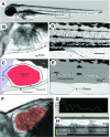
High-resolution cardiovascular function confirms functional orthology of myocardial contractility pathways in zebrafish
- PMID: 20388839
- PMCID: PMC3032279
- DOI: 10.1152/physiolgenomics.00206.2009
High-resolution cardiovascular function confirms functional orthology of myocardial contractility pathways in zebrafish
Abstract
Phenotype-driven screens in larval zebrafish have transformed our understanding of the molecular basis of cardiovascular development. Screens to define the genetic determinants of physiological phenotypes have been slow to materialize as a result of the limited number of validated in vivo assays with relevant dynamic range. To enable rigorous assessment of cardiovascular physiology in living zebrafish embryos, we developed a suite of software tools for the analysis of high-speed video microscopic images and validated these, using established cardiomyopathy models in zebrafish as well as modulation of the nitric oxide (NO) pathway. Quantitative analysis in wild-type fish exposed to NO or in a zebrafish model of dilated cardiomyopathy demonstrated that these tools detect significant differences in ventricular chamber size, ventricular performance, and aortic flow velocity in zebrafish embryos across a large dynamic range. These methods also were able to establish the effects of the classic pharmacological agents isoproterenol, ouabain, and verapamil on cardiovascular physiology in zebrafish embryos. Sequence conservation between zebrafish and mammals of key amino acids in the pharmacological targets of these agents correlated with the functional orthology of the physiological response. These data provide evidence that the quantitative evaluation of subtle physiological differences in zebrafish can be accomplished at a resolution and with a dynamic range comparable to those achieved in mammals and provides a mechanism for genetic and small-molecule dissection of functional pathways in this model organism.
Figures


Similar articles
-
Laser-scanning velocimetry: a confocal microscopy method for quantitative measurement of cardiovascular performance in zebrafish embryos and larvae.BMC Biotechnol. 2007 Jul 10;7:40. doi: 10.1186/1472-6750-7-40. BMC Biotechnol. 2007. PMID: 17623073 Free PMC article.
-
Rapid three-dimensional imaging and analysis of the beating embryonic heart reveals functional changes during development.Dev Dyn. 2006 Nov;235(11):2940-8. doi: 10.1002/dvdy.20926. Dev Dyn. 2006. PMID: 16921497
-
Cardio-respiratory control during early development in the model animal zebrafish.Acta Histochem. 2009;111(3):230-43. doi: 10.1016/j.acthis.2008.11.005. Epub 2009 Jan 3. Acta Histochem. 2009. PMID: 19121852 Review.
-
A multi-endpoint in vivo larval zebrafish (Danio rerio) model for the assessment of integrated cardiovascular function.J Pharmacol Toxicol Methods. 2014 Jan-Feb;69(1):30-8. doi: 10.1016/j.vascn.2013.10.002. Epub 2013 Oct 16. J Pharmacol Toxicol Methods. 2014. PMID: 24140389
-
In vivo biofluid dynamic imaging in the developing zebrafish.Birth Defects Res C Embryo Today. 2004 Sep;72(3):277-89. doi: 10.1002/bdrc.20019. Birth Defects Res C Embryo Today. 2004. PMID: 15495183 Review.
Cited by
-
The Role of MAPRE2 and Microtubules in Maintaining Normal Ventricular Conduction.Circ Res. 2024 Jan 5;134(1):46-59. doi: 10.1161/CIRCRESAHA.123.323231. Epub 2023 Dec 14. Circ Res. 2024. PMID: 38095085 Free PMC article.
-
Pharmacological HIF2α inhibition improves VHL disease-associated phenotypes in zebrafish model.J Clin Invest. 2015 May;125(5):1987-97. doi: 10.1172/JCI73665. Epub 2015 Apr 13. J Clin Invest. 2015. PMID: 25866969 Free PMC article.
-
Loss of zebrafish atp6v1e1b, encoding a subunit of vacuolar ATPase, recapitulates human ARCL type 2C syndrome and identifies multiple pathobiological signatures.PLoS Genet. 2021 Jun 18;17(6):e1009603. doi: 10.1371/journal.pgen.1009603. eCollection 2021 Jun. PLoS Genet. 2021. PMID: 34143769 Free PMC article.
-
Identification of a new modulator of the intercalated disc in a zebrafish model of arrhythmogenic cardiomyopathy.Sci Transl Med. 2014 Jun 11;6(240):240ra74. doi: 10.1126/scitranslmed.3008008. Sci Transl Med. 2014. PMID: 24920660 Free PMC article.
-
Zebrafish as tools for drug discovery.Nat Rev Drug Discov. 2015 Oct;14(10):721-31. doi: 10.1038/nrd4627. Epub 2015 Sep 11. Nat Rev Drug Discov. 2015. PMID: 26361349 Review.
References
-
- Ahmed M, Ishiguro M, Nagatomo T. Molecular modeling of SWR-0342SA, a beta3-selective agonist, with beta1- and beta3-adrenoceptor. Life Sci 78: 2019–2023, 2006. - PubMed
-
- Arrighi JA, Dilsizian V, Perrone-Filardi P, Diodati JG, Bacharach SL, Bonow RO. Improvement of the age-related impairment in left ventricular diastolic filling with verapamil in the normal human heart. Circulation 90: 213–219, 1994. - PubMed
-
- Bagatto B, Burggren W. A three-dimensional functional assessment of heart and vessel development in the larva of the zebrafish (Danio rerio). Physiol Biochem Zool 79: 194–201, 2006. - PubMed
-
- Behr B, Hoffmann C, Ottolina G, Klotz KN. Novel mutants of the human beta1-adrenergic receptor reveal amino acids relevant for receptor activation. J Biol Chem 281: 18120–18125, 2006. - PubMed
Publication types
MeSH terms
Grants and funding
LinkOut - more resources
Full Text Sources
Molecular Biology Databases

