. 2010 Apr 13;3(117):ra29.
doi: 10.1126/scisignal.2000594.

Identification of the miR-106b~25 microRNA cluster as a proto-oncogenic PTEN-targeting intron that cooperates with its host gene MCM7 in transformation

Affiliations

Identification of the miR-106b~25 microRNA cluster as a proto-oncogenic PTEN-targeting intron that cooperates with its host gene MCM7 in transformation

Laura Poliseno et al. Sci Signal. .

Erratum in

  • Sci Signal. 2010;3(123):er6

Abstract

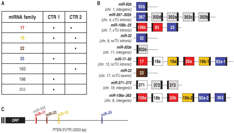
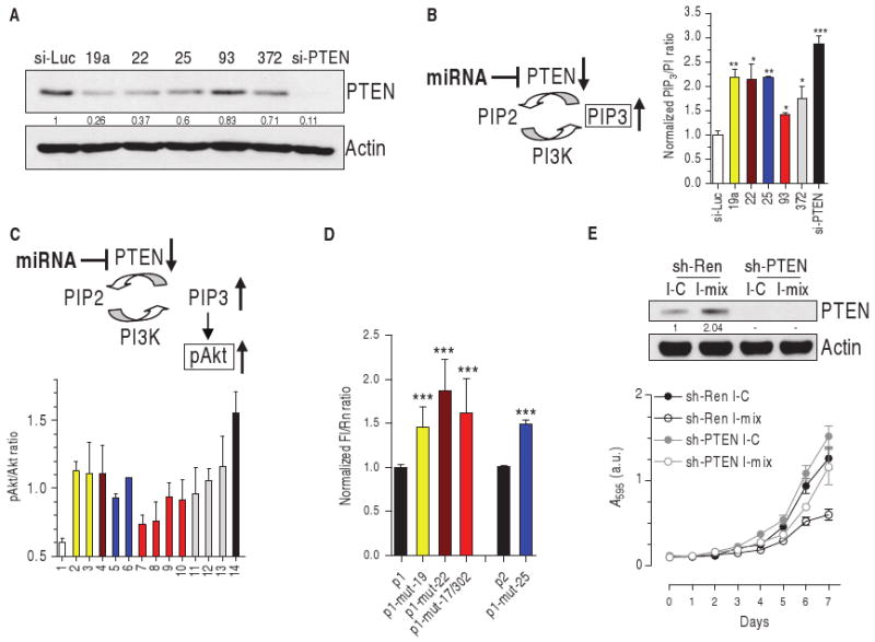
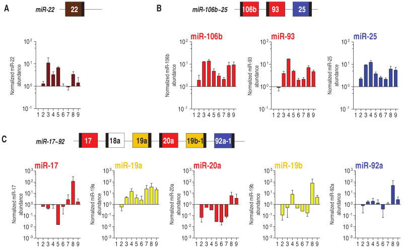
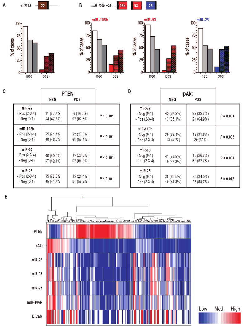
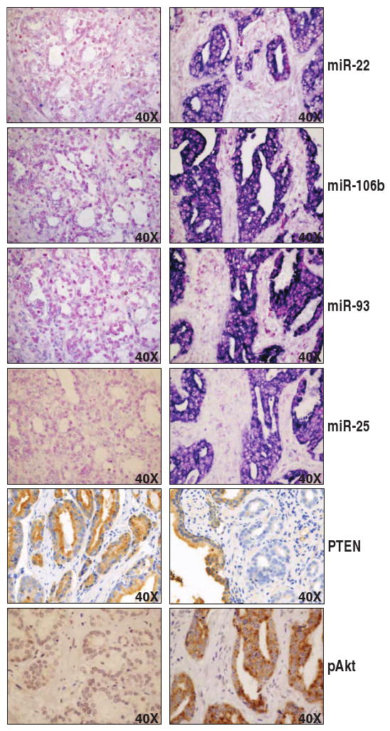
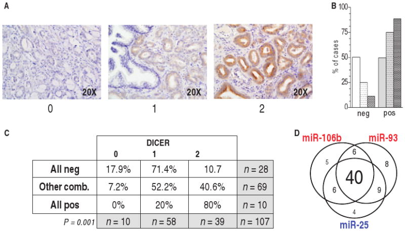
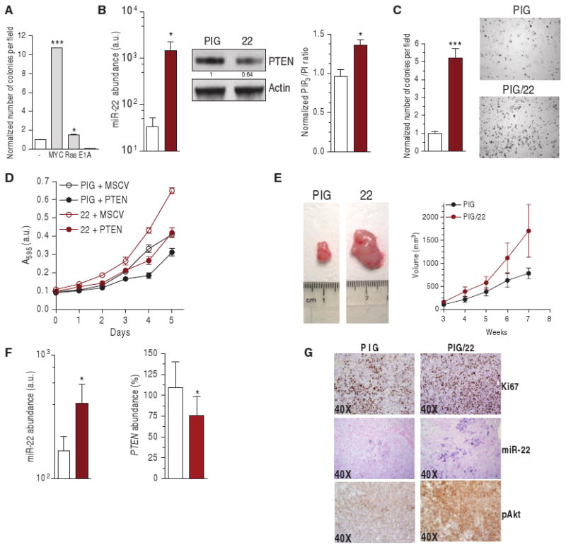
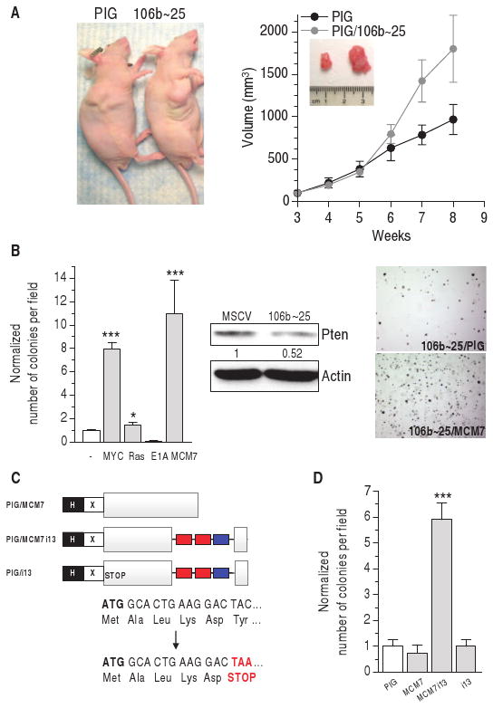
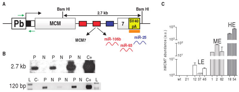
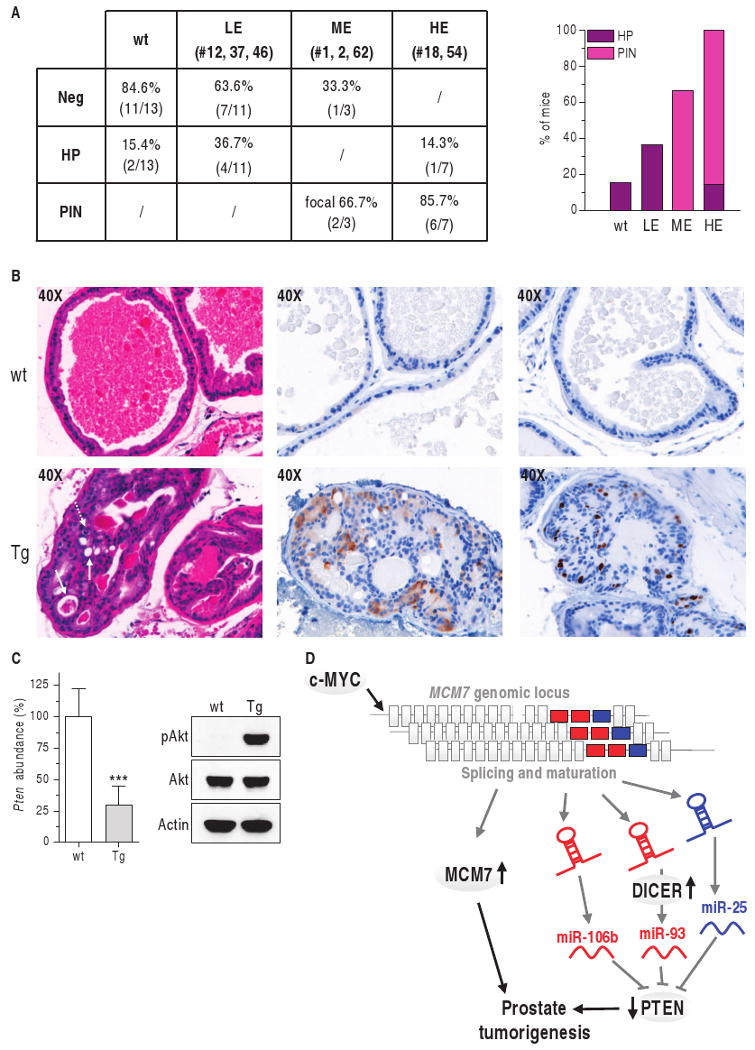
PTEN (phosphatase and tensin homolog deleted on chromosome 10) is a tumor suppressor that antagonizes signaling through the phosphatidylinositol 3-kinase-Akt pathway. We have demonstrated that subtle decreases in PTEN abundance can have critical consequences for tumorigenesis. Here, we used a computational approach to identify miR-22, miR-25, and miR-302 as three PTEN-targeting microRNA (miRNA) families found within nine genomic loci. We showed that miR-22 and the miR-106b~25 cluster are aberrantly overexpressed in human prostate cancer, correlate with abundance of the miRNA processing enzyme DICER, and potentiate cellular transformation both in vitro and in vivo. We demonstrated that the intronic miR-106b~25 cluster cooperates with its host gene MCM7 in cellular transformation both in vitro and in vivo, so that the concomitant overexpression of MCM7 and the miRNA cluster triggers prostatic intraepithelial neoplasia in transgenic mice. Therefore, the MCM7 gene locus delivers two simultaneous oncogenic insults when amplified or overexpressed in human cancer. Thus, we have uncovered a proto-oncogenic miRNA-dependent network for PTEN regulation and defined the MCM7 locus as a critical factor in initiating prostate tumorigenesis.


Conflict of interest statement

Competing interests: The authors declare that they do not have any competing financial, personal, or professional interest.

Figures

Fig. 1
Fig. 1
PTEN-targeting miR-17, 19, 22, 25, and 302 families. (A) List of the seven miRNA families that passed either of our selection criteria (CTR) (table S2). (B) Genomic organization of miRNAs belonging to miR-17 (red), miR-19 (yellow), miR-22 (brown), miR-25 (blue), and miR-302 (gray) families. miRNAs that do not target PTEN are in white. Large boxes, pre-miRNAs; small black boxes, mature miRNAs. The miRNA clusters are named with the most upstream and the most downstream miRNA connected by a tilde (∼) (54). The chromosomes (chr.) in which the miRNA genes are located are indicated below the names. It is also specified whether the miRNA genes are independent TUs (intergenic) or are located in introns or exons of coding or noncoding TUs. (C) Schematic representation of the seed matches of miR-17, 19, 22, and 25 (TargetScan) and 302 (PicTar) families along the PTEN 3′UTR. The miR-17 and miR-302 families overlap in binding to the six-nucleotide oligomer Alu 5′-GCACTT-3′ motif (55). ORF, open reading frame.
Fig. 2
Fig. 2
miR-17, 19, 22, 25, and 302 families decrease PTEN abundance and activate the Akt pathway. (A) Western blot of DU145 cells transiently transfected with control si-Luc, the indicated si-miRNAs, or the siRNA directed against human PTEN (si-PTEN). Quantification of PTEN protein is reported. (B) PIP3 detection in DU145 cells transiently transfected as above, serum-starved, and stimulated with 200 nM insulin for 5 min. (C) pAkt/Akt ratio in PWR-1E cells after the transient transfection of 1, si-Luc (white); 2 to 13, si-miRNAs: 19a, 19b (yellow, miR-19 family), 22 (brown), 25, 92a (blue, miR-25 family), 17, 20a, 93, 106b (red, miR-17 family), 302a, 372, 373 (gray, miR-302 family); 14, si-PTEN (black). (D) Wild-type or mutant p1 and p2 reporter plasmids (fig. S1, B and C) were transfected into DU145 cells. Twenty-four hours later, the luciferase activity of the mutant plasmids was higher than that of the corresponding wild-type plasmids, indicating that the introduced mutations in the seed matches impair miRNA binding to PTEN 3′UTR. ***P < 0.001. (E) DU145 cells were stably infected with a retroviral vector expressing a control shRNA directed against Renilla luciferase (sh-Ren) or an shRNA directed against human PTEN (sh-PTEN) and then transiently transfected with a control miRNA inhibitor (I-C) or a mix of the inhibitors against miR-19a/22/25/93/106b (I-mix). Top: Western blot showing PTEN 96 hours after transfection. Bottom: growth curve. See text for details.
Fig. 3
Fig. 3
Presence of miR-22, miR-106b∼25, and miR-17∼92 PTEN-targeting miRNAs in human prostate cell lines. Mature miRNAs [miR-22 (A), miR-106b, miR-93, miR-25 (B), miR-17, miR-19a, miR-20a, miR-19b, and miR-92a (C)] were detected by real-time PCR analysis in the cell lines derived from the following (numbered 1 to 9 in order): RWPE-1 (normal prostate epithelium immortalized), PWR-1E (normal prostate epithelium immortalized), Ca-HpV-10 (primary prostate carcinoma), 22Rv1 (xenograft of a primary carcinoma), DU145, LnCaP, MDA-PCa-2b, PC3, and VCap (prostate carcinoma metastasized to distal organs). The ratio of the miRNA being tested to that of RNU24 (internal standard) in RWPE-1 was taken as 1. miR-302 family members belonging to the miR-371∼373 and miR-367∼302b clusters (Fig. 1B) were not detectable in all the cell lines analyzed.
Fig. 4
Fig. 4
Presence of miR-22 and miR-106b∼25 miRNAs in human prostate tumor samples. (A and B) Score of the fractional presence of miR-22 (A) and miR-106b, miR-93 and miR-25 (B) in peritumoral tissue (left bars), PIN (middle bars), and prostate cancer (right bars), as detected by ISH. 0 to 1, negative (neg); 2 to 4: positive (pos). (C) Correlation between the lack of PTEN and the presence of miR-22 and miR-106b∼25 cluster miRNAs. (D) Correlation between the presence of pAkt and that of miR-22 and miR-106b∼25 cluster miRNAs. (E) Heat map of PTEN, pAkt, DICER, and miR-22/25/93/106b status in tumor samples. The heat map shows an inverse correlation between PTEN abundance and that of DICER, the miRNAs, and pAkt. The heat map was generated with TIBCO Spotfire Metrics (version 3.0) software.
Fig. 5
Fig. 5
Representative pictures of the prostate TMA. A tumor specimen negative for miRNAs and pAkt and positive for PTEN (left) and a tumor specimen positive for miRNAs and pAkt and negative for PTEN (right) are shown.
Fig. 6
Fig. 6
Correlation of DICER abundance in prostate tumor samples with that of miR-22 and miR-106b∼25 miRNAs. (A) TMA samples were considered negative (score, 0) or positive (score, 1 to 2) for DICER by IHC. (B) Presence of DICER in peritumoral tissues (left bars), PIN (middle bars), and prostate cancer (right bars). 0, negative; 1 to 2, positive. (C) Correlation between DICER presence and that of miR-22/miR-25/miR-93/miR-106b. (D) Venn diagram of tumor samples positive for one, two, or all three miRNAs in the miR-106b25 cluster. In most of the cases, the three miRNAs of the cluster were co-overexpressed.
Fig. 7
Fig. 7
miR-22 proto-oncogenic activity. (A) Transformation assay performed in wild-type MEF by co-infecting MSCV/miR-22 and PIG, PIG/c-MYC, PIG/RasV12, or PIG/E1A. The number of colonies formed by MEFs double–infected with PIG and MSCV empty plasmids is taken as 1. (B) Abundance of mature miR-22 (left, real-time PCR), PTEN abundance (middle, Western blot) and PIP3 production (right) in stably infected DU145 cells. *P < 0.05. (C) Number of colonies formed in soft agar. ***P < 0.001. (D) Growth curve of DU145 cells stably infected with PIG/22 and MSCV-neo-Flag-PTEN plasmids. PTEN encoded by MSCV-neo-Flag-PTEN contains the entire PTEN open reading frame, but lacks the 3′UTR. (E) Representative tumor (left) and increase in tumor volume (right) formed after subcutaneous injection into nude mice (n = 6). (F and G) Analysis of the tumors: mature miR-22 (F, left) and PTEN mRNA (F, right) detection by real-time PCR. *P < 0.05. (G) IHC staining of the proliferation marker Ki67; ISH of miR-22; IHC of pAkt. (B, C, and F) White bars, PIG; brown bars, PIG/22.
Fig. 8
Fig. 8
miR-106b∼25 cluster proto-oncogenic activity. (A) Representative tumor (left) and increase in tumor volume (right) after subcutaneous injection into nude mice of DU145 stably infected with the miR-106b25 cluster (n = 6). (B) Left: transformation assay performed in wt MEF by co-infecting MSCV/miR-106b∼25 and PIG empty, PIG/c-MYC, PIG/RasV12, PIG/E1A, or PIG/MCM7 plasmids. The number of colonies formed by MEFs double-infected with PIG and MSCV empty plasmids is taken as 1. Center: decrease in Pten abundance caused by the cluster as measured by Western blot. Right: representative fields of miR-106b∼25/PIG and miR-106b∼25/MCM7 colonies. ***P < 0.001. (C) Schematic representation of the PIG/MCM7, PIG/MCM7i13, and PIG/i13 plasmids. PIG/MCM7 plasmid expresses only MCM7; PIG/MCM7i13 expresses both MCM7 and the three miRNAs belonging to the miR-106b25 cluster, as a result of the splicing and maturation of the recombinant intron 13; PIG/i13 is derived from PIG/MCM7i13 by insertion of a point mutation that transforms the sixth amino acid of MCM7 (TAC, Tyr) into a stop codon (TAA, STOP). Therefore, this plasmid expresses only the intronic miRNAs. H, HA tag; X, Xpress tag; gray box, MCM7 coding sequence; red/blue boxes, miR-106b/93 and miR-25 genes. (D) Transformation assay performed in wt MEFs with the plasmids shown in (C). ***P < 0.001.
Fig. 9
Fig. 9
Pb/MCM7i13 transgenic construct. (A) Pb, prostate-specific rat probasin promoter ARR2PB; black box, HA tag; white box, Xpress tag; gray boxes, human MCM7 coding sequence; red and blue boxes, miR-93/106b and miR-25 genes; yellow box, SV40 poly(A) (SV40 pA); orange box, probe used for Southern analysis of transgenic lines; green arrows, primers used for PCR analysis of transgenic lines and for genotyping. (B) Top: Southern blot analysis. Genomic DNA extracted from tails was digested with Bam HI and hybridized with a probe complementary to the SV40 poly(A) region as shown in (A). The Bam HI–digested Pb/MCM7i13 plasmid was used as positive control (C+). Bottom: PCR analysis. A forward primer complementary to the probasin promoter and a reverse primer complementary to the tag were used, as shown in (A). Empty Pb and Pb/MCM7i13 plasmids were used as negative (C−) and positive (C+) controls. L, 100-bp ladder. Three representative positive (P) and negative (N) mice are shown. (C) Abundance of the transgenic construct measured by real-time PCR in nine different lines. A primer pair specific for human MCM7 coding sequence was used. LE, ME, HE: low, medium, and high expressors, respectively. Human MCM7 was not detectable in wild-type (wt) mice, confirming the specificity of the primers. See also fig. S9A.
Fig. 10
Fig. 10
The MCM7 locus can initiate prostate tumorigenesis. (A) Incidence of hyperplasia (HP) and PIN in the DLP of 1-year-old transgenic mice. (B) Analysis of 1-year-old transgenic mice belonging to HE line 18. H&E staining (left) shows PIN in the DLP, as indicated by loss of cell polarity, abnormal proliferation, presence of mitotic figures (dotted arrow), and intraepithelial lumen formation (straight arrows). PIN glands are characterized by aberrant pS6 phosphorylation (middle, brown staining) and by an increased number of Ki67-positive cells (right, brown staining). (C) Decreased Pten mRNA (real-time PCR) is associated with increased Akt phosphorylation (Western blot). ***P < 0.001. (D) Model of MCM7 locus oncogenicity in prostate cancer. The MCM7 locus contains two oncogenic and cooperating elements: MCM7 (gray) and the PTEN-targeting miR-106b∼25 miRNAs in intron 13. This locus undergoes genomic amplification in prostate cancer. c-MYC increases transcription of both MCM7 and the miR-106b∼25 miRNAs and increased DICER abundance leads to increased miRNA maturation.

References

    1. Nomenclature: PTEN, human protein; PTEN, human mRNA and gene; Pten, mouse protein; Pten, mouse mRNA and gene; DICER, human protein; DICER, human mRNA or gene; MCM7, human protein; MCM7, human mRNA and gene; miR-X, mature miRNA; miR-X, miRNA gene.

    1. Kim VN, Han J, Siomi MC. Biogenesis of small RNAs in animals. Nat Rev Mol Cell Biol. 2009;10:126–139. - PubMed
    1. Kumar MS, Lu J, Mercer KL, Golub TR, Jacks T. Impaired microRNA processing enhances cellular transformation and tumorigenesis. Nat Genet. 2007;39:673–677. - PubMed
    1. Chiosea S, Jelezcova E, Chandran U, Acquafondata M, McHale T, Sobol RW, Dhir R. Up-regulation of Dicer, a component of the microRNA machinery, in prostate adenocarcinoma. Am J Pathol. 2006;169:1812–1820. - PMC - PubMed
    1. Baskerville S, Bartel DP. Microarray profiling of microRNAs reveals frequent coexpression with neighboring miRNAs and host genes. RNA. 2005;11:241–247. - PMC - PubMed

Publication types