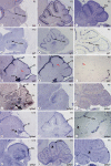
Differential modulation of Sonic-hedgehog-induced cerebellar granule cell precursor proliferation by the IGF signaling network
- PMID: 20389077
- PMCID: PMC2866582
- DOI: 10.1159/000274458
Differential modulation of Sonic-hedgehog-induced cerebellar granule cell precursor proliferation by the IGF signaling network
Abstract
The molecular mechanisms regulating organ growth and size remain unclear. Sonic hedgehog (SHH) signaling is a major player in the regulation of cerebellar development: SHH is secreted by Purkinje neurons and acts on the proliferation of granule cell precursors (GCPs) in the external germinal layer. These then become postmitotic and form the internal granular layer but do so in the presence of SHH ligand, begging the question of how the proliferative response to SHH signaling is downregulated in differentiating GCPs. Here, we have determined the precise cellular localization of the expression of insulin-like growth factor (IGF) network components in the developing mouse cerebellum and show that this network modulates the proliferative effects of SHH signaling on GCPs. IGF1 and IGF2 are potent mitogens for GCPs and both synergize with SHH in inducing GCP proliferation. Whereas the proliferative activity of IGF1 or IGF2 on GCPs does not require intact SHH signaling, aspects of SHH activity on GCP proliferation require signaling through the IGF receptor 1. Moreover, we find that 3 of the IGF-binding proteins, IGFBP2, IGFBP3 and IGFBP5, inhibit IGF1/2-induced cell proliferation, whereas IGFBP5 also inhibits SHH-induced GCPs proliferation. This novel function of IGFBP5 that we have uncovered demonstrates the exquisite regulation of SHH signaling by different components of the IGF network.
Copyright 2010 S. Karger AG, Basel.
Figures


Similar articles
-
Insulin receptor substrate 1 is an effector of sonic hedgehog mitogenic signaling in cerebellar neural precursors.Development. 2008 Oct;135(19):3291-300. doi: 10.1242/dev.022871. Epub 2008 Aug 28. Development. 2008. PMID: 18755774 Free PMC article.
-
Pituitary adenylate cyclase-activating polypeptide and sonic hedgehog interact to control cerebellar granule precursor cell proliferation.J Neurosci. 2002 Nov 1;22(21):9244-54. doi: 10.1523/JNEUROSCI.22-21-09244.2002. J Neurosci. 2002. PMID: 12417650 Free PMC article.
-
The effect of cyclic phosphatidic acid on the proliferation and differentiation of mouse cerebellar granule precursor cells during cerebellar development.Brain Res. 2015 Jul 21;1614:28-37. doi: 10.1016/j.brainres.2015.04.013. Epub 2015 Apr 17. Brain Res. 2015. PMID: 25896936
-
Sonic hedgehog patterning during cerebellar development.Cell Mol Life Sci. 2016 Jan;73(2):291-303. doi: 10.1007/s00018-015-2065-1. Epub 2015 Oct 24. Cell Mol Life Sci. 2016. PMID: 26499980 Free PMC article. Review.
-
Caught in the matrix: how vitronectin controls neuronal differentiation.Trends Neurosci. 2001 Dec;24(12):680-2. doi: 10.1016/s0166-2236(00)02058-0. Trends Neurosci. 2001. PMID: 11718852 Review.
Cited by
-
Design, synthesis, and biological evaluation of estrone-derived hedgehog signaling inhibitors.Tetrahedron. 2011 Dec 30;67(52):10261-10266. doi: 10.1016/j.tet.2011.10.028. Tetrahedron. 2011. PMID: 22199406 Free PMC article.
-
microRNA-9 and -29a regulate the progression of diabetic peripheral neuropathy via ISL1-mediated sonic hedgehog signaling pathway.Aging (Albany NY). 2020 Jun 16;12(12):11446-11465. doi: 10.18632/aging.103230. Epub 2020 Jun 16. Aging (Albany NY). 2020. PMID: 32544883 Free PMC article.
-
Identification of a neuronal transcription factor network involved in medulloblastoma development.Acta Neuropathol Commun. 2013 Jul 11;1:35. doi: 10.1186/2051-5960-1-35. Acta Neuropathol Commun. 2013. PMID: 24252690 Free PMC article.
-
Requirement of Smurf-mediated endocytosis of Patched1 in sonic hedgehog signal reception.Elife. 2014 Jun 12;3:e02555. doi: 10.7554/eLife.02555. Elife. 2014. PMID: 24925320 Free PMC article.
-
Delineating the factors and cellular mechanisms involved in the survival of cerebellar granule neurons.Cerebellum. 2015 Jun;14(3):354-9. doi: 10.1007/s12311-015-0646-z. Cerebellum. 2015. PMID: 25596943 Review.
References
-
- Goldowitz D, Hamre K. The cells and molecules that make a cerebellum. Trends Neurosci. 1998;21:375–382. - PubMed
-
- Hatten ME, Heintz N. Mechanisms of neural patterning and specification in the developing cerebellum. Annu Rev Neurosci. 1995;18:385–408. - PubMed
-
- Sotelo C. Cellular and genetic regulation of the development of the cerebellar system. Prog Neurobiol. 2004;72:295–339. - PubMed
-
- Wang VY, Zoghbi HY. Genetic regulation of cerebellar development. Nat Rev Neurosci. 2001;2:484–491. - PubMed
-
- Smeyne RJ, Chu T, Lewin A, Bian F, Sanlioglu SC, Kunsch C, Lira SA, Oberdick J. Local control of granule cell generation by cerebellar Purkinje cells. Mol Cell Neurosci. 1995;6:230–251. - PubMed
Publication types
MeSH terms
Substances
Grants and funding
LinkOut - more resources
Full Text Sources
Other Literature Sources
Miscellaneous

