

Gene correction by homologous recombination with zinc finger nucleases in primary cells from a mouse model of a generic recessive genetic disease
- PMID: 20389291
- PMCID: PMC2889743
- DOI: 10.1038/mt.2010.57
Gene correction by homologous recombination with zinc finger nucleases in primary cells from a mouse model of a generic recessive genetic disease
Abstract
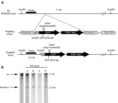
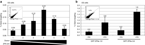
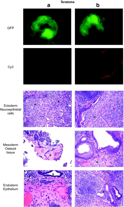
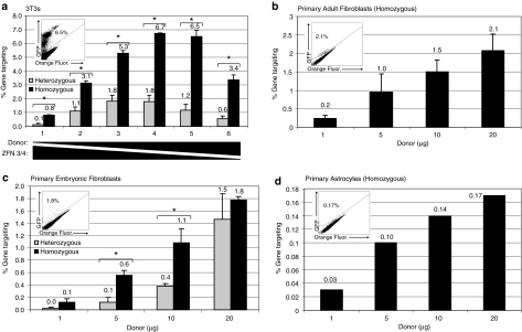
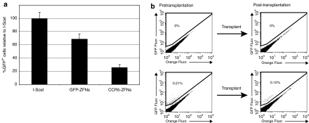
Zinc Finger nucleases (ZFNs) have been used to create precise genome modifications at frequencies that might be therapeutically useful in gene therapy. We created a mouse model of a generic recessive genetic disease to establish a preclinical system to develop the use of ZFN-mediated gene correction for gene therapy. We knocked a mutated GFP gene into the ROSA26 locus in murine embryonic stem (ES) cells and used these cells to create a transgenic mouse. We used ZFNs to determine the frequency of gene correction by gene targeting in different primary cells from this model. We achieved targeting frequencies from 0.17 to 6% in different cell types, including primary fibroblasts and astrocytes. We demonstrate that ex vivo gene-corrected fibroblasts can be transplanted back into a mouse where they retained the corrected phenotype. In addition, we achieved targeting frequencies of over 1% in ES cells, and the targeted ES cells retained the ability to differentiate into cell types from all three germline lineages. In summary, potentially therapeutically relevant frequencies of ZFN-mediated gene targeting can be achieved in a variety of primary cells and these cells can then be transplanted back into a recipient.
Figures
Similar articles
-
Gene targeting to the ROSA26 locus directed by engineered zinc finger nucleases.Nucleic Acids Res. 2012 Apr;40(8):3741-52. doi: 10.1093/nar/gkr1214. Epub 2011 Dec 14. Nucleic Acids Res. 2012. PMID: 22169954 Free PMC article.
-
Gene targeting by homologous recombination in mouse zygotes mediated by zinc-finger nucleases.Proc Natl Acad Sci U S A. 2010 Aug 24;107(34):15022-6. doi: 10.1073/pnas.1009424107. Epub 2010 Aug 4. Proc Natl Acad Sci U S A. 2010. PMID: 20686113 Free PMC article.
-
Gene targeting of a disease-related gene in human induced pluripotent stem and embryonic stem cells.Cell Stem Cell. 2009 Jul 2;5(1):97-110. doi: 10.1016/j.stem.2009.05.023. Epub 2009 Jun 18. Cell Stem Cell. 2009. PMID: 19540188 Free PMC article.
-
Gene targeting using zinc finger nucleases.Nat Biotechnol. 2005 Aug;23(8):967-73. doi: 10.1038/nbt1125. Nat Biotechnol. 2005. PMID: 16082368 Review.
-
Zinc finger nucleases: custom-designed molecular scissors for genome engineering of plant and mammalian cells.Nucleic Acids Res. 2005 Oct 26;33(18):5978-90. doi: 10.1093/nar/gki912. Print 2005. Nucleic Acids Res. 2005. PMID: 16251401 Free PMC article. Review.
Cited by
-
Zinc-finger nuclease-mediated gene correction using single AAV vector transduction and enhancement by Food and Drug Administration-approved drugs.Gene Ther. 2013 Jan;20(1):35-42. doi: 10.1038/gt.2011.211. Epub 2012 Jan 19. Gene Ther. 2013. PMID: 22257934 Free PMC article.
-
A survey of ex vivo/in vitro transduction efficiency of mammalian primary cells and cell lines with Nine natural adeno-associated virus (AAV1-9) and one engineered adeno-associated virus serotype.Virol J. 2013 Mar 6;10:74. doi: 10.1186/1743-422X-10-74. Virol J. 2013. PMID: 23497173 Free PMC article.
-
Gene knockout and knockin by zinc-finger nucleases: current status and perspectives.Cell Mol Life Sci. 2013 Aug;70(16):2969-83. doi: 10.1007/s00018-012-1204-1. Epub 2012 Nov 17. Cell Mol Life Sci. 2013. PMID: 23161061 Free PMC article. Review.
-
Current technology in the diagnosis of developmentally related lung disorders.Neonatology. 2012;101(4):353-9. doi: 10.1159/000337356. Epub 2012 Jun 1. Neonatology. 2012. PMID: 22940625 Free PMC article. Review.
-
Zinc-finger nuclease mediated disruption of Rag1 in the LEW/Ztm rat.BMC Immunol. 2012 Nov 8;13:60. doi: 10.1186/1471-2172-13-60. BMC Immunol. 2012. PMID: 23136839 Free PMC article.
References
-
- Hacein-Bey-Abina S, Le Deist F, Carlier F, Bouneaud C, Hue C, De Villartay JP, et al. Sustained correction of X-linked severe combined immunodeficiency by ex vivo gene therapy. N Engl J Med. 2002;346:1185–1193. - PubMed
-
- Aiuti A, Cattaneo F, Galimberti S, Benninghoff U, Cassani B, Callegaro L, et al. Gene therapy for immunodeficiency due to adenosine deaminase deficiency. N Engl J Med. 2009;360:447–458. - PubMed
-
- Bainbridge JW, Smith AJ, Barker SS, Robbie S, Henderson R, Balaggan K, et al. Effect of gene therapy on visual function in Leber's congenital amaurosis. N Engl J Med. 2008;358:2231–2239. - PubMed
-
- Cartier N, Hacein-Bey-Abina S, Bartholomae CC, Veres G, Schmidt M, Kutschera I, et al. Hematopoietic stem cell gene therapy with a lentiviral vector in X-linked adrenoleukodystrophy. Science. 2009;326:818–823. - PubMed
Publication types
MeSH terms
Substances
Grants and funding
LinkOut - more resources
Full Text Sources
Other Literature Sources
Medical
