Review
. 2010 May;11(5):301-15.
doi: 10.1038/nrn2834. Epub 2010 Apr 14.

Emerging roles for G protein-gated inwardly rectifying potassium (GIRK) channels in health and disease

Affiliations
Review

Emerging roles for G protein-gated inwardly rectifying potassium (GIRK) channels in health and disease

Christian Lüscher et al. Nat Rev Neurosci. 2010 May.

Abstract

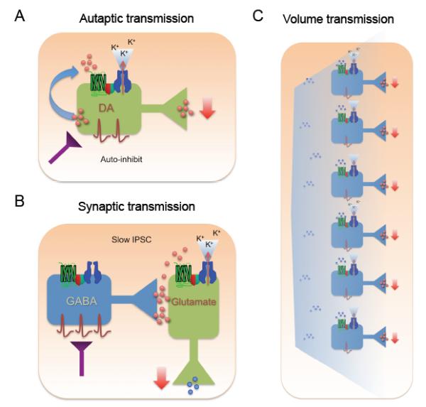
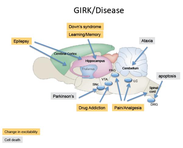
G protein-gated inwardly rectifying potassium (GIRK) channels hyperpolarize neurons in response to activation of many different G protein-coupled receptors and thus control the excitability of neurons through GIRK-mediated self-inhibition, slow synaptic potentials and volume transmission. GIRK channel function and trafficking are highly dependent on the channel subunit composition. Pharmacological investigations of GIRK channels and studies in animal models suggest that GIRK activity has an important role in physiological responses, including pain perception and memory modulation. Moreover, abnormal GIRK function has been implicated in altering neuronal excitability and cell death, which may be important in the pathophysiology of diseases such as epilepsy, Down's syndrome, Parkinson's disease and drug addiction. GIRK channels may therefore prove to be a valuable new therapeutic target.


Figures

Figure 1
Figure 1. Physiology of GIRK channels in the mammalian brain
A, Hypothetical example of inwardly rectifying potassium currents through GIRK channels. Current (I) is plotted as a function of voltage (V) (I-V plot). With physiological levels of extracellular K, the current reverses (zero current potential) near −90 mV at the equilibrium potential for K (EK). The basal current (red) and agonist-induced (blue) currents show inward rectification (large inward (−) and small outward current (+)). The outward flow of current near the resting potential of the cell (large arrow) hyperpolarizes the neuron’s membrane potential. With higher extracellular potassium (e.g. 20 mM KCl), a concentration commonly used to study GIRK channels, the I-V relation shifts to the right to the new EK (dashed line), demonstrating a universal property of inwardly rectifying K channels. B, Three primary GIRK subunits exist in brain and form heterotetramers (GIRK1-GIRK2, GIRK2-GIRK3, GIRK1-GIRK3) and homotetramers (GIRK2-GIRK2),-. Less is known about GIRK4 in the brain. C, Generation of the slow IPSC. Two levels of synaptic activity are shown. Low stimulation of a GABA-ergic neuron releases GABA at levels sufficient to only activate GABAA channels (left panel). Higher stimulation (right panel) releases more GABA that accumulates in the synaptic cleft and diffuses to neighboring GABAB/GIRK complexes, located on the shaft and dendritic spine-. The resulting hypothetical fast and slow IPSCs from these two scenarios are shown below. D, Electrophysiological recording of the fast IPSC (GABAA) and slow IPSC (GABAB-GIRK) from a hippocampal slice (taken with permission from ref119). The relationship between the GABAA and GABAB response is non-linear (A2), suggesting accumulation of GABA is needed to activate GABAB receptors. E, The GABAB receptor dependent sIPSC is absent in the hippocampus of mice lacking GIRK2 channels (taken with permission from ref27).
Figure 2
Figure 2. Structural insights into gating and the formation of a macromolecular GIRK signaling complex
A, A structure (ribbon model superimposed with space filled model) of the Kirbac1.3/GIRK1 chimeric channel is shown in a typical lipid bilayer (‘out’: extracellular) This structure contains the cytoplasmic domains (N- and C-termini) of GIRK1, and the transmembrane domains (M1, M2) and pore region of the bacterial inward rectifier (Kirbac1.3) channel (PDB:2QKS). The structure of the GIRK1 cytoplasmic domain in this full-length chimeric protein is similar to those of other inward rectifiers -. The K+ selectivity filter is the site of the weaver mutation,,. The regions implicated in Na+ and PIP2 association are shown-,,. Two channel gates formed by the M2 transmembrane domain and cytoplasmic G-loop form a physical barrier to ion permeation,,,-. Note the constriction formed by the G-loop gate. B, Modulation sites are shown from a side-view perspective in two adjacent subunits. The regions implicated in Gβγ activation (light and dark green β-strands; H69, L273 and L344: Yellow),-, Gαi/o association (red and pink β-strands, G318, C321 and A323 - blue),,, and ethanol-dependent activation (N-terminus:blue; βD-βE sheet: yellow; βL-βM sheet: green; L257:red) are shown. The sites are mapped on structure of GIRK2 (PDB:2E4F) C), Schematic shows a macromolecular signaling complex that contains a GPCR, which couples to PTX-sensitive Gαi/o G proteins, a GIRK channel, an RGS protein, SNX27, and possibly PSD95. Gβγ opens GIRK channels (+). Modulators of GIRK channels are also shown, including tyrosine kinase, CaMKII kinase, PKA/PKC kinases, and PP1 phosphatase-,,,. A second GPCR that couples to the Gq pathway is shown. Activation of this pathway stimulates PLC, which leads to activation of PKC, and depletion of PIP2, both of which reduce (−) GIRK channel activity,,. Pertussis toxin (PTX) inhibits (−) activation of GIRK channels through Gαi/o G proteins.
Figure 3
Figure 3. Three types of neuronal signaling pathways for GIRK channels in the brain
A) In autaptic synapses, neurotransmitters are released by and bind to the same neuron, stimulating GPCRs and activating GIRK channels. The net effect of such an autapic synapse is a reduction in the release of neurotransmitter (negative feedback). B) The slow IPSC. High stimulation triggers release of GABA that spills over to neighboring GABAB /GIRK complexes, located on the dendritic spine and shaft. C) Network modulation. GIRK channels involved in volume transmission. When a neurotransmitter is released from many neurons, the ambient concentration rises and diffuses to activate GIRK channels on target neurons. This leads to reduced network activity.
Figure 4
Figure 4. GIRK channels are implicated in different disease states
Schematic shows saggital view of a rat brain highlighting regions where GIRK channels have been implicated in brain neuronal disorders or diseases due to changes in excitability (yellow) or cell death (grey). Changes in GIRK activity throughout the brain may contribute to: Epilepsy: GIRK2 knockout mice develop spontaneous convulsions and show a propensity for generalized seizures. Similar pro-convulsive effects are observed with GIRK channel inhibitor tertiapin when administered intrathecally.]. Down’s Syndrome & Learning and Memory: Two mouse models for Down’s syndrome, the Ts65Dn and the Ts1Cje DS, contain a gene duplication for Girk2 and show larger slow IPSC mediated by GABAB receptors, impairments in LTP and enhancement of LTD151. Although all neurons are affected, this phenotype may involve mostly hippocampal functions. Drug Addiction: GIRK knockouts show impairment in self-administration of cocaine, altered response to GHB, less severe withdrawal from sedatives and reduced conditioned place taste aversion for ethanol. Changes in the function of the mesolimbic system, involving the VTA, are thought to underlie these changes. Pain: Removing GIRK2/GIRK3 reduces the potency (coupling efficiency), but leaves efficacy (maximal response) of opioid-analgesia intact,, effects most likely involving the peri-acqueductal grey and spinal cord. Chronic neuropathic pain can lead to tyrosine phosphorylation of GIRK1 subunits, which reduces basal GIRK activity. Ataxia/Parkinson’s: In the weaver mouse, GIRK2 channels contain a mutation that eliminates K+ selectivity and leads to degeneration of midbrain SNc DA neurons and cerebellar granular neurons. The gain-of-function phenotype in DA neurons is of clinical interest due to its similarity to the degeneration in Parkinson’s disease. In dorsal root ganglion (DRG) cells, activation of the neurotrophin receptor p75 NTR increases levels of PIP2, which activates GIRK2 channels and signals to promote programmed cell death via K+ efflux-induced apoptosis.

References

    1. Trevelyan AJ, Watkinson O. Does inhibition balance excitation in neocortex? Progress in Biophysics and Molecular Biology. 2005;87:109–143. - PubMed
    1. Lujan R, Maylie J, Adelman JP. New sites of action for GIRK and SK channels. Nat Rev Neurosci. 2009;10:475–480. - PubMed
    1. Yamada M, Inanobe A, Kurachi Y. G protein regulation of potassium ion channels. Pharmacol Rev. 1998;50:723–757. - PubMed
    1. Pfaffinger PJ, Martin JM, Hunter DD, Nathanson NM, Hille B. GTP-binding proteins couple cardiac muscarinic receptors to a K channel. Nature. 1985;317:536–538. - PubMed
    1. Logothetis DE, Kurachi Y, Galper J, Neer EJ, Clapham DE. The βγ subunits of GTP-binding proteins activate the muscarinic K+ channel in heart. Nature. 1987;325:321–326. - PubMed

Publication types

MeSH terms

Substances

LinkOut - more resources