

Altered TGFbeta signaling and cardiovascular manifestations in patients with autosomal recessive cutis laxa type I caused by fibulin-4 deficiency
- PMID: 20389311
- PMCID: PMC2987390
- DOI: 10.1038/ejhg.2010.45
Altered TGFbeta signaling and cardiovascular manifestations in patients with autosomal recessive cutis laxa type I caused by fibulin-4 deficiency
Abstract
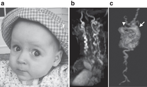
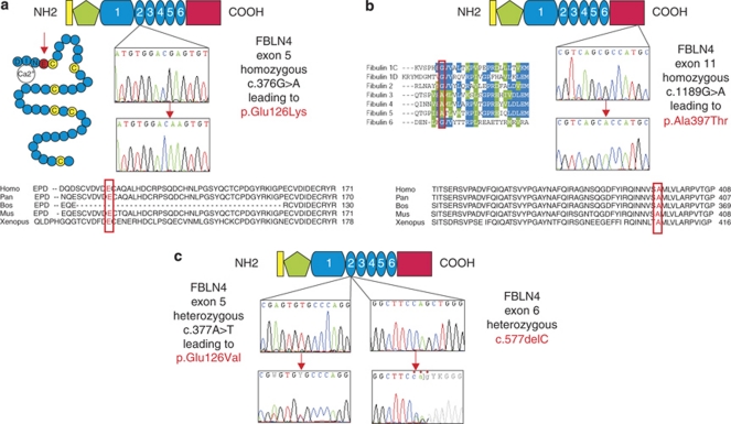
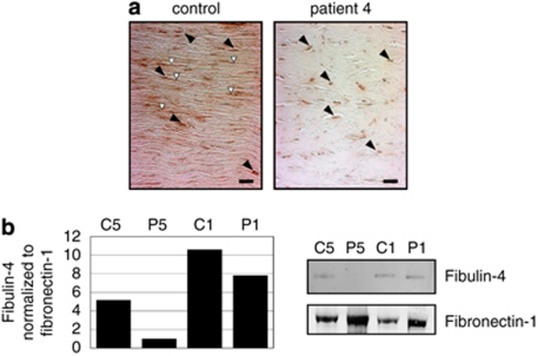
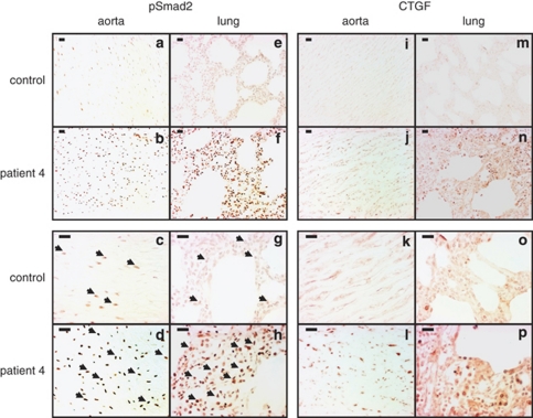
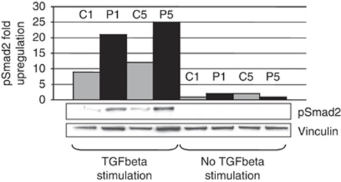
Fibulin-4 is a member of the fibulin family, a group of extracellular matrix proteins prominently expressed in medial layers of large veins and arteries. Involvement of the FBLN4 gene in cardiovascular pathology was shown in a murine model and in three patients affected with cutis laxa in association with systemic involvement. To elucidate the contribution of FBLN4 in human disease, we investigated two cohorts of patients. Direct sequencing of 17 patients with cutis laxa revealed no FBLN4 mutations. In a second group of 22 patients presenting with arterial tortuosity, stenosis and aneurysms, FBLN4 mutations were identified in three patients, two homozygous missense mutations (p.Glu126Lys and p.Ala397Thr) and compound heterozygosity for missense mutation p.Glu126Val and frameshift mutation c.577delC. Immunoblotting analysis showed a decreased amount of fibulin-4 protein in the fibroblast culture media of two patients, a finding sustained by diminished fibulin-4 in the extracellular matrix of the aortic wall on immunohistochemistry. pSmad2 and CTGF immunostaining of aortic and lung tissue revealed an increase in transforming growth factor (TGF)beta signaling. This was confirmed by pSmad2 immunoblotting of fibroblast cultures. In conclusion, patients with recessive FBLN4 mutations are predominantly characterized by aortic aneurysms, arterial tortuosity and stenosis. This confirms the important role of fibulin-4 in vascular elastic fiber assembly. Furthermore, we provide the first evidence for the involvement of altered TGFbeta signaling in the pathogenesis of FBLN4 mutations in humans.
Figures
Similar articles
-
Cutis laxa: intersection of elastic fiber biogenesis, TGFβ signaling, the secretory pathway and metabolism.Matrix Biol. 2014 Jan;33:16-22. doi: 10.1016/j.matbio.2013.07.006. Epub 2013 Aug 16. Matrix Biol. 2014. PMID: 23954411 Free PMC article. Review.
-
Fibulin-4 E57K Knock-in Mice Recapitulate Cutaneous, Vascular and Skeletal Defects of Recessive Cutis Laxa 1B with both Elastic Fiber and Collagen Fibril Abnormalities.J Biol Chem. 2015 Aug 28;290(35):21443-59. doi: 10.1074/jbc.M115.640425. Epub 2015 Jul 15. J Biol Chem. 2015. PMID: 26178373 Free PMC article.
-
Fibulin-4 is essential for maintaining arterial wall integrity in conduit but not muscular arteries.Sci Adv. 2017 May 3;3(5):e1602532. doi: 10.1126/sciadv.1602532. eCollection 2017 May. Sci Adv. 2017. PMID: 28508064 Free PMC article.
-
Fibulin-5 mutations: mechanisms of impaired elastic fiber formation in recessive cutis laxa.Hum Mol Genet. 2006 Dec 1;15(23):3379-86. doi: 10.1093/hmg/ddl414. Epub 2006 Oct 11. Hum Mol Genet. 2006. PMID: 17035250
-
Severe aortopathy due to fibulin-4 deficiency: molecular insights, surgical strategy, and a review of the literature.Eur J Pediatr. 2014 May;173(5):671-5. doi: 10.1007/s00431-013-2217-y. Epub 2013 Nov 26. Eur J Pediatr. 2014. PMID: 24276535 Review.
Cited by
-
Educational paper. Connective tissue disorders with vascular involvement: from gene to therapy.Eur J Pediatr. 2013 Aug;172(8):997-1005. doi: 10.1007/s00431-012-1773-x. Epub 2012 Jul 17. Eur J Pediatr. 2013. PMID: 22801769 Free PMC article. Review.
-
The SMAD-binding domain of SKI: a hotspot for de novo mutations causing Shprintzen-Goldberg syndrome.Eur J Hum Genet. 2015 Feb;23(2):224-8. doi: 10.1038/ejhg.2014.61. Epub 2014 Apr 16. Eur J Hum Genet. 2015. PMID: 24736733 Free PMC article.
-
Comprehensive clinical and molecular analysis of 12 families with type 1 recessive cutis laxa.Hum Mutat. 2013 Jan;34(1):111-21. doi: 10.1002/humu.22165. Epub 2012 Aug 13. Hum Mutat. 2013. PMID: 22829427 Free PMC article.
-
Cutis laxa: intersection of elastic fiber biogenesis, TGFβ signaling, the secretory pathway and metabolism.Matrix Biol. 2014 Jan;33:16-22. doi: 10.1016/j.matbio.2013.07.006. Epub 2013 Aug 16. Matrix Biol. 2014. PMID: 23954411 Free PMC article. Review.
-
Genes in thoracic aortic aneurysms/dissections - do they matter?Ann Cardiothorac Surg. 2013 Jan;2(1):73-82. doi: 10.3978/j.issn.2225-319X.2012.12.01. Ann Cardiothorac Surg. 2013. PMID: 23977562 Free PMC article. No abstract available.
References
-
- Nakamura T, Lozano PR, Ikeda Y, et al. Fibulin-5/DANCE is essential for elastogenesis in vivo. Nature. 2002;415:171–175. - PubMed
-
- Sasaki T, Gohring W, Miosge N, Abrams WR, Rosenbloom J, Timpl R. Tropoelastin binding to fibulins, nidogen-2 and other extracellular matrix proteins. FEBS Lett. 1999;460:280–284. - PubMed
-
- Yanagisawa H, Davis EC, Starcher BC, et al. Fibulin-5 is an elastin-binding protein essential for elastic fibre development in vivo. Nature. 2002;415:168–171. - PubMed
-
- Giltay R, Timpl R, Kostka G. Sequence, recombinant expression and tissue localization of two novel extracellular matrix proteins, fibulin-3 and fibulin-4. Matrix Biol. 1999;18:469–480. - PubMed
Publication types
MeSH terms
Substances
Grants and funding
LinkOut - more resources
Full Text Sources
Molecular Biology Databases
Miscellaneous
