

Femtosecond protein nanocrystallography-data analysis methods
- PMID: 20389587
- PMCID: PMC4038330
- DOI: 10.1364/OE.18.005713
Femtosecond protein nanocrystallography-data analysis methods
Abstract
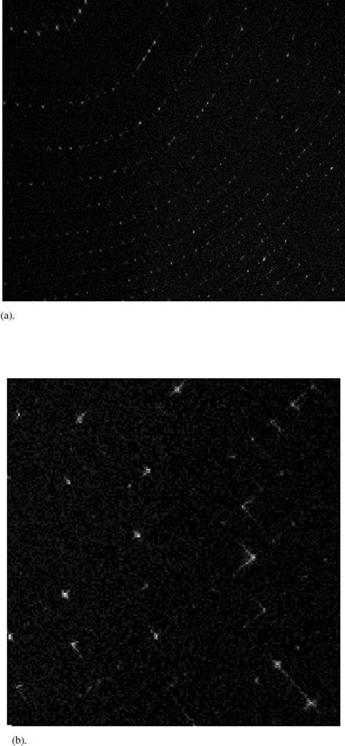
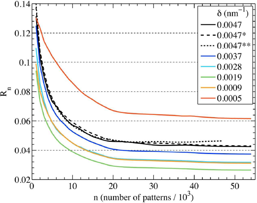
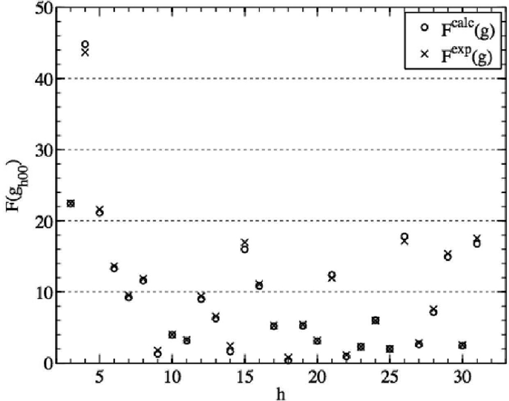
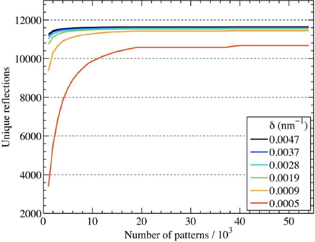
X-ray diffraction patterns may be obtained from individual submicron protein nanocrystals using a femtosecond pulse from a free-electron X-ray laser. Many "single-shot" patterns are read out every second from a stream of nanocrystals lying in random orientations. The short pulse terminates before significant atomic (or electronic) motion commences, minimizing radiation damage. Simulated patterns for Photosystem I nanocrystals are used to develop a method for recovering structure factors from tens of thousands of snapshot patterns from nanocrystals varying in size, shape and orientation. We determine the number of shots needed for a required accuracy in structure factor measurement and resolution, and investigate the convergence of our Monte-Carlo integration method.
Figures
References
-
- Chapman HN. X-ray imaging beyond the limits. Nat. Mater. 2009;8(4):299–301. - PubMed
-
- Howells MR, Beetz T, Chapman HN, Cui C, Holton JM, Jacobsen CJ, Kirz J, Lima E, Marchesini S, Miao H, Sayre D, Shapiro DA, Spence JCH, Starodub D. An assessment of the resolution limitation due to radiation-damage in X-ray diffraction microscopy. J. Electron Spectrosc. Relat. Phenom. 2009;170(1–3):4–12. - PMC - PubMed
-
- Ravelli RBG, Garman EF. Radiation damage in macromolecular cryocrystallography. Curr. Opin. Struct. Biol. 2006;16(5):624–629. - PubMed
-
- Strüder L, Epp S, Rolles D, Hartmann R, Holl P, Lutz G, Soltau H, Eckart R, Reich C, Heinzinger K, Thamm C, Rudenko A, Krasniqi F, Kühnel K-U, Bauer C, Schröter C-D, Moshammer R, Techert S, Miessner D, Porro M, Hälker O, Meidinger N, Kimmel N, Andritschke R, Schopper F, Weidenspointner G, Ziegler A, Pietschner D, Herrmann S, Pietsch U, Walenta A, Leitenberger W, Bostedt C, Möller T, Rupp D, Adolph M, Graafsma H, Hirsemann H, Gärtner K, Richter R, Foucar L, Shoeman RL, Schlichting I, Ullrich J. Large-format, high-speed, X-ray pnCCDs combined with electron and ion imaging spectrometers in a multipurpose chamber for experiments at 4th generation light sources. Nucl. Instrum. Methods. in press.
-
- Denes P, Doering D, Padmore HA, Walder JP, Weizeorick J. A fast, direct x-ray detection charge-coupled device. Rev. Sci. Instrum. 2009;80(8):083302. - PubMed
Publication types
MeSH terms
Substances
Grants and funding
LinkOut - more resources
Full Text Sources
Other Literature Sources
