

Endophthalmitis: Pathogenesis, clinical presentation, management, and perspectives
- PMID: 20390032
- PMCID: PMC2850824
- DOI: 10.2147/opth.s6461
Endophthalmitis: Pathogenesis, clinical presentation, management, and perspectives
Abstract
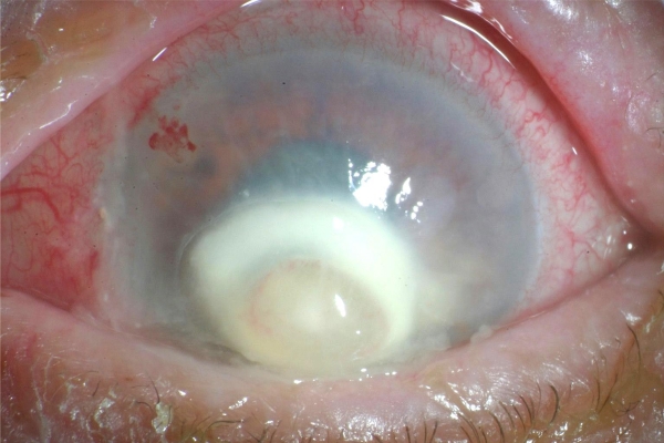
Endophthalmitis is a rare but sight-threatening complication that can occur after ocular surgery or trauma or as a consequence of systemic infection. To optimize visual outcome, early diagnosis and treatment are essential. Over recent decades, advances in hygienic standards, improved microbiologic and surgical techniques, development of powerful antimicrobial drugs, and the introduction of intravitreal antibiotic therapy have led to a decreased incidence and improved management of endophthalmitis. However, endophthalmitis still represents a serious clinical problem. This review focuses on current principles and techniques for evaluation and treatment of endophthalmitis. In addition, it addresses recent developments regarding antimicrobial treatment and prophylaxis of infectious endophthalmitis.
Keywords: antibiotics; caspofungin; endophthalmitis; intravitreal; moxifloxacin; victrectomy; voriconazole.
Figures
References
-
- Mamalis N. Endophthalmitis. J Cataract Refract Surg. 2002;28(5):729–730. - PubMed
-
- Essman TF, Flynn HW, Jr, Smiddy WE, et al. Treatment outcomes in a 10-year study of endogenous fungal endophthalmitis. Ophthalmic Surg Lasers. 1997;28(3):185–194. - PubMed
-
- Jackson TL, Eykyn SJ, Graham EM, Stanford MR. Endogenous bacterial endophthalmitis: 17-year prospective series and review of 267 reported cases. Surg Ophthalmol. 2003;48(4):403–423. - PubMed
-
- Okada AA, Johnson RP, Liles WC, D’Amico DJ, Baker AS. Endogenous bacterial endophthalmitis. Report of a ten-year retrospective study. Ophthalmology. 1994;101(5):832–838. - PubMed
-
- Rao NA, Hidayat AA. Endogenous mycotic endophthalmitis: Variations in clinical and histopathologic changes in candidiasis compared with aspergillosis. Am J Ophthalmol. 2001;132(2):244–251. - PubMed
LinkOut - more resources
Full Text Sources
Other Literature Sources
Medical
