Review
. 2010 Jun;13(2):161-73.
doi: 10.1007/s10456-010-9165-1.

Paramagnetic and fluorescent liposomes for target-specific imaging and therapy of tumor angiogenesis

Affiliations
Review

Paramagnetic and fluorescent liposomes for target-specific imaging and therapy of tumor angiogenesis

Gustav J Strijkers et al. Angiogenesis. 2010 Jun.

Abstract

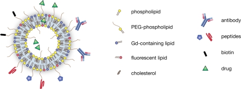
Angiogenesis is essential for tumor growth and metastatic potential and for that reason considered an important target for tumor treatment. Noninvasive imaging technologies, capable of visualizing tumor angiogenesis and evaluating the efficacy of angiostatic therapies, are therefore becoming increasingly important. Among the various imaging modalities, magnetic resonance imaging (MRI) is characterized by a superb spatial resolution and anatomical soft-tissue contrast. Revolutionary advances in contrast agent chemistry have delivered versatile angiogenesis-specific molecular MRI contrast agents. In this paper, we review recent advances in the preclinical application of paramagnetic and fluorescent liposomes for noninvasive visualization of the molecular processes involved in tumor angiogenesis. This liposomal contrast agent platform can be prepared with a high payload of contrast generating material, thereby facilitating its detection, and is equipped with one or more types of targeting ligands for binding to specific molecules expressed at the angiogenic site. Multimodal liposomes endowed with contrast material for complementary imaging technologies, e.g., MRI and optical, can be exploited to gain important preclinical insights into the mechanisms of binding and accumulation at angiogenic vascular endothelium and to corroborate the in vivo findings. Interestingly, liposomes can be designed to contain angiostatic therapeutics, allowing for image-supervised drug delivery and subsequent monitoring of therapeutic efficacy.


Figures

Fig. 1
Fig. 1
Schematic representation of a target-specific multimodal liposome for combined angiogenesis imaging and therapy. Generally, the liposome contains a high payload of Gd containing lipids and fluorescent lipids for MRI and fluorescence microscopy, respectively. The liposome can be equipped with single or multiple populations of antibodies or peptides to introduce target specificity, biotin for an avidin-induced clearance strategy, as well as with drugs in the lumen, in the bilayer or by covalent binding to the distal ends of the PEG chains
Fig. 2
Fig. 2
In vivo visualization of tumor angiogenesis by application of αvβ3 integrin-targeted paramagnetic and fluorescent liposomes. a MR images of three slices through a mouse with a xenograft human LS174T colon carcinoma in a subcutaneous location on the right flank. The images were taken 35 min after injection of RGD-conjugated liposomes, which target the αvβ3 integrins expressed on the angiogenic tumor endothelium. Pixels in the tumor that were significantly enhanced by the presence of the paramagnetic liposomes were highlighted and color coded according to the scale on the right. Enhancement was mainly found at the rim of the tumor in correspondence with the spatial distribution of angiogenic blood vessels. The percentage of enhanced pixels serves as a noninvasive readout of angiogenic activity. b MR images of a mouse 35 min after injection with nonspecific RAD-conjugated liposomes. Nonspecific liposomes distributed more evenly throughout the whole tumor. c MR images of a mouse 35 min after pretreatment with nonparamagnetic RGD-conjugated liposomes to block the αvβ3 integrin followed by injection with paramagnetic RGD-conjugated liposomes. Only a small number of pixels showed signal enhancement proving specificity of the αvβ3 targeting concept. Adapted from Ref. [22] with permission
Fig. 3
Fig. 3
Fluorescence microscopy of dissected tumors of mice, which were injected with paramagnetic, fluorescent, RGD- or RAD-conjugated liposomes. a, b In the rim of the tumors, the red rhodamine fluorescence originating from the RGD-conjugated liposomes revealed circular and longitudinal distribution patterns associated with blood vessels. Cell nuclei were stained with DAPI (blue fluorescence). c A slice through the middle of the tumor revealed no fluorescence from RGD-conjugated liposomes, in agreement with a lack of angiogenic blood vessels in this location. d A diffuse pattern of fluorescence was observed in tumors of mice injected with nonspecific RAD-conjugated liposomes, indicative for nonspecific distribution beyond the blood vessels throughout the whole tumor. Adapted from Ref. [22] with permission
Fig. 4
Fig. 4
In vivo assessment of angiostatic therapy efficacy by targeted multimodal liposomes. Mice with a B16F10 melanoma tumor in a subcutaneous position on the right flank were treated for 3 or 14 days with either anginex or endostatin, which both are angiostatic compounds. a Microvessel density (MVD, mean number of vessels per 0.25 mm2) was determined by ex vivo staining of tumor blood vessels with CD31 antibody and counting their density. (b) The percentage of enhanced pixels in the tumor on T1-weighted MRI after injection with paramagnetic RGD-conjugated liposomes served as a noninvasive in vivo readout of angiogenic activity (see also Fig. 2). MVD revealed a significant treatment effect for anginex (3 and 14 days treatment) and endostatin (3 days treatment). Importantly, the in vivo MRI readout of angiogenic activity closely reflected the treatment effects as deduced from the ex vivo analyses, proving that the multimodal liposomes can be used to noninvasively monitor angiostatic therapy. Adapted from Ref. [24] with permission
Fig. 5
Fig. 5
Improved MR imaging of tumor angiogenesis by avidin-induced clearance of nonbound liposomes. a, b T2-weighted (T2-w, left) and T1-weighted MR images (right) of a mouse with a B16F10 melanoma tumor in a subcutaneous position on the right flank (arrow). a Images of a mouse 90 and 150 min after intravenous injection of liposomes conjugated with both RGD peptide as well as biotin (RGD-biotin-liposomes). The significantly enhanced pixels are color coded according to the scale on the right. b Images of a mouse after intravenous injection of RGD-biotin-liposomes. Between the 90- and 150-min time points, nonbound liposomes were removed by an intravenous infusion of avidin, which binds with high affinity to the biotin on the liposomes and induces rapid blood clearance. c Time dependence of the percentage of enhanced pixels after injection with RGD-biotin-liposomes or liposomes with biotin only (biotin-liposomes). d Time dependence of the percentage of enhanced pixels after injection with RGD-biotin-liposomes and biotin-liposomes. After 100 min of circulation, the mice received and intravenous infusion of avidin over a period of approximately 15 min (gray bar) to induce clearance of nonbound liposomes. Figure adapted from Ref. [71] with permission
Fig. 6
Fig. 6
Synergistic targeting of αvβ3 integrin and galectin-1 with heteromultivalent paramagnetic liposomes for combined imaging and treatment of angiogenesis. a Schematic representation of the incubation schemes of in vitro human endothelial cells, for the following conditions: 1. Culture medium [Control]; 2. Nontargeted liposomes [Bare-L]; 3. Anx-conjugated liposomes [Anx-L]; 4. RGD-conjugated liposomes [RGD-L]; 5. Anx and RGD dual-conjugated liposomes containing high concentration of peptides [Anx/RGD L (H)]; 6. Anx and RGD dual-conjugated liposomes containing low concentration of peptides [Anx/RGD-L (L)]; 7. Mixture of Anx-L and RGD-L containing high concentration of peptides [mixed Anx-L + RGD-L (H)]; 8. Mixture of Anx-L and RGD-L containing low concentration of peptides [mixed Anx-L + RGD-L (L)]. High concentration of peptides (H) were 10 μg/μmol lipid of Anx and 3 μg/μmol lipid of RGD, while low concentration (L) corresponded to 5 μg/μmol lipid of Anx and 1.5 μg/μmol lipid of RGD. b Gd uptake levels achieved with the different targeting strategies. Highest uptake of liposomal Gd was achieved for condition 5, demonstrating the synergistic effect of the two targeting ligands. c Fluorescence microscopy images of human endothelial cells incubated under the different conditions. Cell nuclei were stained with DAPI (blue fluorescence) and red fluorescence originated from the rhodamine-labeled liposomes. Highest cell associated red fluorescence was again observed for Anx and RGD dual-conjugated liposomes containing high concentration of peptides (condition 5). Adapted from Ref. [78] with permission

References

    1. Kerbel RS. Tumor angiogenesis. N Engl J Med. 2008;358:2039–2049. doi: 10.1056/NEJMra0706596. - DOI - PMC - PubMed
    1. Ferrara N, Kerbel RS. Angiogenesis as a therapeutic target. Nature. 2005;438:967–974. doi: 10.1038/nature04483. - DOI - PubMed
    1. Kim KJ, Li B, Winer J, et al. Inhibition of vascular endothelial growth factor-induced angiogenesis suppresses tumour growth in vivo. Nature. 1993;362:841–844. doi: 10.1038/362841a0. - DOI - PubMed
    1. Beliveau R, Gingras D, Kruger EA, et al. The antiangiogenic agent neovastat (AE-941) inhibits vascular endothelial growth factor-mediated biological effects. Clin Cancer Res. 2002;8:1242–1250. - PubMed
    1. O’Reilly MS, Boehm T, Shing Y, et al. Endostatin: an endogenous inhibitor of angiogenesis and tumor growth. Cell. 1997;88:277–285. doi: 10.1016/S0092-8674(00)81848-6. - DOI - PubMed

Publication types