. 2010 May 17;49(10):4405-19.
doi: 10.1021/ic901751z.

Five- and six-coordinate adducts of nitrosamines with ferric porphyrins: structural models for the Type II interactions of nitrosamines with ferric cytochrome P450

Affiliations

Five- and six-coordinate adducts of nitrosamines with ferric porphyrins: structural models for the Type II interactions of nitrosamines with ferric cytochrome P450

Nan Xu et al. Inorg Chem. .

Abstract

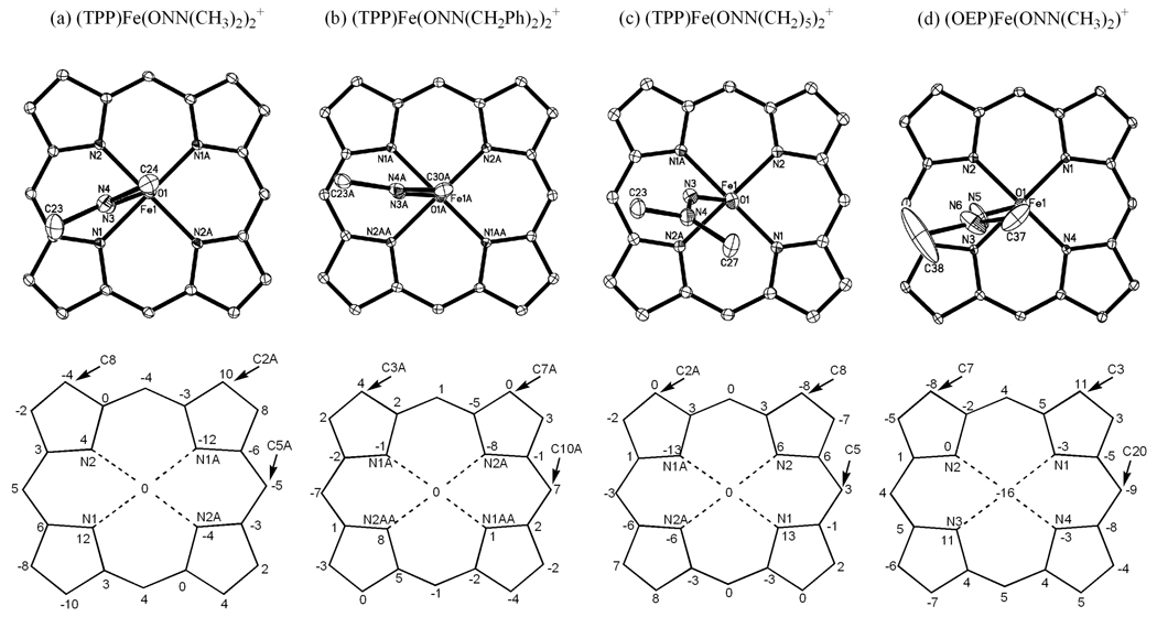
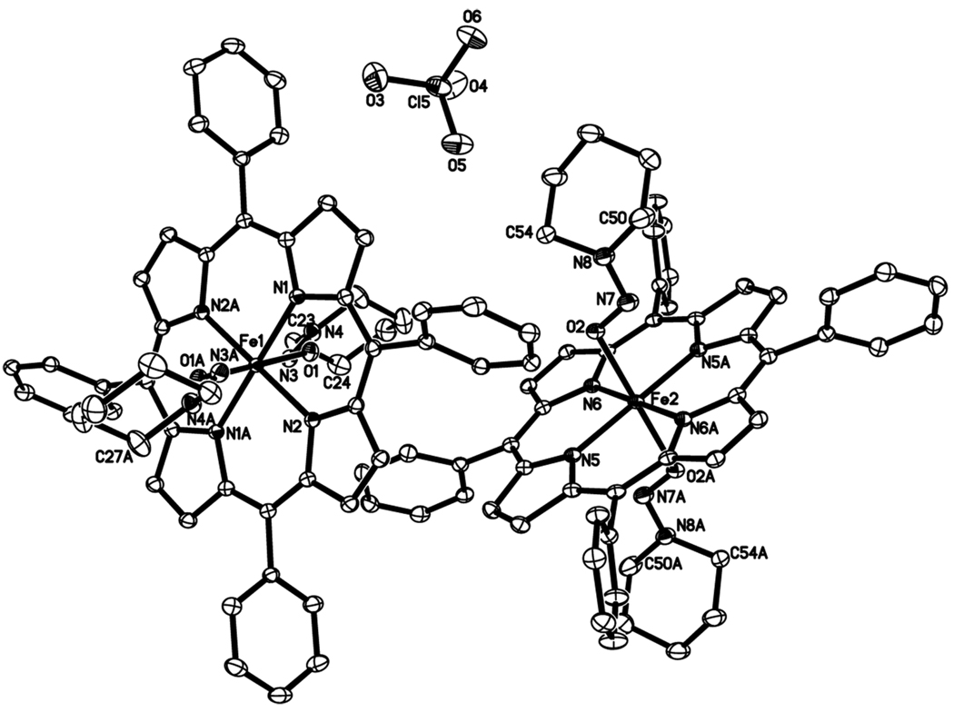
Nitrosamines are well-known for their toxic and carcinogenic properties. The metabolic activation of nitrosamines occurs via interaction with the heme-containing cytochrome P450 enzymes. We report the preparation and structural characterization of a number of nitrosamine adducts of synthetic iron porphyrins. The reactions of the cations [(por)Fe(THF)(2)]ClO(4) (por = TPP, TTP, OEP) with dialkylnitrosamines (R(2)NNO; R(2) = Me(2), Et(2), (cyclo-CH(2))(4), (cyclo-CH(2))(5), (PhCH(2))(2)) in toluene generate the six-coordinate high-spin (S = 5/2) [(por)Fe(ONNR(2))(2)]ClO(4) compounds and a five-coordinate intermediate-spin (S = 3/2) [(OEP)Fe(ONNMe(2))]ClO(4) derivative in 57-72% yields (TPP = 5,10,15,20-tetraphenylporphyrinato dianion, TTP = 5,10,15,20-tetra-p-tolylporphyrinato dianion, OEP = 2,3,7,8,12,13,17,18-octaethylporphyrinato dianion). The N-O and N-N vibrations of the coordinated nitrosamine groups in [(por)Fe(ONNR(2))(2)]ClO(4) occur in the 1239-1271 cm(-1) range. Three of the six-coordinate [(por)Fe(ONNR(2))(2)]ClO(4) compounds and one five-coordinate [(OEP)Fe(ONNMe(2))]ClO(4) compound have been characterized by single crystal X-ray crystallography. All the nitrosamine ligands in these complexes bind to the ferric centers via a sole eta(1)-O binding mode. No arylnitrosamine adducts were obtained from the reactions of the precursor compounds [(por)Fe(THF)(2)]ClO(4) with three arylnitrosamines (Ph(2)NNO, Ph(Me)NNO, Ph(Et)NNO). However, prolonged exposure of [(por)Fe(THF)(2)]ClO(4) to these arylnitrosamines resulted in the formation of the known five-coordinate (por)Fe(NO) derivatives. The latter (por)Fe(NO) compounds were obtained more readily by the reactions of the three arylnitrosamines with the four-coordinate (por)Fe(II) precursors.


Figures

Figure 1
Figure 1
The Type I and Type II interactions of nitrosamines with the active site of cytochrome P450.
Figure 2
Figure 2
Crystallographically determined binding modes of nitrosamines to metal centers.
Figure 3
Figure 3
IR spectra of the unlabeled (top) and nitroso 15N-labeled (bottom) diethylnitrosamine complex [(TPP)Fe(ONNEt2)2]ClO4 as dried samples on NaCl plates.
Figure 4
Figure 4
Dipolar resonance contributions of nitrosamines.
Figure 5
Figure 5
N-binding and O-binding modes of nitrosoarenes to iron porphyrins.
Figure 6
Figure 6
The proposed N-binding of nitrosamines to ferrous porphyrins.
Figure 7
Figure 7
IR spectra of the products from the reactions of the unlabeled and 15N-labeled [(TPP)Fe(ONNEt2)2]ClO4 complexes with NO, as dried samples on NaCl plates.
Figure 8
Figure 8
Structure of the cation of [(TPP)Fe(ONNMe2)2]ClO4. Hydrogen atoms have been omitted for clarity.
Figure 9
Figure 9
Structure of the cation of [(TPP)Fe(ONN(CH2Ph)2)2][(TPP)Fe(OClO3)2]. Hydrogen atoms have been omitted for clarity.
Figure 10
Figure 10
Structure of one of the two independent cations of [(TPP)Fe(ONN(cyclo-CH2)5)2]ClO4. Hydrogen atoms have been omitted for clarity.
Figure 11
Figure 11
(Top) View of the nitrosamine orientations relative to the porphyrin cores, with the view along the axial O-Fe bonds. Nitrosamine substituents have been removed for clarity. (Bottom) Perpendicular atom displacements (in units of 0.01 Å) of the porphyrin cores from the 24-atom mean porphyrin planes.
Figure 12
Figure 12
View of the cation and anion of [(TPP)Fe(ONN(CH2Ph))2][(TPP)Fe(OClO3)2] showing their orientation to each other. Hydrogen atoms have been omitted for clarity.
Figure 13
Figure 13
(a) Structure of [(TPP)Fe(OClO3)2]. H atoms have been omitted for clarity. (b) View of the ClO4 orientation relative to the porphyrin core, with the view alone the O–Fe bond. (c) Perpendicular atom displacements (in units of 0.01 Å) of the porphyrin core from the 24-atom mean porphyrin plane.
Figure 14
Figure 14
View of the two cations of [(TPP)Fe(ONN(cyclo-CH2)5)2]ClO4 showing their orientation with respect to the perchlorate anion.
Figure 15
Figure 15
(Left) 5K solution EPR spectrum (black) of 3 mM [(TPP)Fe(ONNMe2)2]ClO4 in toluene in the presence of 200 equiv. N-nitrosodimethylamine, and simulated spectrum (red). Fit parameters: S = 5/2; D = 10 cm−1, E/D = 0.020; gx = 2.050, gy = 1.950, gz = 2.010; 9.429 GHz. (Right) 5K solid-state EPR spectrum of [(TPP)Fe(ONNMe2)2]ClO4 (black) and simulated spectrum (red). Fit parameters: S = 5/2; D = 10, E/D = 0.025; gx = 1.950, gy = 1.950, gz = 2.010; 9.424 GHz. (See also Table 4).
Figure 16
Figure 16
(a) Structure of the cation of [(OEP)Fe(ONNMe2)]ClO4. Hydrogen atoms have been omitted for clarity. (b) View of the Me2NNO orientation relative to the porphyrin core, with the view along the O–Fe bond. (c) Perpendicular atom displacements (in units of 0.01 Å) of the porphyrin core from the 24-atom mean porphyrin plane.
Figure 17
Figure 17
View of the π-π stacking involving the cation of [(OEP)Fe(ONNMe2)]ClO4, showing (top) the 3.3 Å separation of the porphyrin planes, and (b) the slippage of the porphyrin rings relative to each other. Hydrogen atoms have been omitted for clarity.
Figure 18
Figure 18
Sketch of the geometric parameters for the OEP-OEP π stacking.
Figure 19
Figure 19
(Bottom) 5K solid-state EPR spectrum of [(OEP)Fe(ONNMe2)]ClO4 (black line) and the simulated spectrum ( red line) based on the sum of the spectra of three different species as shown on the top. (Top) Simulation of three individual species present in the solid-state EPR spectrum of [(OEP)Fe(ONNMe2)]ClO4. The ratio of the three species blue : red : black is 1 : 0.002 : 0.0001. Fit parameters (9.424 GHz): Blue: S = 3/2; D = 10 cm−1, E/D = 0.030; J = 2.0 cm−1; gx = 2.070, gy = 2.070, gz = 2.070; Red: S = 3/2; D = 10 cm−1, E/D = 0.030; J = 0.7 cm−1; gx = 2.000, gy = 2.150, gz = 2.150; Black: S = 5/2; D = 10 cm−1, E/D = 0.010; gx = 1.952, gy = 1.950, gz = 2.010. All fit parameters are listed in Table 4.
Figure 20
Figure 20
(Left) 5K frozen-solution EPR spectrum of 3 mM [(OEP)Fe(ONNMe2)]ClO4 in toluene with 1 eq. of Me2NNO present (top, black trace). The red trace shows the simulated spectrum of the six-coordinate high-spin (S = 5/2) complex, and the final difference spectrum (black – red) is shown in blue in the bottom panel. (Right) 5K frozen-solution EPR spectrum of 3 mM [(OEP)Fe(ONNMe2)]ClO4 in toluene in the presence of 200 eq. of Me2NNO (black trace, top). The red trace shows the simulated spectrum of the six-coordinate high-spin (S = 5/2) complex, and the final difference spectrum (black – red) is shown in blue in the bottom panel. In both cases, the signal of the six-coordinate high-spin (S = 5/2) bis-nitrosamine complex does not completely cancel out in the difference spectrum (blue traces), because the simulation does not fully reproduce the experimental shape of the frozen-solution EPR spectrum of the high-spin species as evident from Figure 15, left.

References

    1. Loeppky RN, Michejda CJ, editors. Nitrosamines and Related N-Nitroso Compounds. Chemistry and Biochemistry. Vol. 553. Washington, D.C.: American Chemical Society; 1994.
    1. Hill MJ, editor. Nitrosamines: Toxicology and Microbiology. Chichester, England: VCH Ellis Horwood Ltd.; 1988.
    1. Lijinsky W. Chemistry and Biology of N-Nitroso Compounds. Cambridge: Cambridge University Press; 1992.
    1. Tannenbaum SR, Archer MC, Wishnok JS, Bishop WW. J. Nat. Cancer Inst. 1978;60:251–253. - PubMed
    1. Lintas C, Clark A, Fox J, Tannenbaum SR, Newberne PM. Carcinogenesis. 1982;3:161–165. - PubMed

Publication types