Review
. 2010 Mar:1192:153-60.
doi: 10.1111/j.1749-6632.2009.05349.x.

Bone turnover across the menopause transition : The role of gonadal inhibins

Affiliations
Review

Bone turnover across the menopause transition : The role of gonadal inhibins

Kristy M Nicks et al. Ann N Y Acad Sci. 2010 Mar.

Abstract

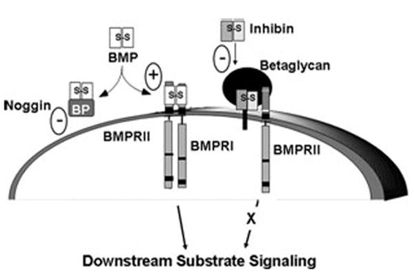
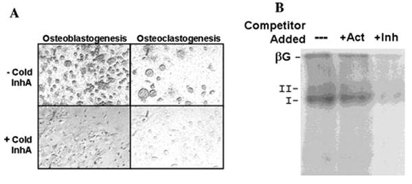
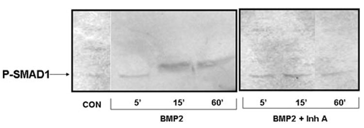
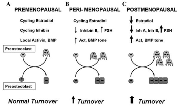
Accumulating evidence demonstrates increasing bone turnover and bone loss in women prior to menopause and decreases in serum estradiol levels. Increased follicle-stimulating hormone levels have been correlated with some of these peri-menopausal changes. However, decreases in gonadal inhibins of the transforming growth factor (TGF)-beta superfamily strongly correlate with increases in bone formation and resorption markers across the menopause transition and predict lumbar bone mass in peri-menopausal women, likely as a result of direct inhibin suppression of osteoblastogenesis and osteoclastogenesis. Inhibins bind specifically to cells during osteoblastogenesis and osteoclastogenesis. They can block bone morphogenetic protein (BMP)-stimulated osteoblast and osteoclast development as well as BMP-stimulated SMAD1 phosphorylation, likely via inhibin-beta-glycan sequestration of BMP Type II receptor (BMPRII). Interestingly, continuous in vivo exposure to inhibin A is anabolic and protective against gonadectomy-induced bone loss in mice, suggesting that inhibins contribute to the endocrine regulation of bone metabolism via a bimodal mechanism of action whereby cycling inhibin exposure suppresses bone turnover and continuous exposure to inhibins is anabolic.


Conflict of interest statement

Conflict of interest The authors declare no conflicts of interest.

Figures

Figure 1
Figure 1
Inhibin antagonism of receptor serine kinase signaling via β-glycan (transforming growth factor [TGF]-β RIII). Bone morphogenetic protein (BMP) signals through the heteromeric type II and type I receptor serine kinases. BMP binds BMPRII, which in turn recruits and phosphorylates BMPRI to stimulate cytoplasmic downstream substrate signaling, primarily through phosphorylation of SMADs 1, 5, and 8. This action can be blocked by soluble BMP antagonists, such as noggin, and enhanced by BMP–β-glycan interaction. Inhibins can also antagonize receptor serine kinase signaling. Inhibin binds with high affinity to β-glycan, allowing for a complex formation with BMPRII. Inhibin–β-glycan–BMPRII interactions sequester BMPRII, preventing its subsequent recruitment of BMPRI and downstream substrate signaling, such as SMAD phosphorylation.
Figure 2
Figure 2
125I inhibin-A (InhA) binds specifically to murine bone marrow cells during cell differentiation. (A) Adult murine bone marrow cells were cultured for 20 days to stimulate osteoblastogenesis in α modified Eagle’s medium (αMEM), 15% fetal bovine serum(FBS), 50 mmol/L ascorbate, and 10 nmol/L β-glycerolphosphate (left) or cultured for 10 days to stimulate osteoclastogenesis in αMEM, 15% FBS, and 10 nmol/L 1.25 (OH)2 Vitamin D3 (right). Subsequently, cells were incubated for 3 h at room temperature in blocking buffer: Dulbecco’s Modified Eagle Medium: Nutrient Mixture F-12 (1:1), 20 mmol/L HEPES, 0.05% cytochrome c, 0.3% bovine serum albumin, 0.01 mg/mL phenylmethylsulfonyl fluoride, 0.01% bacitracin, and 0.4 mg/mL leupeptin. Slides were then incubated at room temperature overnight in the same buffer containing 40 pmol/L lactoperoxidase-labeled 125I-rh (recombinant human) InhA, alone (top) or in the presence of 40 nmol/L InhA, to define a nonspecific background (bottom). Cells were then washed with buffer to remove unbound label, and slides were dipped in emulsion and left to expose in the dark for 1 week. Following development, photographs were taken using the 20× phase objective without counterstaining. (B) Adult murine bone marrow cells were cultured for 20 days to stimulate osteoblastogenesis and then exposed to 125I-InhA prepared as in (A) for 30 min on ice in the absence or presence of 50-fold molar excess of activin A (Act) or InhA (Inh). Cells were cross-linked with disuccidimidyl suberate, lysed in radioimmunoprecipitation assay buffer, and the affinity-labeled complexes analyzed by SDS-PAGE and autoradiography. 125I-InhA formed high-affinity complexes with proteins corresponding to the sizes for β-glycan (βG; type III TGF-β receptor), type II, and type I receptors that were competed for by Inh but not Act.
Figure 3
Figure 3
InhA blocks BMP2-induced SMAD1 phosphorylation during osteoblastogenesis of human mesenchymal cells. Adult human mesenchymal stem cells (Lonza, Walkersville, MD) were used to initiate osteoblast cultures according to themanufacturer’s protocol. Cultures were treated with BMP2 or BMP2 + InhA to determine the ability of InhA to antagonize BMP signaling. Cells were harvested at 5, 15, and 60 min following treatment and protein isolated and analyzed by immunoblot. The presence of SMAD1 phosphorylation was determined using a phospho-specific antibody (R&D Systems, Inc., Minneapolis, MN) as a measure of the initiation of BMP signaling. BMP2 treatment of osteoblast cultures led to increased SMAD1 phosphorylation by 15 min that could still be observed with 60 min of treatment, which was blocked in the presence of 50 ng/mL InhA as early as 15 min of treatment. Neither BMP2 nor InhA had any effect on total SMAD1 protein content (data not shown).
Figure 4
Figure 4
Premenopausal women (A) demonstrate normal cycling hormones and autocrine-paracine levels of activin/BMP, resulting in normal adult bone cell differentiation and bone turnover. (B) Peri-menopausal decreases in inhibin B (InhB) increase circulating FSH and local activin/BMP tone, increasing cell differentiation and turnover. (C) Postmenopausal decreases in InhA and estradiol amplify increases in cell differentiation and dramatically increase bone turnover. FSH, follicle-stimulating hormone.

References

    1. Albright F, Bloomberg E, Smith PH. Postmenopausal osteoporosis. Trans Assoc Am Phys. 1940;55:298–305.
    1. Manolagas SC. Birth and death of bone cells: basic regulatory mechanisms and implications for the pathogenesis and treatment of osteoporosis. Endocr Rev. 2000;21:115–137. - PubMed
    1. Martin TJ, Sims NA. Osteoclast-derived activity in the coupling of bone formation to resorption. Trends Mol Med. 2005;11:76–81. - PubMed
    1. Manolagas SC, Kousteni S, Jilka RL. Sex steroids and bone. Recent Prog Horm Res. 2002;57:385–409. - PubMed
    1. Riggs BL. Endocrine causes of age-related bone loss and osteoporosis. Novartis Found Symp. 2002;242:247–259. discussion 260–4. - PubMed