Review
. 2010 Mar;1191(1):42-61.
doi: 10.1111/j.1749-6632.2010.05445.x.

What does the amygdala contribute to social cognition?

Affiliations
Review

What does the amygdala contribute to social cognition?

Ralph Adolphs. Ann N Y Acad Sci. 2010 Mar.

Abstract

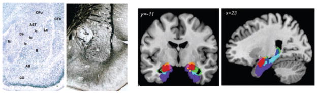
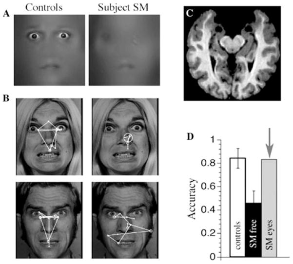
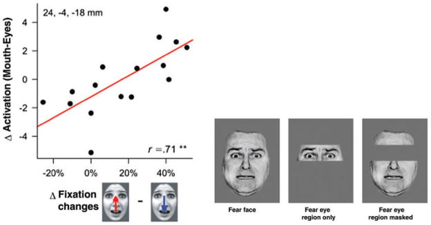
The amygdala has received intense recent attention from neuroscientists investigating its function at the molecular, cellular, systems, cognitive, and clinical level. It clearly contributes to processing emotionally and socially relevant information, yet a unifying description and computational account have been lacking. The difficulty of tying together the various studies stems in part from the sheer diversity of approaches and species studied, in part from the amygdala's inherent heterogeneity in terms of its component nuclei, and in part because different investigators have simply been interested in different topics. Yet, a synthesis now seems close at hand in combining new results from social neuroscience with data from neuroeconomics and reward learning. The amygdala processes a psychological stimulus dimension related to saliency or relevance; mechanisms have been identified to link it to processing unpredictability; and insights from reward learning have situated it within a network of structures that include the prefrontal cortex and the ventral striatum in processing the current value of stimuli. These aspects help to clarify the amygdala's contributions to recognizing emotion from faces, to social behavior toward conspecifics, and to reward learning and instrumental behavior.


Conflict of interest statement

Conflicts of interest

The author declares no conflicts of interest.

Figures

Figure 1
Figure 1
The amygdala and its nuclei. On the left is a Nissl stain, which stains cell bodies, and a silver stain that stains fibers coursing through the amygdala, showing the rat amygdala. On the right are a coronal and parasaggital MRI scan onto which probabilistic locations of amygdala nuclei in humans have been mapped. From Refs. and . Abbreviations of amygdala areas: AB, accessory basal; B, basal nucleus; Ce, central nucleus; itc, intercalated cells; La, lateral nucleus; M, medial nucleus; CO, cortical nucleus. Nonamygdala areas: AST, amygdalo-striatal transition area; CPu, caudate putamen; CTX, cortex. Copyright acknowledgment: two lefthand panels: reproduced from Ref. with permission from Elsevier; two righthand panels: reproduced from Ref. with permission from Springer Science+Business Media.
Figure 2
Figure 2
Social approach behavior following amygdala lesions. (A): Monkeys with amygdala lesions show less fear towards predators and less timidity towards humans. One measure quantifying this is that they spend more time at the front of the cage when there is an unfamiliar person standing there. (B): Approach behavior in patient SM (red bar) compared to control subjects (purple bars) in relation to the experimenter (black bar). Whereas SM had a preferred interpersonal distance of 0.34 meters (C), controls had a distance of 0.64 meters (D). Copyright acknowledgment: A: reproduced from Ref. with permission from the American Psychological Association; (B–D): modified from Ref. with permission from Nature Publishing Group.
Figure 3
Figure 3
The brain and face processing of patient SM. Bilateral amygdala lesions impair the use of the eyes and gaze to the eyes during emotion judgment. (A) A patient with bilateral damage to the amygdala made significantly less use of information from the eye region of faces when judging emotion. (B) While looking at whole faces, the patient (right column of images) exhibited abnormal face gaze, making far fewer fixations to the eyes than did controls (left column of images). This was observed across emotions (free viewing, emotion judgment, gender discrimination). (C) MRI scan of the patient’s brain, whose lesion was relatively restricted to the entire amygdala, a very rare lesion in humans. The two round black regions near the top middle of the image are the lesioned amygdalae. (D) When the subject was instructed to look at the eyes (“SM eyes”) in a whole face, she could do this, resulting in a remarkable recovery in ability to recognize the facial expression of fear. The findings show that an apparent role for the amygdala in processing fearful facial expressions is in fact more abstract, and involves the detection and attentional direction onto features that are socially informative. Modified from Ref. . Copyright acknowledgment: reproduced from Ref. with permission from Nature Publishing Group.
Figure 4
Figure 4
The amygdala helps direct gaze to eyes in faces. Left: data from an fMRI study that plots the response in the amygdala as a function of how often people shift their gaze from the mouth up to the eyes. The amygdala is not activated so much as a consequence of fixating the eyes, but rather in preparation for fixating the eyes. A similar conclusion was obtained in another fMRI study, which found equivalent amygdala activation to fear faces even when the eye region itself was erased (right). Copyright acknowledgment: reproduced from Ref. with permission from the Society for Neuroscience (left), and from Ref. with permission from Elsevier (right).

Similar articles

Cited by

References

    1. Amaral DG, Price JL, Pitkanen A, Carmichael ST. In: The Amygdala: Neurobiological Aspects of Emotion, Memory, and Mental Dysfunction. Aggleton JP, editor. Wiley-Liss; New York, NY: 1992. pp. 1–66.
    1. Swanson LW, Petrovich GD. What is the amygdala? TINS. 1998;21:323–331. - PubMed
    1. Aggleton J. The Amygdala. A Functional Analysis. Oxford University Press; New York, NY: 2000.
    1. Whalen P, Phelps EA. The Human Amygdala. Oxford University Press; New York, NY: 2009.
    1. Costafreda SG, Brammer MJ, David AS, Fu CHY. Predictors of amygdala activation during the processing of emotional stimuli: a meta-analysis of 385 PET and fMRI studies. Brain Res Rev. 2008;58:57–70. - PubMed

Publication types