Review
. 2010;12 Suppl 1(Suppl 1):S2.
doi: 10.1186/ar2883. Epub 2010 Apr 14.

Genetic associations in type I interferon related pathways with autoimmunity

Affiliations
Review

Genetic associations in type I interferon related pathways with autoimmunity

Angélica M Delgado-Vega et al. Arthritis Res Ther. 2010.

Abstract

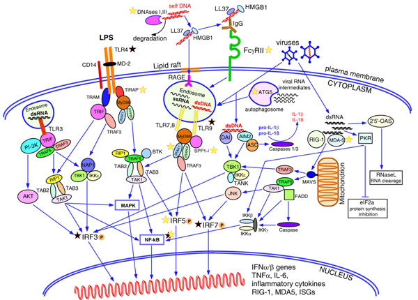
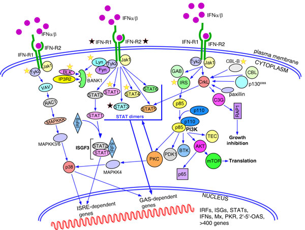
Type I interferons play an outstanding role in innate and adaptive immunity by enhancing functions of dendritic cells, inducing differentiation of monocytes, promoting immunoglobulin class switching in B cells and stimulating effector functions of T cells. The increased production of IFNα/β by plasmacytoid dendritic cells could be responsible for not only efficient antiviral defence, but it also may be a pathological factor in the development of various autoimmune disorders. The first evidence of a genetic link between type I interferons and autoimmune diseases was the observation that elevated IFNα activity is frequently detected in the sera of patients with systemic lupus erythematosus, and that this trait shows high heritability and familial aggregation in their first-degree healthy relatives. To date, a number of genes involved in interferon signalling have been associated with various autoimmune diseases. Patients with systemic lupus erythematosus, Sjögren's syndrome, dermatomyositis, psoriasis, and a fraction of patients with rheumatoid arthritis display a specific expression pattern of interferon-dependent genes in their leukocytes, termed the interferon signature. Here, in an attempt to understand the role of type I interferons in the pathogenesis of autoimmunity, we review the recent advances in the genetics of autoimmune diseases focusing on the association of genes involved in type I interferon pathways.


Figures

Figure 1
Figure 1
Pathways leading to the type I interferon production. Among the family of Toll-like receptors (TLRs), TLR3, TLR4, TLR7/8 and TLR9 are known to induce production of type I interferons in various cells. Surface TLR4 recognizes lipopolysaccharides (LPS) from bacterial cell walls and trasmit the signal downstream via MyD88-dependent or MyD88-independent pathways resulting in phosphorylation, dimerization and nuclear translocation of IRF5 and IRF3, and activation of NF-κB and mitogen-activated protein kinase (MAPK) pathways. Intracellular TLR3, TLR7/8 and TLR9 residing in the endosomes are activated by viral double-stranded (ds)RNA, single-stranded RNA and unmethylated dsDNA, respectively. TLR3 signals via adaptor TRIF and activates IRF3, NF-κB and MAPK pathways. TLR7/8 and TLR9 transmit the signal via the adaptor molecule MyD88. The intracellular form of osteopontin 1 (SPP1) binds to MyD88 upon ligation of TLR9 with unmethylated CpG oligonucleotides and promotes induction of IFNα genes in mouse plasmacytoid dendritic cells (pDCs). TLR7 and TLR9 are the only receptors expressed in pDCs, while other cells contain other TLRs as well. Detection of nucleic acids by TLRs in intracellular endosomes prevents immune responses to the host self-DNA. Normally, nucleic acids released by dying necrotic or apoptotic cells undergoing rapid degradation by nucleases, DNaseI and DNaseIII (TREX1), while bacterial or viral nucleic acids are protected by the cell wall or viral capsid and could be detected by TLRs only after penetrating the cell. Breach of tolerance to self-DNA and activation of pDCs could happen if self-DNA remains undegraded due to defective function of the nucleases and meet endosomal TLR9. Cationic antimicrobial peptide LL37 and high-mobility group box 1 protein (HMGB1) released by damaged or infected cells, mainly keratinocytes and neutrophils, bind DNA making it resistant to degradation and facilitate endocytosis of DNA through the lipid rafts and receptor for advanced glycation end-products (RAGE), delivering it to TLR9. DNA/DNA-protein aggregates could be recognized by anti-DNA/anti-RNA-binding proteins (anti-RNP) antibodies produced by the autoreactive B cells. Binding of these immune complexes to the low-affinity Fcγ receptors II leads to their internalization and translocation to the endosomes containing TLR9. Viral DNAs residing in the cytoplasm could be detected by two cytoplasmic DNA sensors, DNA-dependent activator of interferon regulatory factors (DAI) and absent in melanoma 2 (AIM2), which trigger induction of type I interferon genes through TBK1-mediated and IRF3-mediated signalling. AIM2 also activates caspases 1 and 3 by recruiting adaptor ASC (apoptosis-associated speck-like protein containing a CARD) and forming an inflammasome that promotes release of IL-1β and IL-18. Two RNA helicases, retinoic acid-inducible gene 1 (RIG-1) and melanoma differentiation-associated gene 5 (MDA-5), detect viral RNAs in the cytoplasm. Activated RIG-1 and MDA-5 interact with adaptor protein MAVS anchored by its C-terminal domain to a mitochondrion. This interaction triggers signalling through TRAF3 and TRAF6 adaptors and results in activation of IRF3, IRF7 and NF-κB pathway. Autophagosomes can engulf the replicating viral RNAs and, after fusion with endosomes, present it to the TLR7/8. Viral RNAs can induce a common antiviral defence mechanism aimed at blocking viral replication through total inhibition of cellular transcription and translation. Thus, dsRNAs activate 2',5'-oligoadenylate synthase (OAS) producing 2',5'-oligoadenylates, which in turn activate the latent nuclease RNase L, resulting in the degradation of all cytoplasmic RNAs. Another pathway targets protein synthesis machinery by protein kinase dsRNA-dependent serine-threonine kinase (PKR), which inactivates the alpha subunit of initiation factor eIF2, resulting in rapid inhibition of protein translation. The latter two pathways may induce apoptosis of the infected cell. Yellow stars, genes with strong evidence for association with autoimmune diseases; black stars, genes with inconsistent association. ISG, interferon stimulated genes; PI3K, phosphoinositide 3-kinase.
Figure 2
Figure 2
Pathways activated by type I interferons. Engagement of interferons by interferon receptors activates Jak-signal transducer and activator of transcription (STAT), mitogen-activated protein kinase (MAPK) and phosphoinositide 3-kinase (PI3K) signalling pathways. Jak1-Tyk2-mediated phosphorylation preferentially activates STAT1 and STAT2, which make either homodimers that induce genes with IFNγ-activated site (GAS)-dependent promoters, or heterodimers that bind IRF9 and regulate expression of the genes with interferon-stimulated response elements (ISRE). Other STAT molecules have a more restricted pattern of expression and could be activated by interferons in a cell-specific manner. p38 MAPK is activated in a series of signalling events initiated by IFNα/β and is necessary for induction of genes with both ISRE and GAS-dependent promoters. Type I interferons induce phosphorylation of a number of adaptor proteins, including members of the insulin receptor substrates (IRS1, IRS2, IRS3, and IRS4), growth-factor-receptor-bound protein 2 (GRB2)-associated binding protein 1 and 2 (GAB1 and GAB2) and members of the CRK family (CRKL, and CRK I and CRK II). Phosphorylated CRKL binds through its SH2 domain with STAT5 and activates GAS-dependent genes. Other tyrosine kinase substrates, such as Casitas B-lineage lymphoma (CBL), CBL-b, p130cas and paxillin, also bind to CRKL through the SH2 domain. The guanine-exchange factor C3G interacts with the SH3 domain of CRKL and activates small GTPase RAP 1, which participates in the regulation of cell growth, proliferation and differentiation. Activated IRS adaptors provide binding sites for the p85 regulatory subunit of PI3K, which results in the activation of the catalytic function of the p110 subunit. PI3K is known to activate a number of downstream signalling molecules affecting all aspects of cell biology. Thus, tissue-specific isoforms of protein kinase C (PKC) family, PKCδ, PKCε, PKCθ and PKCη, phosphorylate serine residues in the STAT factors and p38 MAPK. Pharmacological inhibitors that block the activity of distinct PKCs affect the expression of the interferon-responsive genes. The PI3K-AKT signalling cascade mediates survival signals in a cell-type-restricted manner, inducing both anti-apoptopic and pro-apoptotic pathways, and translation of cap-dependent transcripts. Type I interferons activate two members of Src family of kinases, Fyn in T cells and Lyn in B cells. Lyn kinase in its turn phosphorylates B-cell-specific adaptor protein BANK1, facilitating formation of a complex between BANK1, BLK kinase and IP3 receptor 2 (IP3R2). Yellow stars, genes with strong evidence for association with autoimmune diseases; black stars, genes with inconsistent association. ISG, interferon stimulated genes; OAS, 2',5'-oligoadenylate synthase; PKR, protein kinase dsRNA-dependent serine-threonine kinase.

Similar articles

Cited by

References

    1. Pestka S, Krause CD, Walter MR. Interferons, interferon-like cytokines, and their receptors. Immunol Rev. 2004;202:8–32. doi: 10.1111/j.0105-2896.2004.00204.x. - DOI - PubMed
    1. Jaks E, Gavutis M, Uze G, Martal J, Piehler J. Differential receptor subunit affinities of type I interferons govern differential signal activation. J Mol Biol. 2007;366:525–539. doi: 10.1016/j.jmb.2006.11.053. - DOI - PubMed
    1. Diaz MO, Pomykala HM, Bohlander SK, Maltepe E, Malik K, Brownstein B, Olopade OI. Structure of the human type-I interferon gene cluster determined from a YAC clone contig. Genomics. 1994;22:540–552. doi: 10.1006/geno.1994.1427. - DOI - PubMed
    1. Luft T, Pang KC, Thomas E, Hertzog P, Hart DN, Trapani J, Cebon J. Type I IFNs enhance the terminal differentiation of dendritic cells. J Immunol. 1998;161:1947–1953. - PubMed
    1. Gallucci S, Lolkema M, Matzinger P. Natural adjuvants: endogenous activators of dendritic cells. Nat Med. 1999;5:1249–1255. doi: 10.1038/15200. - DOI - PubMed

Publication types

MeSH terms