

Review
doi: 10.1186/ar2885.
Epub 2010 Apr 14.
Type 1 interferons and myositis
Affiliations
- PMID: 20392291
- PMCID: PMC2991777
- DOI: 10.1186/ar2885
Item in Clipboard
Review
Type 1 interferons and myositis
Arthritis Res Ther.
2010.
Abstract
Recent studies suggest a mechanistic role for molecules induced by type 1 interferons in the pathogenesis of some forms of myositis. For dermatomyositis, evidence that these molecules injure myofibers seems especially strong. In the group of disorders known as polymyositis, the study of blood samples suggests a potential role. It is unknown what drives the sustained presence of type 1 interferon-inducible molecules in these diseases, as the type 1 interferons themselves have not been specifically detected along with their downstream biomarkers. Therapeutic development for blockade of IFNα is in progress aided by the identification of blood genomic biomarkers.
Figures
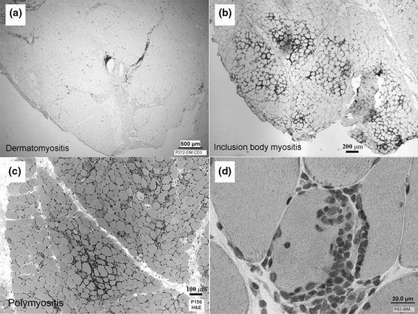
Differing pathologies in myositis subtypes. The distribution of immune system cells differs among myositis subtypes. (a) In dermatomyositis, immune system cells are predominantly in the regions of connective tissue that lie between muscle fascicles and include medium-sized and large blood vessels. In (b) inclusion body myositis and (c) polymyositis, immune cells surround myofibers. (d) Especially in inclusion body myositis, these may sometimes invade myofibers.
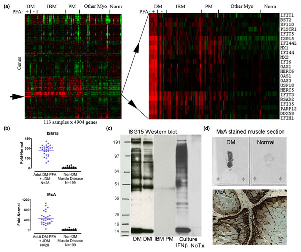
Genomic identification of type 1 interferon-inducible pathway activation in dermatomyositis muscle. (a) Analysis of 22,283 gene transcript probe sets (4,904 shown after filtering; one per row) in 113 muscle biopsy samples (one per column) disclosed a cluster of type 1 interferon-inducible genes specifically and highly upregulated in dermatomyositis (DM) with perifascicular atrophy (PFA) (thin red stripe marked by an arrow). Enlargement of this arrowed region shown on the right. Red and green indicate high and low expression. IBM, inclusion body myositis; Myo, myopathies; PM, polymyositis. (b) High expression of transcripts for interferon-stimulated gene 15 (ISG15) and myxovirus resistance protein A (MxA) are extraordinarily specific to DM muscle. JDM, juvenile DM. (c) Examples of ISG15 western blots show free ISG15 (approximately 15 kDa band) and multiple ISG15 conjugated proteins (discrete bands and smear shown at higher molecular weights) in DM but not other muscle biopsy samples. Cultured human skeletal muscle cells exposed to IFNβ develop the same pattern of free and conjugated ISG15 as occurs in DM. NoTx, no treatment. (d) MxA staining of DM muscle is sometimes so impressive that it is evident on glass slides viewed without the aid of a microscope. MxA is preferentially located in perifascicular myofibers and in blood vessel walls. Adapted from [2] with permission.
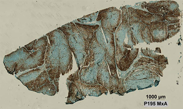
Myxovirus resistance protein A expression in dermatomyositis muscle. Image of whole muscle section stained for myxovirus resistance protein A (MxA) showing abundant myofiber protein expression (brown) preferentially in a perifascicular distribution. Adapted from [3] with permission.
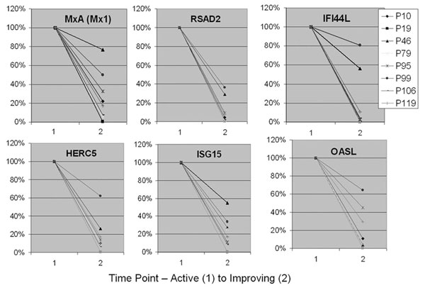
Blood type 1 interferon-inducible gene expression correlation with disease activity. Downregulation of six type 1 interferon inducible genes in eight patients, correlating with improvement in clinical disease from time point 1 (active) to time point 2 (improving). Adapted from [19] with permission.
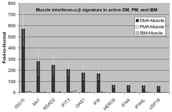
Distinct muscle expression of type 1 interferon-inducible genes in inflammatory myopathies. Distinct muscle expression of type 1 interferon-inducible genes in dermatomyositis (DM) compared with polymyositis (PM) and inclusion body myositis (IBM). Muscle microarray data shown for 20 patients (five each with active dermatomyositis (DMA), active polymyositis (PMA), untreated IBM, and normal) with plotted mean values and error bars for mean ± standard error for each group. Highly expressed genes in DM muscle are orders of magnitude greater than in PM and IBM. Adapted from [19] with permission.
References
-
- Salajegheh M, Kong SW, Pinkus JL, Walsh RJ, Liao A, Nazareno R, Amato AA, Krastins B, Morehouse C, Higgs BW, Jallal B, Yao Y, Sarracino DA, Parker KC, Greenberg SA. Interferon-stimulated gene 15 (ISG15) conjugates proteins in dermatomyositis muscle with perifascicular atrophy. Ann Neurol. 2009;67:53–63. doi: 10.1002/ana.21805. - DOI - PMC - PubMed
Publication types
MeSH terms
Substances
Grants and funding
LinkOut - more resources
Full Text Sources
Other Literature Sources
