Comparative Study
. 2010 Sep;13(8):1089-101.
doi: 10.1017/S1461145710000301. Epub 2010 Apr 15.

Decreased expression of Freud-1/CC2D1A, a transcriptional repressor of the 5-HT1A receptor, in the prefrontal cortex of subjects with major depression

Affiliations
Comparative Study

Decreased expression of Freud-1/CC2D1A, a transcriptional repressor of the 5-HT1A receptor, in the prefrontal cortex of subjects with major depression

Bernadeta Szewczyk et al. Int J Neuropsychopharmacol. 2010 Sep.

Abstract

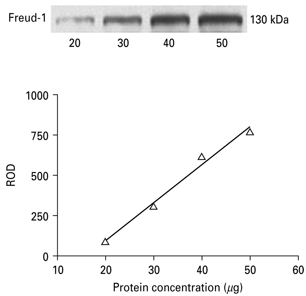
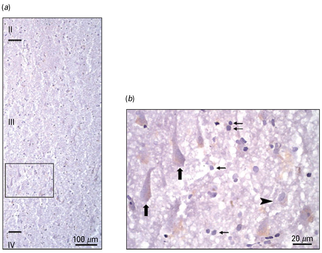
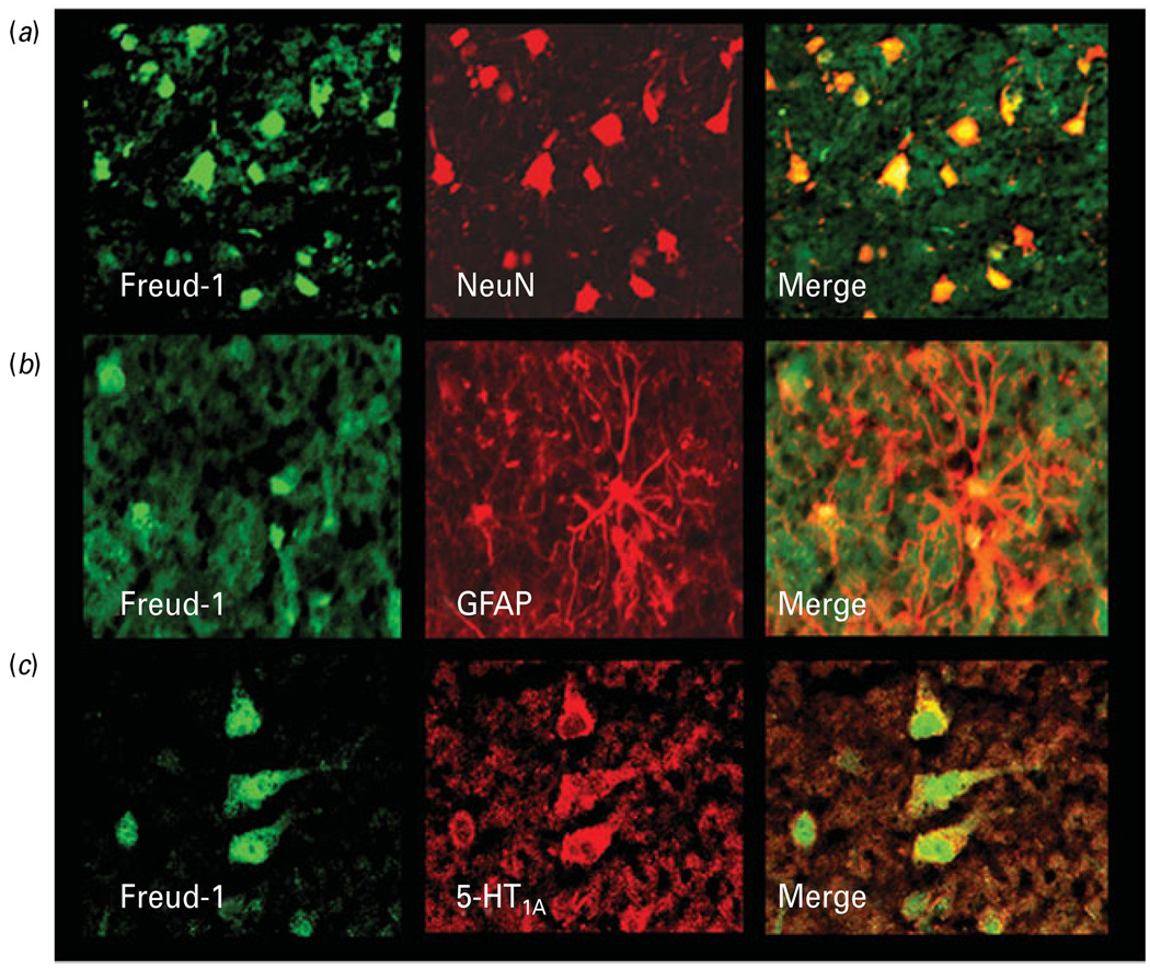
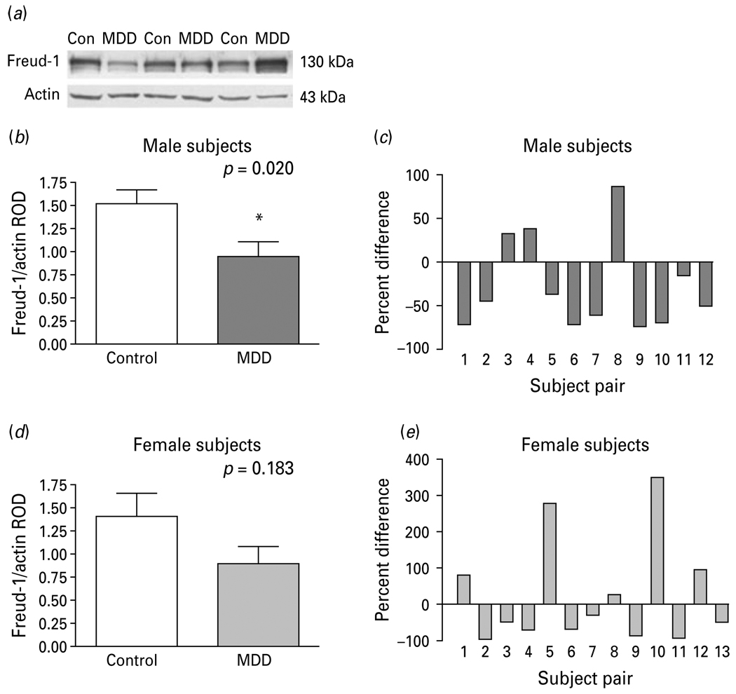
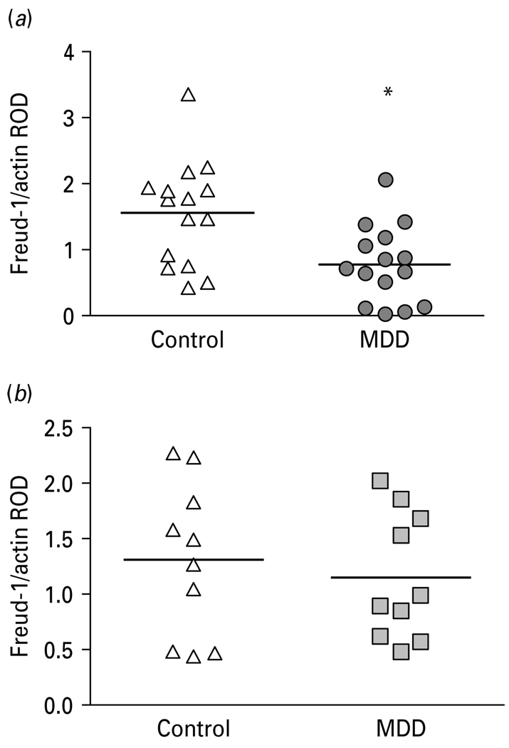
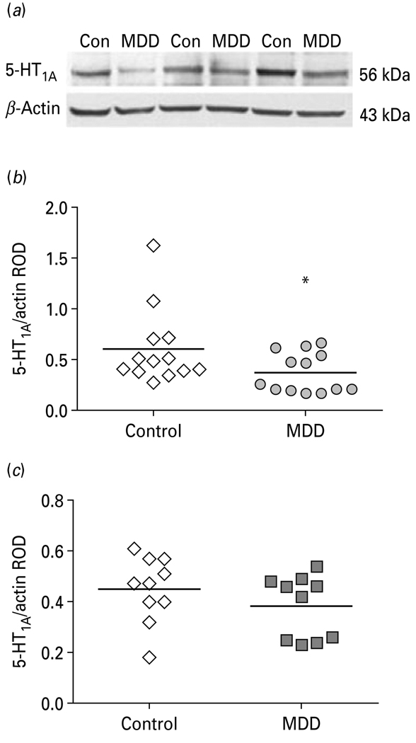
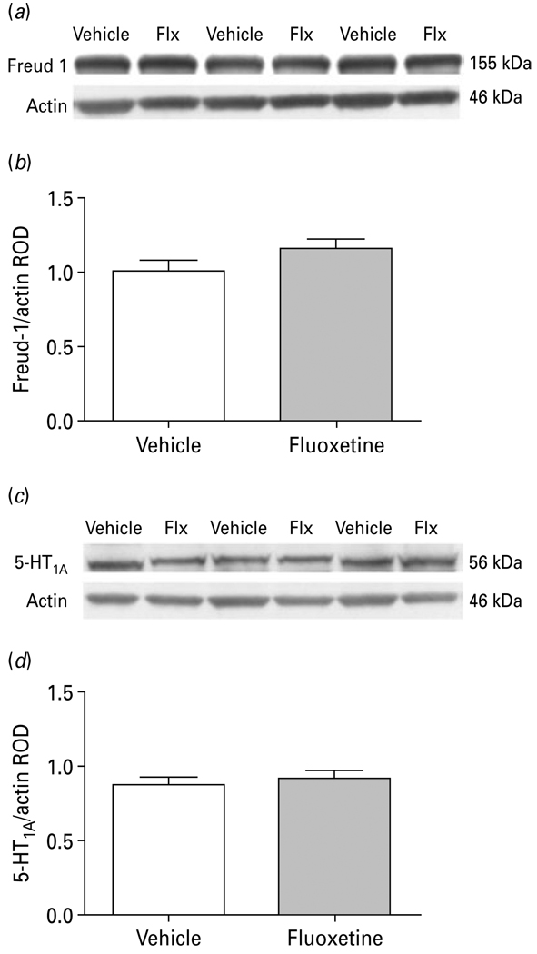
Serotonin1A (5-HT(1A)) receptors are reported altered in the brain of subjects with major depressive disorder (MDD). Recent studies have identified transcriptional regulators of the 5-HT(1A) receptor and have documented gender-specific alterations in 5-HT(1A) transcription factor and 5-HT(1A) receptors in female MDD subjects. The 5' repressor element under dual repression binding protein-1 (Freud-1) is a calcium-regulated repressor that negatively regulates the 5-HT(1A) receptor gene. This study documented the cellular expression of Freud-1 in the human prefrontal cortex (PFC) and quantified Freud-1 protein in the PFC of MDD and control subjects as well as in the PFC of rhesus monkeys chronically treated with fluoxetine. Freud-1 immunoreactivity was present in neurons and glia and was co-localized with 5-HT(1A) receptors. Freud-1 protein level was significantly decreased in the PFC of male MDD subjects (37%, p=0.02) relative to gender-matched control subjects. Freud-1 protein was also reduced in the PFC of female MDD subjects (36%, p=0.18) but was not statistically significant. When the data was combined across genders and analysed by age, the decrease in Freud-1 protein level was greater in the younger MDD subjects (48%, p=0.01) relative to age-matched controls as opposed to older depressed subjects. Similarly, 5-HT(1A) receptor protein was significantly reduced in the PFC of the younger MDD subjects (48%, p=0.01) relative to age-matched controls. Adult male rhesus monkeys administered fluoxetine daily for 39 wk revealed no significant change in cortical Freud-1 or 5-HT(1A) receptor proteins compared to vehicle-treated control monkeys. Reduced protein expression of Freud-1 in MDD subjects may reflect dysregulation of this transcription factor, which may contribute to the altered regulation of 5-HT(1A) receptors observed in subjects with MDD. These data may also suggest that reductions in Freud-1 protein expression in the PFC may be associated with early onset of MDD.


Figures

Fig. 1
Fig. 1
Relationship between the relative optical density (ROD) values of Freud-1 immunoreactivity and increasing total protein concentrations (20, 30, 40, 50 µg) of human prefrontal cortex.
Fig. 2
Fig. 2
Laminar and cellular localization of Freud-1 immunoreactivity (IR) in the human prefrontal cortex (PFC) (BA 10). (a) Distribution of Freud-1 IR in the upper cortical layers of BA 10. (b) High-power micrograph of Freud-1 IR [box in panel (a)] showing the presence of immunoreactive product in both neurons and the glial cell nuclei located in layer III. Large arrows identify Freud-1 IR localized to pyramidal neurons, small arrows identify Freud-1 IR is localized to glial cells and arrowhead identifies Freud-1 IR localized to non-pyramidal neuron.
Fig. 3
Fig. 3
Colocalization of Freud-1 immunoreactivity (IR) in neurons and glia. (a) Co-labelling of Freud-1 IR (green) with the neuronal marker NeuN (red). Freud-1 protein (yellow) is localized to neuronal nuclei and perinuclear areas. (b) Co-labelling of Freud-1 IR (green) with the astrocytic marker GFAP (red). Freud-1 (yellow) is localized in glial cell nuclei. (c) Colocalization of Freud-1 IR (green) and immunoreactivity for 5-HT1A receptor (red). Freud-1 IR (yellow) is present in the cytoplasm of a majority of cells expressing 5-HT1A receptor immunoreactivity.
Fig. 4
Fig. 4
(a) Representative Western blots showing the immunolabelling of Freud-1 and β-actin in prefrontal cortex (PFC) of three pairs of control (Con) and matched major depressive disorder (MDD) subjects. (b) Freud-1 protein levels in PFC of 12 male (b, c) and 13 female (d, e) control and MDD subjects expressed as the mean ± S.E.M. of Freud-1/actin relative optical density (ROD). Data were analysed using a maximum-likelihood mixed-models test. Male subject groups: control subjects (1.52 ± 0.16) compared to the matched MDD subjects (0.96 ± 0.15, t21=2.53, * p=0.020). Female subject groups: control subjects (1.41 ± 0.25) compared to the matched MDD subjects (0.90 ± 0.18, t21=1.38, p=0.183). Freud-1 levels in PFC of male (c) and female (d) depressive subjects expressed as percent difference from paired control subjects.
Fig. 5
Fig. 5
(a) Scatterplot of Freud-1 protein levels normalized to actin relative optical density (ROD) in prefrontal cortex (PFC) of younger (<58 yr) controls and major depressive disorder (MDD) subjects. (b) Scatterplot of Freud-1 protein levels normalized to actin ROD in PFC of older (≥58 yr) control and MDD subjects. Data were analysed using a maximum-likelihood mixed-models test. Younger subject groups: control subjects (1.56 ± 0.20, mean ± S.E.M.) compared to the matched MDD subjects (0.78 ± 0.15; age <58 yr; t21=2.84, * p=0.010). Older subject groups: control subjects (1.32 ± 0.22) compared to matched MDD subjects (1.15 ± 0.18; age ≥58 yr; t21=0.53, p=0.602).
Fig. 6
Fig. 6
(a) Representative Western blots showing the immunolabelling of 5-HT1A receptor and β-actin in prefrontal cortex (PFC) of three pairs of control (Con) and matched MDD subjects. (b) Scatterplot of 5-HT1A receptor protein levels normalized to actin relative optical density (ROD) in PFC of younger (<58 yr) controls and major depressive disorder (MDD) subjects. (c) Scatterplot of 5-HT1A receptor protein levels normalized to actin ROD in PFC of older (≥58 yr) control and MDD subjects. Data were analysed using a maximum-likelihood mixed-models test. Younger subject groups: control subjects (0.55 ± 0.10, mean ± S.E.M.) compared to the matched MDD subjects (0.37 ± 0.07; age <58 yr; t18=2.58, * p=0.019). Older subject groups: control subjects (0.45 ± 0.06) compared to matched MDD subjects (0.39 ± 0.05; age ≥58 yr; t18=0.76, p=0.459).
Fig. 7
Fig. 7
(a) Representative Western blots showing the immunolabelling of Freud-1 and β-actin in prefrontal cortex (PFC) of vehicle- and fluoxetine (Flx)-treated monkeys. (b) Freud-1 protein levels in PFC of six vehicle- and seven fluoxetine-treated monkeys expressed as the mean ± S.E.M. of Freud-1/actin relative optical density (ROD). (c) Representative Western blots showing the immunolabelling of 5-HT1A receptor and β-actin in PFC of vehicle and fluoxetine-treated monkeys. (d) 5-HT1A receptor protein levels in PFC of six vehicle- and seven fluoxetine-treated monkeys expressed as the mean ± S.E.M. of 5-HT1A/actin ROD.

References

    1. Albert PR, Lemonde S. 5-HT1A receptors, gene repression, and depression: guilt by association. Neuroscientist. 2004;10:575–593. - PubMed
    1. APA. Diagnostic and Statistical Manual of Mental Disorders. 4th edn. Washington, DC: American Psychiatric Association; 1994.
    1. Arango V, Underwood MD, Gubbi AV, Mann JJ. Localized alterations in pre and postsynaptic serotonin binding sites in the ventrolateral prefrontal cortex of suicide victims. Brain Research. 1995;688:121–133. - PubMed
    1. Arranz B, Eriksson A, Mellerup E, Plenge P, et al. Brain 5-HT1A, 5-HT1D, and 5-HT2 receptors in suicide victims. Biological Psychiatry. 1994;35:457–463. - PubMed
    1. Bhagwagar Z, Rabiner EA, Sargent PA, Grasby PM, et al. Persistent reduction in brain serotonin 1A receptor binding in recovered depressed men measured by positron emission tomography with [11C]WAY-100635. Molecular Psychiatry. 2004;9:386–392. - PubMed

Publication types

MeSH terms

Substances