Review
. 2010 Mar;27(1-2):1-8.
doi: 10.1017/S0952523810000076. Epub 2010 Apr 15.

Six different roles for crossover inhibition in the retina: correcting the nonlinearities of synaptic transmission

Affiliations
Review

Six different roles for crossover inhibition in the retina: correcting the nonlinearities of synaptic transmission

Frank S Werblin. Vis Neurosci. 2010 Mar.

Abstract

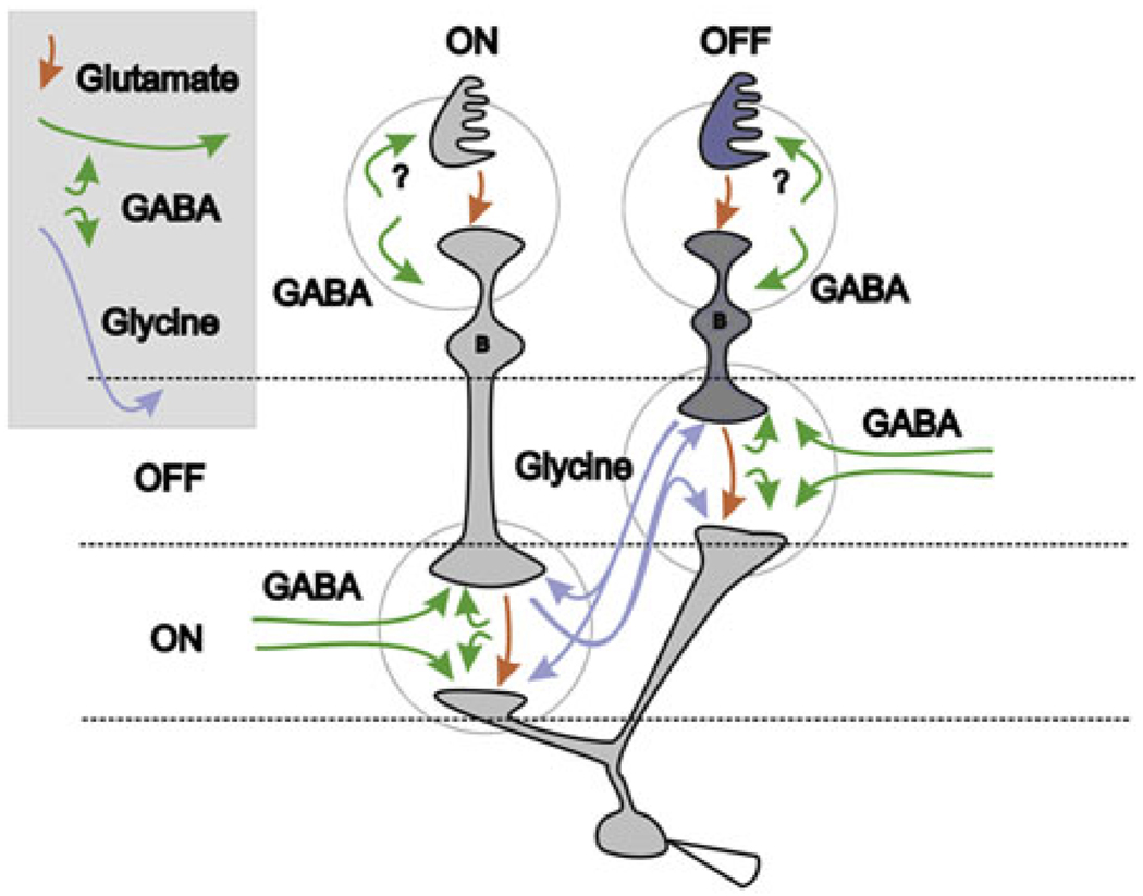
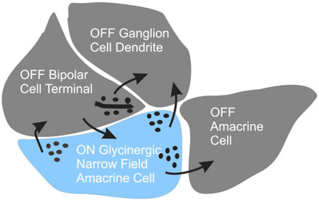
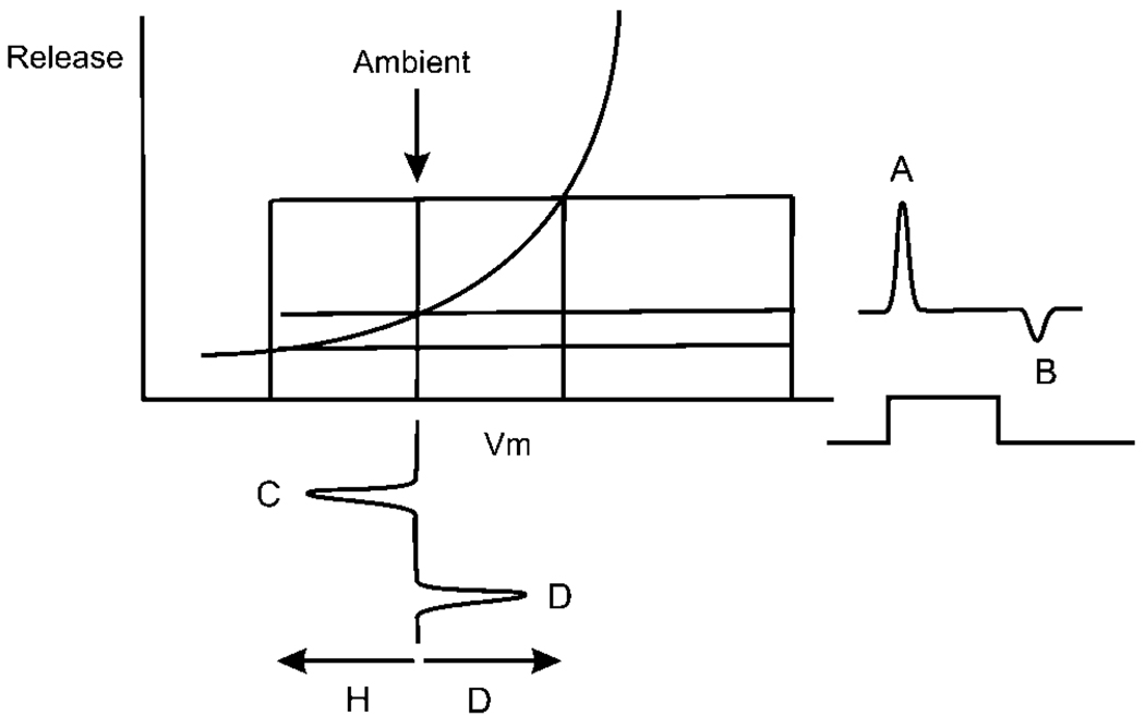
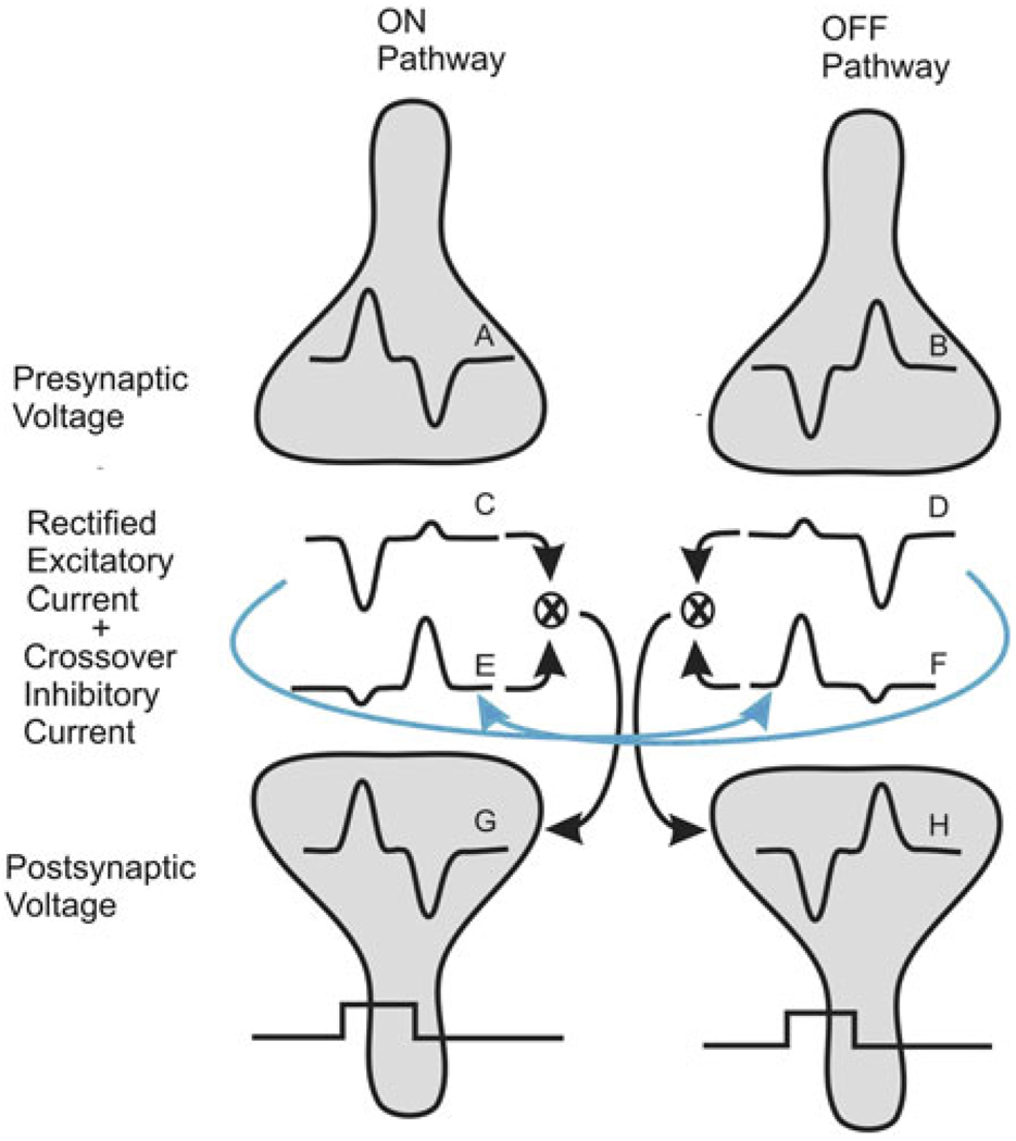
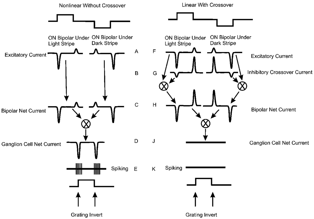
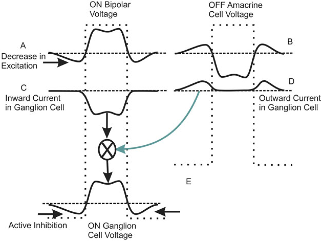
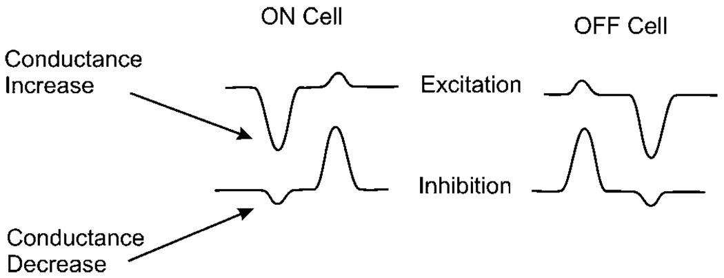
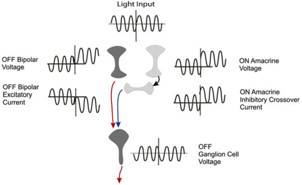
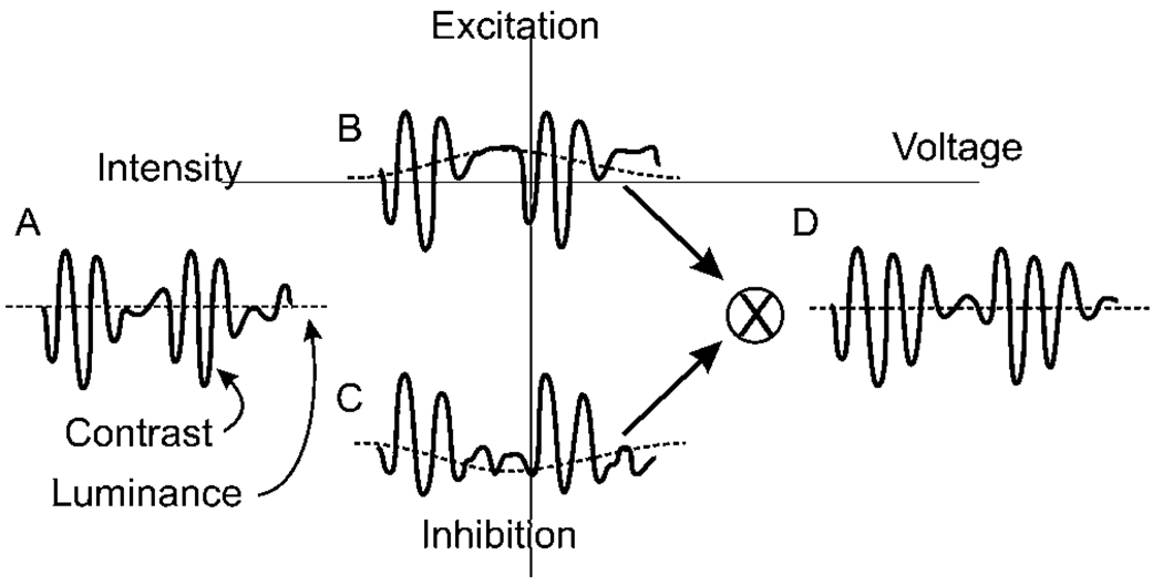
Early retinal studies categorized ganglion cell behavior as either linear or nonlinear and rectifying as represented by the familiar X- and Y-type ganglion cells in cat. Nonlinear behavior is in large part a consequence of the rectifying nonlinearities inherent in synaptic transmission. These nonlinear signals underlie many special functions in retinal processing, including motion detection, motion in motion, and local edge detection. But linear behavior is also required for some visual processing tasks. For these tasks, the inherently nonlinear signals are "linearized" by "crossover inhibition." Linearization utilizes a circuitry whereby nonlinear ON inhibition adds with nonlinear OFF excitation or ON excitation adds with OFF inhibition to generate a more linear postsynaptic voltage response. Crossover inhibition has now been measured in most bipolar, amacrine, and ganglion cells. Functionally crossover inhibition enhances edge detection, allows ganglion cells to recognize luminance-neutral patterns with their receptive fields, permits ganglion cells to distinguish contrast from luminance, and maintains a more constant conductance during the light response. In some cases, crossover extends the operating range of cone-driven OFF ganglion cells into the scotopic levels. Crossover inhibition is also found in neurons of the lateral geniculate nucleus and V1.


Figures

Fig. 1
Fig. 1
Crossover inhibition between bipolar, amacrine, and ganglion cells. The blue arrows show the pathways for crossover inhibition acting at bipolar and ganglion cells. ON bipolar, amacrine, and ganglion cells receive glycinergic OFF inhibition and OFF bipolar, amacrine, and ganglion cells receive glycinergic ON inhibition (blue arrows). Narrow field ON and OFF GABAergic amacrine cells (short green arrows) receive glycinergic inhibition. Wide field ON–OFF amacrine cells (long lateral green arrows) receive no inhibition. GABAergic amacrine cells (all green arrows) feedback to bipolar cells and forward to ganglion cells but not to other amacrine cells. Red arrows indicated excitatory pathways. This circuitry is verified by measurements of excitation and pharmacological block of inhibition in each cell type in many previous studies. All crossover signals could interact at the diad at the bipolar terminal as suggested in Fig. 2.
Fig. 2
Fig. 2
Summary sketch of electron micrograph of a synaptic terminal of an OFF bipolar cell terminal diad showing the synaptic pathways typically found in these images. OFF bipolar cell drives an OFF ganglion cell and an OFF amacrine cell. An ON amacrine cell (blue), driven by an ON bipolar cell, feeds back to the OFF bipolar cell and forward to the OFF ganglion cell. The amacrine cell also inhibits a neighboring OFF amacrine cell. A complementary set of connections would exist for the ON bipolar cell terminal. This sketch suggests all the connections that would be required for crossover inhibition to bipolar, amacrine, and ganglion cells. It is difficult to fit all of the processes around the bipolar cell ribbon in this two-dimensional representation, but all processes could be included in three dimensions.
Fig. 3
Fig. 3
A representation of a typical voltage response as might be measured in an ON depolarizing bipolar cell is shown along the abscissa. At the input, the transient depolarizing and hyperpolarizing response peaks C and D are of similar magnitude. The membrane would typically begin near −40 mV and generate 5–10 mV transients. This voltage response initiates voltage-dependent release A and B generating asymmetrical outward and inward currents in a postsynaptic cell A and B. These current peaks would typically be between 50 and 100 pA.
Fig. 4
Fig. 4
Compensation for nonlinearities mediated by crossover inhibition. Scheme for signal flow of crossover inhibition at a generalized synapse in the retina. (A and B) Voltage responses in ON and OFF presynaptic cells to a bright step of light. (C) Excitatory currents generated in the postsynaptic ON cells showing rectification where presynaptic depolarization elicits a large inward current, while presynaptic hyperpolarization elicits a smaller outward current. (D) Excitatory currents generated in a postsynaptic OFF cell. (E) Crossover current to an ON postsynaptic cell derived from the OFF pathway carried by an OFF amacrine cell (blue arrow). (F) Crossover current to an OFF postsynaptic cell. (G) Voltage in an ON postsynaptic cell generated by the addition of ON excitation and OFF crossover inhibition. (H) Postsynaptic voltage in an OFF postsynaptic cell generated by OFF excitation and ON crossover inhibition.
Fig. 5
Fig. 5
Superposition of the excitatory and inhibitory currents in a population of ON ganglion cells leading to null response to inverting grating. Left column: nonlinear responses without crossover. Right column: responses linearized by crossover. The initiation of “dark stripe” activity and “light stripe” activity occurs simultaneously in neighboring spatial regions. The upward and downward steps in the timing graphs at the top of the figure represent the transitions of the contrasting stripes. They overlap in time and are adjacent in space. Here, the left stripe transitions to light, while the right stripe transitions to dark. These two temporally coincident events are shown separately in the figure to illustrate how temporally-simultaneous currents beneath neighboring stripes are integrated by the postsynaptic neuron. Without crossover inhibition, shown in the left panel, the currents at ON and OFF are asymmetrical (C). These two currents add at the ganglion cell membrane. A net inward current is generated at both the onset of the dark transition and the offset of the light transition, leading to a response at each transition of the inverting grating (D). The inward currents at ON and OFF in the nonlinear cells on the left would generate an ON–OFF (E) spiking response characteristic of a Y cell in cat (Richter & Ullman, 1982). With crossover inhibition (right panel), the excitatory currents under each stripe (F) are combined with the inhibitory currents (G) to generate symmetrical currents with each stripe inversion as shown in (H). These currents at (H) are equal and opposite and occur simultaneously, so there is no net current generated in the ganglion cell (J) and no spiking (K).
Fig. 6
Fig. 6
Crossover creates an active antagonistic surround. (A) Original spatial profile of voltage responses for a population of ON bipolar cells in response to a bright stripe. The width of the stripe is shown in the dotted trace. (B) Voltage profile for an array of OFF crossover amacrine cells. (C) Inward current arriving at the ON ganglion cell from the ON bipolar cells. (D) Outward-going currents elicited at the periphery of the light bar. (E) Currents (C) and (D) add to generate an active inhibitory region in the periphery of the light bar in the ON ganglion cells.
Fig. 7
Fig. 7
Crossover enhances low light sensitivity via AII amacrine cells. OFF bipolar cells provide excitation to the OFF ganglion cell. Rod bipolar cells excite AII amacrine cells via a conventional glutamate synapse. AII amacrine cells convey crossover ON inhibition to the OFF ganglion cell to extend the range of this otherwise cone-driven response to include scotopic sensitivity. This figure is abstracted from Manookin et al. (2008).
Fig. 8
Fig. 8
Crossover inhibition reduces conductance changes in the postsynaptic neuron. Traces extracted from Fig. 1. In the ON cell at light ON, the inward excitatory current is associated with a conductance increase, but the inward inhibitory current is associated with a conductance decrease. The net change in conductance is less than the increase due to excitation.
Fig. 9
Fig. 9
Crossover corrects for offsets in retinal circuitry. At the midpoint of the traces the voltages of the OFF bipolar and ON amacrine cells become more positive. At the ganglion cell, the bipolar cell current becomes more inward, but the inhibitory current from the ON amacrine cell becomes more outward. The contrast signals are in phase, but the offset currents cancel so that the OFF ganglion cell voltage is unaffected by the offsets in bipolar and amacrine cell voltage.
Fig. 10
Fig. 10
Crossover inhibition eliminates confusion between contrast and luminance. (A) Fast sinusoidal illumination modulated by a slower frequency sinusoid. (B and C) Rectified currents in postsynaptic ganglion cell. In this case, rectification causes an artifactual shift in the representation of intensity in both the excitatory (B) and the inhibitory (C) currents. The higher frequency components are in phase and additive, but the shift components are out of phase and cancel. (D) Addition of the excitatory and inhibitory currents results in an output voltage in which the high frequency components are preserved, but the artifactual changes in the representation of luminance are suppressed (from Molnar & Werblin, 2007).

Similar articles

Cited by

References

    1. Anderson JS, Carandini M, Ferster D. Orientation tuning of input conductance, excitation, and inhibition in cat primary visual cortex. Journal of Neurophysiology. 2000;84:909–926. - PubMed
    1. Arkin MS, Miller RF. Bipolar origin of synaptic inputs to sustained OFF-ganglion cells in the mudpuppy retina. Journal of Neurophysiology. 1988;60:1122–1142. - PubMed
    1. Baccus SA, Olveczky BP, Manu M, Meister M. A retinal circuit that computes object motion. The Journal of Neuroscience. 2008;28:6807–6817. - PMC - PubMed
    1. Barlow HB, Levick WR. The mechanism of directionally selective units in rabbit’s retina. The Journal of Physiology. 1965;178:477–504. - PMC - PubMed
    1. Belgum JH, Dvorak DR, McReynolds JS. Light-evoked sustained inhibition in mudpuppy retinal ganglion cells. Vision Research. 1982;22:257–260. - PubMed