. 2010 May;137(10):1625-33.
doi: 10.1242/dev.046797. Epub 2010 Apr 14.

Interdependence of macrophage migration and ventral nerve cord development in Drosophila embryos

Affiliations

Interdependence of macrophage migration and ventral nerve cord development in Drosophila embryos

Iwan R Evans et al. Development. 2010 May.

Abstract

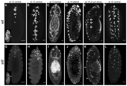
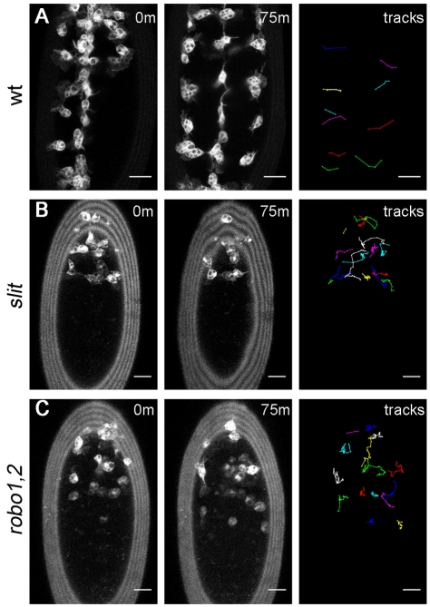
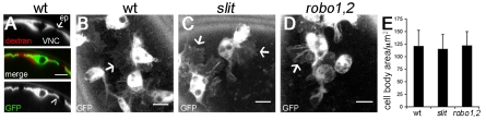
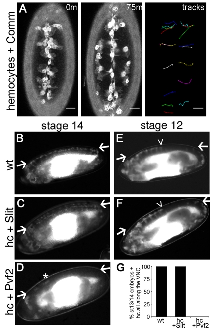
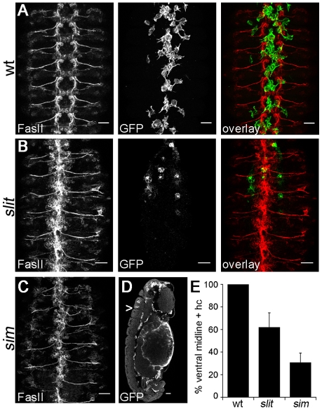
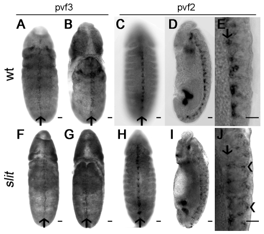
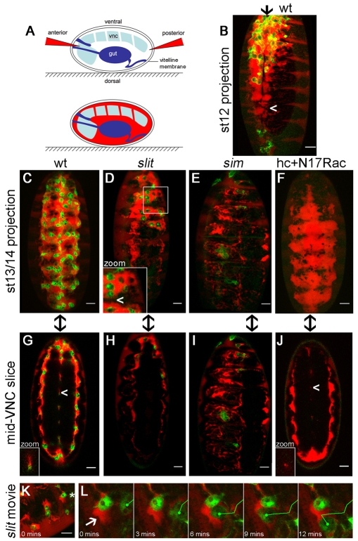
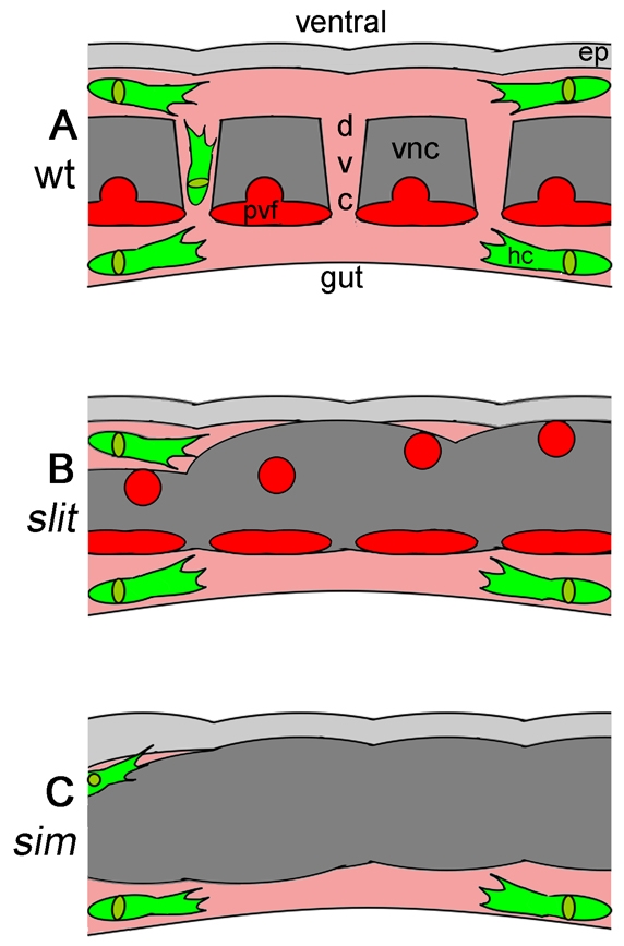
During embryonic development, Drosophila macrophages (haemocytes) undergo a series of stereotypical migrations to disperse throughout the embryo. One major migratory route is along the ventral nerve cord (VNC), where haemocytes are required for the correct development of this tissue. We show, for the first time, that a reciprocal relationship exists between haemocytes and the VNC and that defects in nerve cord development prevent haemocyte migration along this structure. Using live imaging, we demonstrate that the axonal guidance cue Slit and its receptor Robo are both required for haemocyte migration, but signalling is not autonomously required in haemocytes. We show that the failure of haemocyte migration along the VNC in slit mutants is not due to a lack of chemotactic signals within this structure, but rather to a failure in its detachment from the overlying epithelium, creating a physical barrier to haemocyte migration. This block of haemocyte migration in turn disrupts the formation of the dorsoventral channels within the VNC, further highlighting the importance of haemocyte migration for correct neural development. This study illustrates the important role played by the three-dimensional environment in directing cell migration in vivo and reveals an intriguing interplay between the developing nervous system and the blood cells within the fly, demonstrating that their development is both closely coupled and interdependent.


Figures

Fig. 1.
Fig. 1.
Haemocyte developmental migrations are perturbed in slit mutants. (A-L) Wild-type (w;srp-Gal4,UAS-GFP;crq-Gal4,UAS-GFP; wt) (A-F) and slit mutant (w;slitGA178;crq-Gal4,UAS-GFP) (G-L) Drosophila embryos were immunostained for GFP to show haemocyte dispersal. Ventral views of wild-type (A) and slit mutant (G) embryos reveal that haemocyte progression along the midline is only moderately delayed in the latter at stage 12. Lateral views of the same embryos (insets) show the extent of germ band retraction and the position of the posterior-most haemocyte along the ventral midline (arrowhead). By stage 13, haemocytes occupy the full length of the ventral midline in wild types (B), but make little progress posteriorly in slit mutant embryos (H). Dorsal views indicate that haemocytes reach other destinations equally well in each genotype (C,I). Lateral views at stage 14 reveal that whereas haemocytes continue to occupy the ventral midline in wild types (D), they are absent from large regions of the ventral surface of the VNC in slit mutants (J). Single confocal slices through the midline show haemocytes along both sides of the VNC and within dorsoventral channels in wild types (arrows in E), whereas only the dorsal side of the VNC is fully occupied in slit mutants (the region between arrowheads lacks haemocytes on the ventral side of the VNC) (K). By stage 15, three parallel lines of haemocytes are visible ventrally in both wild-type (F) and slit mutant (L) embryos (arrows indicate the midline), but haemocytes remain absent from much of the ventral side of the VNC in the latter. Anterior is up. Scale bars: 20 μm.
Fig. 2.
Fig. 2.
Dynamic imaging reveals a failure in haemocyte progression along the ventral midline in slit and robo1,2 mutants. (A-C) GFP-expressing haemocytes in stage 13/14 wild-type (w;srp-Gal4,UAS-GFP;crq-Gal4,UAS-GFP) (A), slit (w;slitGA178;crq-Gal4,UAS-GFP) (B) and robo1,2 mutant (w;robo1,2;crq-Gal4,UAS-GFP) (C) Drosophila embryos were subjected to live imaging for 75 minutes as they migrated along the ventral midline. Positions of haemocytes at 0 and 75 minutes are shown. For clarity, only the laterally migrating wild-type tracks are shown, whereas all cell trajectories are shown for slit and robo1,2 mutants. Whereas haemocytes migrate laterally in each segment in wild-type embryos (A), haemocytes fail to cover the length of the ventral midline in slit (B) and robo1,2 (C) mutants and, consequently, lateral migration does not occur in every segment. Anterior is up. See Movies 1-3 in the supplementary material for full videos. Scale bars: 20 μm.
Fig. 3.
Fig. 3.
Haemocyte morphology is unaffected in slit and robo1,2 mutants. (A) Rhodamine-dextran (red in merged image) was injected into wild-type Drosophila embryos (w;srp-Gal4,UAS-GFP;crq-Gal4,UAS-GFP) with GFP-labelled haemocytes (green in merge) to label the extracellular spaces between the VNC and epidermis (ep). Orthogonal projections of haemocytes at the ventral midline reveal their thin lamellipodial protrusions (arrowhead). (B-D) z-projections show that haemocytes on the ventral side of the VNC in wild-type embryos (w;;crq-Gal4,UAS-GFP) (B) are morphologically indistinct from those containing the same Gal4 driver and UAS reporter in slit (C) and robo1,2 (D) mutants, extending large lamellipodial protrusions (arrows) from their phagosome-containing cell bodies. (E) No significant difference in haemocyte cell body area was found between wild types, slit and robo1,2 mutants (41, 54 and 35 haemocytes were measured, respectively). Error bars indicate s.d. Scale bars: 10 μm.
Fig. 4.
Fig. 4.
Slit does not directly regulate haemocyte migration. (A) GFP- and Comm-expressing haemocytes in stage 13/14 Drosophila embryos were subjected to live imaging for 75 minutes as they migrated on the ventral midline. The positions of haemocytes at 0 and 75 minutes and their associated tracks as they leave the midline reveal that Comm expression has no effect on haemocyte progression down the midline or on lateral migration. For the full video, see Movie 4 in the supplementary material. Anterior is up. Scale bars: 20 μm. (B-F)To verify that Slit does not directly regulate haemocyte migration, srp-Gal4 and crq-Gal4 were used to drive haemocyte-specific expression of UAS-GFP alone (wt), UAS-GFP and UAS-slit (hc + Slit), or UAS-GFP and UAS-Pvf2 (hc + Pvf2). Haemocytes cover the VNC at stage 14 when wt (B) or expressing Slit (C), whereas Pvf2 expression causes retention in the head (D). Few escapees were observed (asterisk), with D representing the least severe retention observed. Compared with wild types (E), Slit expression did not accelerate exit from the head at stage 12 (F). Lateral views with anterior to the left and ventral up. Arrows indicate VNC; arrowheads indicate the progression of haemocytes along the VNC. (G) Percentage of embryos with haemocytes all along the VNC at stage 13/14 (35, 60 and 78 embryos were scored for wt, hc + Slit and hc + Pvf2, respectively).
Fig. 5.
Fig. 5.
Haemocyte migration and VNC development are perturbed in both slit and sim mutants. (A-D) Stage 13/14 wild-type (A), slit (B) and sim mutant (C,D) Drosophila embryos with GFP-expressing haemocytes were co-immunostained for the neuronal marker FasII (red in overlay) and GFP (green in overlay) to show VNC morphology and haemocyte position, respectively. Only haemocytes on the ventral side of the VNC are shown for clarity. z-projections of FasII staining (ventral view) reveal that the regular repeating structure of the wild-type VNC (A) is collapsed in slit (B) and sim (C) mutants. Although haemocytes are able to migrate along the dorsal surface of the VNC in sim mutants, single 1.5 μm confocal slices of laterally orientated embryos (D) reveal that very few segments contain haemocytes on the ventral side of the VNC at the midline (the arrowhead indicates the posterior-most haemocyte). (E) Quantification of haemocyte distribution along the VNC at stage 13/14. The number of segments containing haemocytes on the ventral side of the VNC is expressed as a percentage of the total number of segments for each embryo. A significant difference (P<0.05, Student's t-test) was found between each genotype when 44 wild-type, 63 slit and 40 sim embryos were scored; error bars indicate s.d. Anterior is up. Scale bars: 20 μm.
Fig. 6.
Fig. 6.
Expression of Pvf ligands is not lost in slit mutants. (A,B,F,G) In situ hybridisations on stage 12 wild-type (A,B) and slit mutant (F,G) Drosophila embryos show that Pvf3 is expressed along the midline on both the ventral (A,F) and dorsal (B,G) sides in both genotypes (arrows indicate the midline). (C,H) In stage 13/14 wild-type (C) and slit mutant (H) embryos, in situ hybridisation indicates that Pvf2 is expressed at the ventral midline (arrows). (D,I) Lateral views at stage 13/14 reveal Pvf2 expression to be restricted to the dorsal side of the VNC in wild-type embryos (D), but displaced from the dorsal aspect of the VNC in slit mutants (I). (E,J) High-magnification images of the ventral surface of embryos from D,I show tight clustering of the Pvf2 signal on the dorsal aspect of the VNC (arrow) in wild types (E), whereas some Pvf2-expressing cells are displaced ventrally (arrowheads) in slit mutants (J). Anterior is up. Scale bars: 20 μm.
Fig. 7.
Fig. 7.
VNC defects create physical barriers to haemocyte migration. (A) Rhodamine-dextran (red) was injected into live Drosophila embryos with GFP-labelled haemocytes (green in B-L) to reveal extracellular space. Dextran was excluded from tissues such as the VNC and gut but spread between the epidermis and VNC and through dorsoventral channels in the VNC. (B,C) Dextran pooled along the ventral midline in stage 12 (B) and 13/14 (C) wild-type embryos, but was more restricted at mid-trunk segments at stage 12 (arrowhead, B). (D)By contrast, VNC-epidermal contacts created physical barriers (arrowhead in inset) to posterior movement of dextran and haemocytes in stage 13/14 slit mutants. (E) The restriction in dextran-accessible space and haemocyte progression was more severe in sim mutants. (F) Haemocytes are not responsible for VNC-epidermal separation, as dextran localised along the entire VNC at stage 13/14 when haemocyte migration was blocked by haemocyte-specific expression of N17Rac. (G-J) Single 1.5 μm confocal sections through the middle of the VNC highlight dorsoventral channels in stage 13/14 wild-type embryos (G), which are absent in slit (H) and sim mutants (I) and greatly reduced in embryos containing N17Rac-expressing haemocytes (J). Arrowheads indicate channels, which are shown at the same scale in insets (G,J). Anterior is up and arrows indicate the ventral midline in B-J. (K) Single image from Movie 7 in the supplementary material, showing the confinement of haemocytes by dextran-inaccessible barriers in the anterior of a stage 13/14 slit mutant embryo. (L) A haemocyte (asterisk in K) is deflected by a physical barrier (arrow) during the movie. Scale bars: 20 μm.
Fig. 8.
Fig. 8.
VNC-epidermal separation and haemocyte migration are developmentally coupled. (A-C) Schematic of the VNC and surrounding embryonic environment at stage 13 in wild-type (A), slit (B) and sim (C) Drosophila embryos. The embryos are viewed laterally at the midline; anterior is to the left. For normal development, the epidermis (light grey) and VNC (dark grey) must separate to enable haemocytes (green) to migrate along the VNC through dextran-accessible space (pink) by following the attractive Pvf ligands secreted by Sim-expressing midline cells (red). This allows haemocytes to reach mid-trunk segments and remodel the developing VNC (A). In slit mutants, despite the presence of Pvfs, a defective VNC results in its failure to separate from the epidermis, blocking haemocyte migration (B). Pvf expression is lost and VNC-epidermal separation defects are enhanced in sim mutants, completely preventing haemocyte progress along the ventral midline (C). ep, epidermis; dvc, dorsoventral channel; vnc, ventral nerve cord; hc, haemocyte.

References

    1. Battye R., Stevens A., Jacobs J. R. (1999). Axon repulsion from the midline of the Drosophila CNS requires slit function. Development 126, 2475-2481 - PubMed
    1. Bruckner K., Kockel L., Duchek P., Luque C. M., Rorth P., Perrimon N. (2004). The PDGF/VEGF receptor controls blood cell survival in Drosophila. Dev. Cell 7, 73-84 - PubMed
    1. Carmeliet P., Tessier-Lavigne M. (2005). Common mechanisms of nerve and blood vessel wiring. Nature 436, 193-200 - PubMed
    1. Cho N. K., Keyes L., Johnson E., Heller J., Ryner L., Karim F., Krasnow M. A. (2002). Developmental control of blood cell migration by the Drosophila VEGF pathway. Cell 108, 865-876 - PubMed
    1. Choi K., Kennedy M., Kazarov A., Papadimitriou J. C., Keller G. (1998). A common precursor for hematopoietic and endothelial cells. Development 125, 725-732 - PubMed

Publication types

MeSH terms