. 2010 Aug;38(15):5177-92.
doi: 10.1093/nar/gkq260. Epub 2010 Apr 14.

Ribosomal protein L35 is required for 27SB pre-rRNA processing in Saccharomyces cerevisiae

Affiliations

Ribosomal protein L35 is required for 27SB pre-rRNA processing in Saccharomyces cerevisiae

Reyes Babiano et al. Nucleic Acids Res. 2010 Aug.

Abstract

Ribosome synthesis involves the concomitance of pre-rRNA processing and ribosomal protein assembly. In eukaryotes, this is a complex process that requires the participation of specific sequences and structures within the pre-rRNAs, at least 200 trans-acting factors and the ribosomal proteins. There is little information on the function of individual 60S ribosomal proteins in ribosome synthesis. Herein, we have analysed the contribution of ribosomal protein L35 in ribosome biogenesis. In vivo depletion of L35 results in a deficit in 60S ribosomal subunits and the appearance of half-mer polysomes. Pulse-chase, northern hybridization and primer extension analyses show that processing of the 27SB to 7S pre-rRNAs is strongly delayed upon L35 depletion. Most likely as a consequence of this, release of pre-60S ribosomal particles from the nucleolus to the nucleoplasm is also blocked. Deletion of RPL35A leads to similar although less pronounced phenotypes. Moreover, we show that L35 assembles in the nucleolus and binds to early pre-60S ribosomal particles. Finally, flow cytometry analysis indicated that L35-depleted cells mildly delay the G1 phase of the cell cycle. We conclude that L35 assembly is a prerequisite for the efficient cleavage of the internal transcribed spacer 2 at site C(2).


Figures
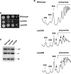
Figure 1.
Figure 1.
Deletion of either RPL35A or RPL35B leads to a deficit in 60S r-subunits. (A) Growth test of the rpl35A and rpl35B null mutants compared to their isogenic wild-type control strain. Strains RBY138 (Δrpl35A), RBY139 (Δrpl35B) and BY4741 (Wild-type) were grown in YPD and diluted to an OD600 of 0.05. A 10-fold series of dilutions was performed for each strain and 5 µl drops were plated on YPD plates. Plates were incubated at 30°C for 4 days. (B) The above strains were grown in YPD at 30°C and harvested at an OD600 of 0.8, cell extracts were prepared and 10 A260 of each extract were resolved in 7–50% sucrose gradients. The A254 was continuously measured. Sedimentation is from left to right. The peaks of free 40S and 60S r-subunits, 80S free couples/monosomes and polysomes are indicated. Half-mers are labelled by arrows. (C) Equivalent amounts of the cell extracts described above were subjected to western blot analysis with antibodies against the r-proteins L35, L1 and S8.
Figure 2.
Figure 2.
Depletion of L35 results in a deficit in 60S r-subunits. (A) Growth comparison of the strains BY4741 (Wild-type) and RBY175 (GAL::RPL35). The cells were grown in YPGal and streaked on YPGal (Gal) and YPD (Glc) plates, which were incubated at 30°C for 3 days. (B) The above strains were grown in YPGal at 30°C and shifted to YPD. Cells were harvested at the indicated times after the shift and cell extracts were prepared. Equivalent amounts of cell extracts were subjected to western blot analysis with antibodies against the r-proteins L35, L1 and S8. (C) 10 A260 of cell extract from the GAL::RPL35 strain grown in YPGal (Gal) or shifted for 6 h (6 h Glc) or 12 h (12 h Glc) to YPD were subjected to polysome analysis in 7–50% sucrose gradients. The peaks of free 40S and 60S r-subunits, 80S free couples/monosomes and polysomes are indicated. Half-mers are labelled by arrows.
Figure 3.
Figure 3.
Depletion of L35 impairs 27S pre-rRNA processing. (A) Wild-type control strain BY4741 (Wild-type) and strain RBY175 (GAL::RPL35) were transformed with YCplac33 and then grown at 30°C in SGal-Ura and shifted for 8 h to SD-Ura. Cells were pulse-labelled with [,6-3H]uracil for 2 min followed by a chase with a large excess of unlabelled uracil for the times indicated. Total RNA was extracted and 20 000 cpm was loaded and separated on (A) a 1.2% agarose-6% formaldehyde gel or (B) a 7% polyacrylamide-8M urea, transferred to nylon membranes and visualized by fluorography. The positions of the different pre-rRNAs and mature rRNA are indicated.
Figure 4.
Figure 4.
Deletion of RPL35A and depletion of L35 affects the steady-state levels of pre-rRNA and mature rRNA species. Strains BY4741 (Wild-type), RBY138 (Δrpl35A) and RBY139 (Δrpl35B) were grown in YPD at 30°C and harvested. BY4741 (Wild-type) and RBY175 (GAL::RPL35) were grown in YPGal at 30°C, shifted to YPD and harvested at the indicated times. Total RNA was extracted of each sample. Equal amount of total RNA (5 µg) was subjected to northern hybridization or primer extension analysis. (A) Northern analysis of high-molecular-mass pre-rRNAs and mature rRNAs. (B) Northern analysis of low-molecular-mass pre-rRNAs and mature rRNAs. Probes, between parentheses, are described in Supplementary Figure S1A. (C) Primer extension analysis with probe g within ITS2 allows detection of 27SA2, 27SA3 and both 27SB pre-rRNAs. Signal intensities were measured by phosphorimager scanning; values (indicated below each lane) were normalized to those obtained for the wild-type control grown either in YPD or YPGal, arbitrarily set at 1.0.
Figure 5.
Figure 5.
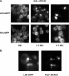
Depletion of L35 leads to nuclear retention of the 60S r-subunit reporter L25-eGFP. (A) RBY175 (GAL::RPL35) cells expressing either L25-eGFP or S2-eGFP were grown in SGal-Ura at 30°C, shifted to SD-Ura for up to 9 h. The GFP signal was analyzed by fluorescence microscopy. (B) Magnified picture of selected GAL::RPL35 cells expressing L25-eGFP and the nucleolar marker Nop1-DsRed, which were depleted for 9 h in SD-Ura. Arrows point to nucleolar fluorescence.
Figure 6.
Figure 6.
L35B assembles in the nucleus as inferred upon inhibition of export of 60S pre-ribosomal particles. (A) Localization of L25-eGFP and L35B-eGFP in a LMB sensitive strain. Either L25-eGFP or L35B-eGFP was expressed in AJY1539 cells, which harbours the LMB sensitive crm1 (T539C) allele. Cells were incubated for 3 h at 30°C in the absence (-LMB) or presence of 200 ng/ml LMB (+LMB). The GFP signal was inspected by fluorescence microscopy. Arrows point to nuclear fluorescence. (B) Localization of L25-eGFP and L35-eGFP upon induction of a nmd3 dominant negative allele. Δrpl35B cells expressing L25-eGFP or L35B-eGFP were transformed with the pRS316-GAL-nmd3Δ100 plasmid and transformants were grown in the presence of raffinose (Raf, SRaf-Leu-Ura medium). Then, galactose was added to fully induce the Nmd3Δ100 protein. The GFP signal was inspected by fluorescence microscopy at the time indicated. Arrows point to nuclear fluorescence.
Figure 7.
Figure 7.
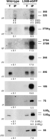
L35B associates with all pre-60S ribosomal particles. L35B-eGFP was affinity purified from total cellular extracts of Δrpl35B cells expressing L35B-eGFP with GFP-Trap®_A beads as indicated in ‘Materials and Methods’ section. Wild-type cells, which express untagged L35B, serves as a negative control. RNA was extracted from the pellets obtained after purification (lanes IP) or from an amount of total extract corresponding to 1/100 of that used for purification (lanes T) and subjected to northern analysis of pre-rRNAs and mature rRNAs. Probes, between parentheses, are described in Figure S1A. Signal intensity was measured by phosphorimager scanning; values (below each IP lane) refer to as the percentage of each RNA recovered after purification.
Figure 8.
Figure 8.
Depletion of L35 leads to a mild delay of the cell cycle at the G1 phase. (A) FACS analysis of unsynchronized cells from the BY4741 (Wild-type) or the RBY175 (GAL::RPL35) strains. Cells were grown in YPGal (Gal) or shifted for up to 12 h to YPD (Glc) at 30°C. 1C and 2C peaks correspond to cells with unreplicated and duplicated genomes, respectively. Numbers refers to the 1C/2C area peak ratios (B) Cell morphology of the wild-type and GAL::RPL35 cells. Cells were stained with DAPI for localization of nuclei and then visualized by fluorescence and visible phase contrast microscopy. Only merged images are shown. Numbers refer to mean percentages of unbudded cells after three independent experiments.

References

    1. Steitz TA. A structural understanding of the dynamic ribosome machine. Nat. Rev. Mol. Cell. Biol. 2008;9:242–253. - PubMed
    1. Schmeing TM, Ramakrishnan V. What recent ribosome structures have revealed about the mechanism of translation. Nature. 2009;461:1234–1242. - PubMed
    1. Spahn CM, Beckmann R, Eswar N, Penczek PA, Sali A, Blobel G, Frank J. Structure of the 80S ribosome from Saccharomyces cerevisiae-tRNA-ribosome and subunit-subunit interactions. Cell. 2001;107:373–386. - PubMed
    1. Chandramouli P, Topf M, Menetret JF, Eswar N, Cannone JJ, Gutell RR, Sali A, Akey CW. Structure of the mammalian 80S ribosome at 8.7 A resolution. Structure. 2008;16:535–548. - PMC - PubMed
    1. Henras AK, Soudet J, Gerus M, Lebaron S, Caizergues-Ferrer M, Mougin A, Henry Y. The post-transcriptional steps of eukaryotic ribosome biogenesis. Cell. Mol. Life Sci. 2008;65:2334–2359. - PMC - PubMed

Publication types

MeSH terms