. 2010 Jun;151(6):2876-85.
doi: 10.1210/en.2009-1282. Epub 2010 Apr 14.

Signal transducer and activator of transcription 5a mediates mammary ductal branching and proliferation in the nulliparous mouse

Affiliations

Signal transducer and activator of transcription 5a mediates mammary ductal branching and proliferation in the nulliparous mouse

Sarah J Santos et al. Endocrinology. 2010 Jun.

Abstract

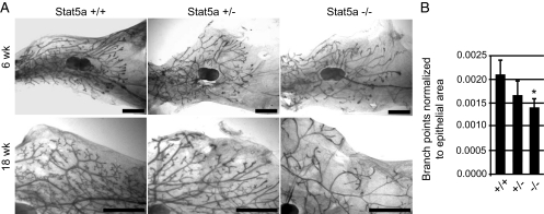
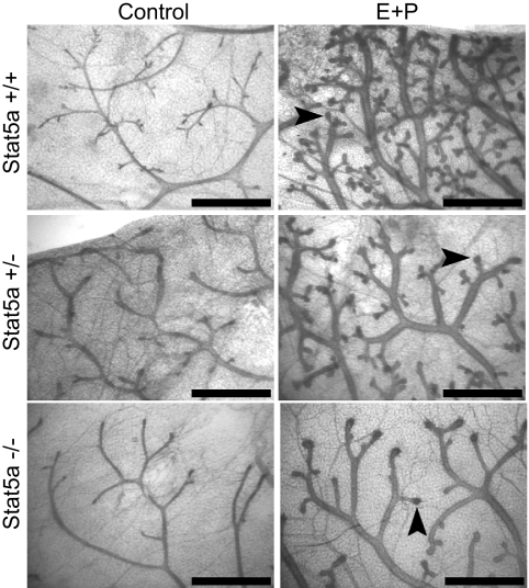
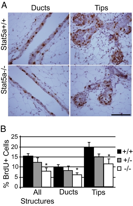
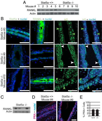
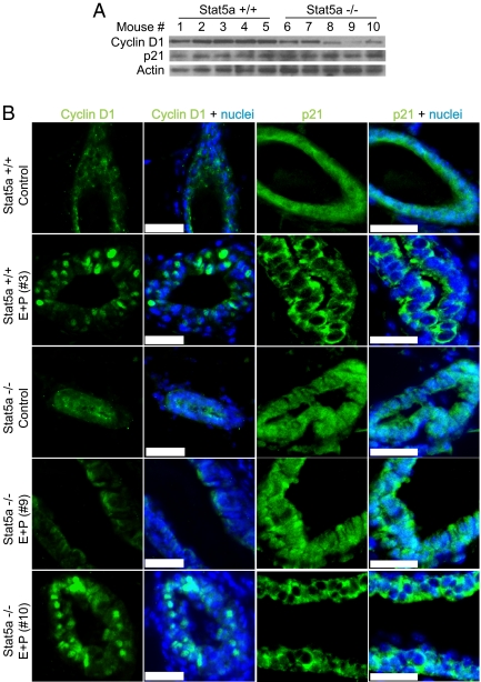
Signal transducer and activator of transcription (Stat)5a is a critical regulator of mammary gland development. Previous studies have focused on Stat5a's role in the late pregnant and lactating gland, and although active Stat5a is detectable in mammary epithelial cells in virgin mice, little is known about its role during early mammary gland development. In this report, we compare mammary gland morphology in pubertal and adult nulliparous wild-type and Stat5a-/- mice. The Stat5a-null mammary glands exhibited defects in secondary and side branching, providing evidence that Stat5a regulates these processes. In addition, Stat5a-/- mammary glands displayed an attenuated proliferative response to pregnancy levels of estrogen plus progesterone (E+P), suggesting that it plays an important role in early pregnancy. Finally, we examined one potential mediator of Stat5a's effects, receptor activator of nuclear factor-kappaB ligand (RANKL). Stat5a-/- mammary glands were defective in inducing RANKL in response to E+P treatment. In addition, regulation of several reported RANKL targets, including inhibitor of DNA binding 2 (Id2), cyclin D1, and the cyclin-dependent kinase inhibitor p21(Waf1/Cip1), was altered in Stat5a-/- mammary cells, suggesting that one or more of these proteins mediate the effects of Stat5a in E+P-treated mammary epithelial cells.


Figures

Figure 1
Figure 1
Whole-mount analysis of pubertal and adult virgin mice. The fourth inguinal mammary glands were removed from Stat5a+/+, Stat+/−, and Stat−/− mice and prepared as whole mounts. A, Mammary glands from 6-wk-old pubertal mice and 18-wk-old mature mice. Scale bars, 2 mm. B, Analysis of primary branch points in 18-wk-old Stat5a+/+, Stat5a+/−, and Stat5a−/− mammary glands. Quantitation was carried out as described in Materials and Methods for three to nine animals per genotype and is shown as the average ± sd. *, P < 0.05 that the number of branch points in Stat5a−/− mammary glands were less than in Stat5a+/+ glands.
Figure 2
Figure 2
Morphological response of mammary glands to hormone treatment. Mammary gland whole mounts were prepared from 18-wk-old OVX Stat5a+/+, Stat5a+/−, and Stat5a−/− mice treated for 5 d with E+P. Arrows indicate enlarged ductal tips where alveoli will form. Scale bar, 1 mm.
Figure 3
Figure 3
Impaired proliferative response to hormones in Stat5a−/− mammary glands. Immunoperoxidase detection of incorporated BrdU was performed on mammary gland sections from 18-wk-old OVX mice treated for 5 d with E+P. A, Representative images from BrdU staining are shown for Stat5a+/+ and Stat5a−/− ducts and alveolar buds. Scale bar, 30 μm. B, Quantitation of the percent BrdU+ luminal epithelial cells. Results are shown for quantitation carried out on all structures, as well as duct and alveolar bud structures independently. The values represent the mean ± sem from five mice per group with a minimum of 1000 cells analyzed per mouse. *, P < 0.05 that there were fewer BrdU+ cells in glands of Stat5a−/− mice compared with Stat5a+/+ glands.
Figure 4
Figure 4
RANKL expression and Id2 localization are downstream targets of Stat5a in the mammary gland. A, Western blot analysis of RANKL levels in whole mammary gland homogenates from OVX 18-wk-old Stat5a+/+ and Stat5a−/− mice treated for 5 d with E+P. Each lane represents a single mammary gland from an individual mouse. B, Immunofluorescent staining for Stat5a, RANKL, and Id2 was performed on mammary gland sections from the mice described in A. Representative images of Stat5a, RANKL, and Id2 staining (green) are shown. For sections stained for Stat5a and RANKL, nuclei were counter-stained with DAPI (blue), whereas nuclei in sections stained for Id2 were counterstained with TO-PRO (blue). Scale bar, 25 μm. Note the nuclear localization of Id2 in mice nos. 3 and 10 (green arrows) and the cytoplasmic localization of Id2 in mouse no. 9 (white arrows). C, Western blot analysis of RANKL levels in whole mammary gland homogenates from OVX 18-wk-old Stat5a−/− mice treated for 5 d with P. Each lane represents a single mammary gland from an individual mouse. D, Immunofluorescent detection of PRA (red) in mammary gland samples from the mice described in A. Nuclei were stained with DAPI (blue). Representative images from sections of Stat5a+/+ and Stat5a−/− mammary glands are shown. Scale bar, 30 μm. E, Quantitation of the percentage of PRA-positive epithelial cells was conducted. The values represent the mean ± sem from five mice per group, with a minimum of 1000 cells analyzed per mouse.
Figure 5
Figure 5
Cyclin D1 expression and p21 localization are altered in mammary glands from Stat5a knockout mice. A, Western blot analysis of cyclin D1 and p21 levels in whole mammary gland homogenates from 18-wk-old Stat5a+/+ and Stat5a−/− mice treated for 5 d with E+P. Each lane represents a single mammary gland from an individual mouse, corresponding to the same mice shown in Fig. 4. B, Immunofluorescent staining for cyclin D1 and p21 was performed on mammary gland sections from the mice described in A. Representative images of cyclin D1 and p21 staining (green) are shown. Nuclei were counterstained with DAPI (blue). Scale bar, 20 μm. Note the strong nuclear expression of cyclin D1 and the cytoplasmic localization of p21 in mice nos. 3 and 10. Additional cyclin D1 and p21 staining is shown in Supplemental Fig. 2.

Similar articles

Cited by

References

    1. Howlin J, McBryan J, Martin F 2006 Pubertal mammary gland development: insights from mouse models. J Mammary Gland Biol Neoplasia 11:283–297 - PubMed
    1. Brisken C, Rajaram RD 2006 Alveolar and lactogenic differentiation. J Mammary Gland Biol Neoplasia 11:239–248 - PubMed
    1. Darnell Jr JE 1997 STATs and gene regulation. Science 277:1630–1635 - PubMed
    1. Ihle JN 1996 STATs: signal transducers and activators of transcription. Cell 84:331–334 - PubMed
    1. Liu X, Robinson GW, Gouilleux F, Groner B, Hennighausen L 1995 Cloning and expression of Stat5 and an additional homologue (Stat5b) involved in prolactin signal transduction in mouse mammary tissue. Proc Natl Acad Sci USA 92:8831–8835 - PMC - PubMed

Publication types