. 2010 Jun 1;21(11):1850-63.
doi: 10.1091/mbc.e09-09-0801. Epub 2010 Apr 14.

Alpha-synuclein delays endoplasmic reticulum (ER)-to-Golgi transport in mammalian cells by antagonizing ER/Golgi SNAREs

Affiliations

Alpha-synuclein delays endoplasmic reticulum (ER)-to-Golgi transport in mammalian cells by antagonizing ER/Golgi SNAREs

Nandhakumar Thayanidhi et al. Mol Biol Cell. .

Abstract

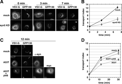
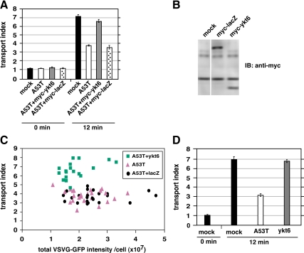
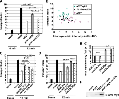
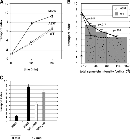
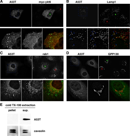
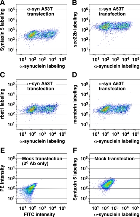
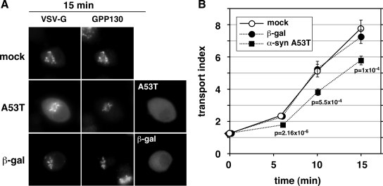
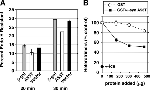
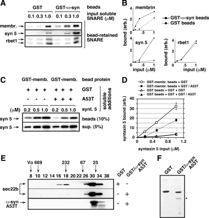
Toxicity of human alpha-synuclein when expressed in simple organisms can be suppressed by overexpression of endoplasmic reticulum (ER)-to-Golgi transport machinery, suggesting that inhibition of constitutive secretion represents a fundamental cause of the toxicity. Whether similar inhibition in mammals represents a cause of familial Parkinson's disease has not been established. We tested elements of this hypothesis by expressing human alpha-synuclein in mammalian kidney and neuroendocrine cells and assessing ER-to-Golgi transport. Overexpression of wild type or the familial disease-associated A53T mutant alpha-synuclein delayed transport by up to 50%; however, A53T inhibited more potently. The secretory delay occurred at low expression levels and was not accompanied by insoluble alpha-synuclein aggregates or mistargeting of transport machinery, suggesting a direct action of soluble alpha-synuclein on trafficking proteins. Co-overexpression of ER/Golgi arginine soluble N-ethylmaleimide-sensitive factor attachment protein receptors (R-SNAREs) specifically rescued transport, indicating that alpha-synuclein antagonizes SNARE function. Ykt6 reversed alpha-synuclein inhibition much more effectively than sec22b, suggesting a possible neuroprotective role for the enigmatic high expression of ykt6 in neurons. In in vitro reconstitutions, purified alpha-synuclein A53T protein specifically inhibited COPII vesicle docking and fusion at a pre-Golgi step. Finally, soluble alpha-synuclein A53T directly bound ER/Golgi SNAREs and inhibited SNARE complex assembly, providing a potential mechanism for toxic effects in the early secretory pathway.


Figures

Figure 1.
Figure 1.
α-Synuclein A53T delays and myc-ykt6 restores ER-to-Golgi Transport. (A) Representative epifluorescent images from several incubation time points displaying VSV-G-GFP and GPP130 in the same cells. The top row of cells were electroporated with buffer only and the bottom row were electroporated with an siRNA for syntaxin 5. (B) Quantitation of transport employing at least 20 randomly chosen cells from each condition (see Materials and Methods). (C) Representative images of cells from 12 min at 32°C on separate coverslips singly transfected with VSV-G-GFP (top row), doubly transfected with VSV-G-GFP and α-synuclein A53T (middle row), and triply transfected with VSV-G-GFP, α-synuclein A53T, and myc-ykt6 (bottom row). Golgi staining with GPP130 is shown for each cell. (D) Quantitation of the experiment in C. Error bars represent SEs.
Figure 2.
Figure 2.
Restoration of ER-to-Golgi transport is specific for coexpression of myc-ykt6 and α-synuclein. (A) NRK cells coexpressing myc-β-galactosidase did not exhibit restored transport. (B) Immunoblot of NRK cell lysates using anti-myc antibody. (C) Scatter plot showing every individual cell from the experiment in part A, as a function of VSV-G-GFP expression and transport index. (D) Transport indices of NRK cells electroporated with either α-synuclein A53T or myc-ykt6 alone (not coexpressed) shows that myc-ykt6 does not itself accelerate transport. Error bars represent SEs.
Figure 3.
Figure 3.
Ykt6 rescues transport more potently than sec22b. (A) Comparison of transport rescue by myc-sec22b and myc-ykt6. (B) Scatter plot showing every cell from the experiment in A, as a function of total cellular α-synuclein A53T staining intensity and transport index. (C) A separate experiment from part A, in which a wide intensity range of myc-sec22b coexpressing cells was imaged, binned according to anti-myc intensity, and transport indices calculated. (D) Another experiment in which anti-myc staining intensities were binned and transport indices plotted for myc-ykt6 and myc-sec22b coexpressing cells. (E) Total anti-myc staining intensities for the bins of cells analyzed in D. (F) Anti-myc immunoblot of NRK cell lysates of duplicate coverslips to those analyzed in D and E. The statistical significance of differences between the indicated mean transport indices are reported as p values from a two-tailed Student‘s t test. Error bars represent SEs.
Figure 4.
Figure 4.
Wild-type α-synuclein delays transport to the same degree, but less potently than A53T. (A) Time course of ER-to-Golgi transport in NRK cells electroporated with wild-type or A53T α-synuclein. Only cells showing strong (approx. top quartile) anti-α-synuclein staining were imaged. (B) A separate experiment in which a wider range of anti-α-synuclein staining intensities were imaged, cells were separated into intensity bins as shown on the x-axis, and their transport indices plotted. Both the 12-min and 0-min timepoints are plotted for mock-transfected cells along the y-axis (values of ∼8.5 and ∼1.2, respectively). (C) Coexpression of myc-ykt6 partially restores transport inhibition caused by wild-type α-synuclein. In some instances, the statistical significance of differences between the indicated mean transport indices are reported as p values from a two-tailed Student‘s t test. Error bars represent SEs.
Figure 5.
Figure 5.
Subcellular distributions of α-synuclein A53T, coexpressed myc-ykt6, and the unperturbed endogenous cellular transport machinery. (A) Comparison between α-synuclein A53T and myc-ykt6 staining patterns. (B) Comparison of α-synuclein A53T and endogenous LAMP-1. Blue arrowheads highlight a few of the α-synuclein-positive particles that are positive for LAMP-1. (C) Comparison of α-synuclein A53T and endogenous rab1. (D) Comparison of α-synuclein A53T and endogenous GPP130. In each block shown, green asterisks mark the nucleus of an α-synuclein A53T-expressing cell and red asterisks mark the nucleus of an untransfected cell for comparison. Shown are individual focal planes of deconvolved widefield images. (E) A53T-transfected NRK cells were extracted with cold 1% Triton X-100 and centrifuged at 10,000 × g for 15 min to generate a supernatant and pellet. Equal proportions of these fractions were analyzed by SDS-PAGE and immunoblotting for α-synuclein and caveolin, a protein known to be in detergent-resistant membrane domains.
Figure 6.
Figure 6.
α-Synuclein A53T overexpression does not significantly affect the expression of ER/Golgi SNAREs. Electroporated NRK cells were allowed to express the α-synuclein A53T construct for 2 d before fixation, permeabilization, and immunofluorescence labeling of α-synuclein (with FITC) and each ER/Golgi SNARE (with PE). Labeled cell suspensions were analyzed by flow cytometry. Dot plots display relationship between α-syn A53T labeling and labeling for syntaxin 5 (A), sec22b (B), rbet1 (C), and membrin (D), where the frequency of cells with similar labeling intensities is indicated by color warmth. (E) Mock-transfected cells in which primary antibodies were omitted. (F) Mock-transfected cells labeled with anti-α-synuclein and anti-syntaxin 5 antibodies.
Figure 7.
Figure 7.
α-Synuclein expression retards ER-to-Golgi transport in neuroendocrine PC12 cells. (A) Representative epifluorescent images from 15 min at 32°C, displaying VSV-G-GFP, GPP130, and either α-synuclein A53T or β-gal driven by pcDNA3.1 transfection. (B) Quantitation of transport employing at least 20 randomly chosen cells from each condition (see Materials and Methods). Bars represent SEs and are shown only when exceeding symbol size. The statistical significance of differences between the A53T and mock transport indices at the indicated time points are reported as p values from a two-tailed Student‘s t test.
Figure 8.
Figure 8.
(A) α-Synuclein inhibits transport before cargo modification by mannosidase II. NRK cells were electroporated with VSV-G-myc DNA together with a construct for either β-galactosidase, α-synuclein A53T, or pcDNA3.1 vector alone. After 24 h, the cells were infected with vaccinia virus vTF7 for 6 h at 40°C to amplify expression (see Materials and Methods). Cells were either lysed directly (data not shown) or shifted to 32°C for 20, 30, or 40 (data not shown) minutes before lysis, digestion with endoglycosidase H, and immunoblotting with anti-myc to detect VSV-G-myc. Histograms show GR as a percentage of GS + GR. Error bars show SE of duplicate gels run on the same cell extracts. The same trends were observed in a completely separate experiment with multiple time points. Supplemental Figure S4 shows one of the immunoblots from which the data were quantified. (B) α-Synuclein A53T directly inhibits COPII vesicle fusion to form pre-Golgi intermediates. Homotypic COPII vesicle fusion measured using the in vitro VSV-G heterotrimer cargo mixing assay. Purified proteins were added after vesicle budding and were incubated with the budded vesicles for 1 h before a 1 h fusion reaction at 32°C. “Ice” indicates heterotrimer signal obtained when the budded vesicles were mixed but left on ice during the fusion incubation. Fusion assay data are presented as means of duplicate determinations with error bars representing SE where larger than symbol size.
Figure 9.
Figure 9.
Soluble α-synuclein A53T protein directly binds ER/Golgi SNAREs and inhibits 4-helix bundle assembly in vitro. (A) Glutathione beads preloaded with GST or GST-α-syn A53T were incubated with one of three concentrations of each individual purified, soluble SNARE as indicated along the top edge of the top panel (18 binding reactions in total). Beads were washed extensively with buffer and then analyzed by SDS-PAGE, ponceau staining, and immunoblotting for bead-retained SNAREs using the antibodies listed along the left edge. Ponceau staining for the same set of blots is shown in Supplemental Figure S5A. (B) Quantitation of the binding experiment from A. (C) Glutathione beads preloaded with GST-membrin were incubated with the soluble proteins listed above the blots (see Materials and Methods for detailed conditions). After a 1-h binding incubation, beads were sedimented and washed extensively. The presence of syntaxin 5 was determined by quantitative immunoblotting of the washed beads (top blot) and the supernatant from the first centrifugation (bottom blot). (D) Quantification of bead-bound syntaxin 5 from two experiments like that shown in C. Binding of syntaxin 5 to control GST beads is also included, but for simplicity was not shown in C. Error bars indicate SE where exceeding symbol size. Ponceau stain of the blot in C can be found in Supplemental Figure S5B. (E) Purified soluble sec22b, syntaxin 5, membrin, and rbet1 were coincubated with either purified soluble GST or GST/α-syn A53T, as indicated with “+” and “−” along right edge. After a 4-h ice incubation with all five purified proteins (see Materials and Methods for detailed conditions), the mixture was fractionated by Superdex 200 chromatography. Selected column fractions, listed along the top edge, were analyzed by SDS-PAGE and immunoblotting using the antibodies listed along the left edge. Positions of globular proteins of known molecular size or blue dextran (“Vo”) are indicated with arrows above the fraction numbers. (F) Coomassie-stained SDS-PAGE gel analysis of GST and GST/α-syn A53T preparations in the same proportions used in the preincubations in parts C and E. An asterisk marks the α-syn A53T protein band.

Similar articles

Cited by

References

    1. Bentley M., Liang Y., Mullen K., Xu D., Sztul E., Hay J. C. SNARE status regulates tether recruitment and function in homotypic COPII vesicle fusion. J. Biol. Chem. 2006;281:38825–38833. - PubMed
    1. Cabin D. E., et al. Synaptic vesicle depletion correlates with attenuated synaptic responses to prolonged repetitive stimulation in mice lacking alpha-synuclein. J. Neurosci. 2002;22:8797–8807. - PMC - PubMed
    1. Cai H., Yu S., Menon S., Cai Y., Lazarova D., Fu C., Reinisch K., Hay J. C., Ferro-Novick S. TRAPPI tethers COPII vesicles by binding the coat subunit Sec23. Nature. 2007;445:941–944. - PubMed
    1. Chandra S., Gallardo G., Fernández-Chacón R., Schlüter O. M., Südhof T. C. alpha-Synuclein cooperates with CSPalpha in preventing neurodegeneration. Cell. 2005;123:383–396. - PubMed
    1. Cookson M. R., van der Brug M. Cell systems and the toxic mechanism(s) of alpha-synuclein. Exp. Neurol. 2008;209:5–11. - PMC - PubMed

Publication types

MeSH terms

LinkOut - more resources