. 2010 Jul;84(13):6810-24.
doi: 10.1128/JVI.00397-10. Epub 2010 Apr 14.

DDX6 (Rck/p54) is required for efficient hepatitis C virus replication but not for internal ribosome entry site-directed translation

Affiliations

DDX6 (Rck/p54) is required for efficient hepatitis C virus replication but not for internal ribosome entry site-directed translation

Rohit K Jangra et al. J Virol. 2010 Jul.

Abstract

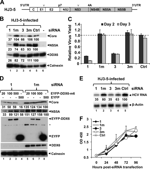
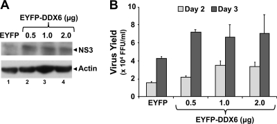
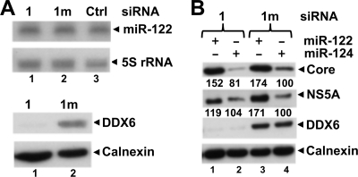
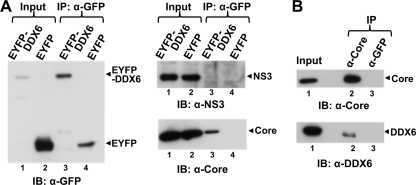
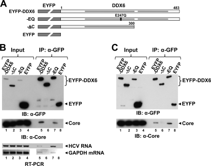
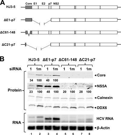
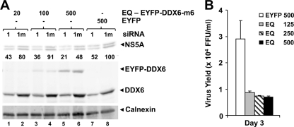
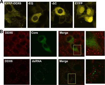
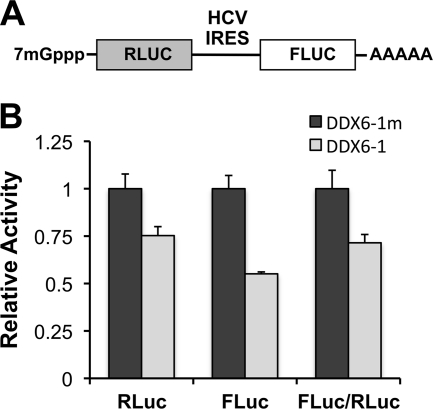
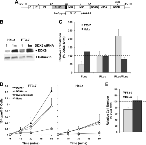
DDX6 (Rck/p54) is an evolutionarily conserved member of the SF2 DEAD-box RNA helicase family that contributes to the regulation of translation and storage and the degradation of cellular mRNAs. It interacts with multiple proteins and is a component of the micro-RNA (miRNA)-induced silencing complex (miRISC). Since miRNA-122 (miR-122) is essential for efficient hepatitis C virus (HCV) replication, we investigated the requirement for DDX6 in HCV replication in cultured hepatoma cells. Small interfering RNA (siRNA)-mediated knockdown of DDX6 and rescue with an siRNA-resistant mutant demonstrated that DDX6 expression is indeed required for optimal HCV replication. However, DDX6 knockdown did not impair miR-122 biogenesis or alter HCV responsiveness to miR-122 supplementation. Overexpression of DDX6 fused to EYFP (EYFP-DDX6) enhanced replication, whereas a helicase-deficient mutant with a substitution in the conserved DEAD-box motif II (DQAD) had a dominant-negative effect, reducing HCV yields. Coimmunoprecipitation experiments revealed an intracellular complex containing DDX6, HCV core protein, and both viral and cellular RNAs, the formation of which was dependent upon the C-terminal domain of DDX6 but not DDX6 helicase activity. However, since DDX6 abundance influenced the replication of subgenomic HCV RNAs lacking core sequence, the relevance of this complex is uncertain. Importantly, DDX6 knockdown caused minimal reductions in cellular proliferation, generally stimulated cellular translation ([(35)S]Met incorporation), and did not impair translation directed by the HCV internal ribosome entry site. Thus, DDX6 helicase activity is essential for efficient HCV replication, reflecting essential roles for DDX6 in HCV genome amplification and/or maintenance of cellular homeostasis.


Figures

FIG. 1.
FIG. 1.
DDX6 knockdown impairs HCV replication. (A) Schematic diagram of genomic organization of chimeric HCV genotype 1a/2a (HJ3-5) RNA that contains core, E1, E2, p7, and NS2 sequence derived from H77s (genotype 1a, white box) and NS3-5B and both UTRs from JFH1 (genotype 2a, gray box). Arrowheads indicate the positions of two adaptive mutations, one each in E1 (Y361H) and NS3 (Q1251L) genes that enhance its replication. (B) FT3-7 cells were transfected with the indicated DDX6-specific or control siRNAs: 1, DDX6-1; 1m, DDX6-1m; 3, DDX6-3; 3m, DDX6-3m; and Ctrl. At 48 h, cells were infected with HJ3-5 HCV at an MOI of 1, and cell lysates prepared 3 days after infection were subjected to immunoblotting. Various band intensities were measured by using AlphaEaseFC Software version 4.0.0 (Alpha Innotech Corp.), normalized to calnexin levels, and represented as a percentage of Ctrl siRNA (Ctrl)-treated cells. (C) Virus supernatants collected 2 and 3 days postinfection were titrated on naive Huh-7.5 cells in triplicate. The results from two independent experiments are presented here as means ± the SD. (D) FT3-7 cells were transfected with 1 (DDX6-1) or 1m (DDX6-1m) siRNA. At 24 h, cells were transfected with DNA encoding siRNA-resistant form of EYFP-DDX6 or pEYFP and were infected with HJ3-5 HCV (MOI = 0.5) on the next day. Cell lysates, prepared 3 days after infection, were immunoblotted. Band densities were quantitated as described above and are represented as percentages of that of DDX6-1m/pEYFP-transfected cells. (E) FT3-7 cells were transfected with the indicated siRNAs and infected with HJ3-5 HCV at an MOI of 1.0 at 48 h posttransfection. Total RNA was isolated 3 days later and subjected to Northern blotting. PhosphorImager quantitations are represented as a percentage of that of Ctrl siRNA-treated cells. (F) FT3-7 cells were transfected with the indicated siRNAs, followed by spectrophotometric measurements of WST-1 activity at 450 nm at the indicated time points (mean optical density at 450 nm ± the SD, n = 2).
FIG. 2.
FIG. 2.
DDX6 overexpression stimulates HCV replication. FT3-7 cells were transfected with pEYFP-DDX6 or pEYFP plasmid DNA. Cells were then infected with HJ3-5 HCV at an MOI of 0.2 at 24 h posttransfection and were fed with fresh media every 24 h. (A) Cell lysates, prepared 3 days postinfection, were immunoblotted. (B) Virus supernatants collected 2 and 3 days postinfection were titrated on naive Huh-7.5 cells, in triplicate. The results from two independent experiments are presented here as means ± the SD.
FIG. 3.
FIG. 3.
DDX6 is not required for miR-122 facilitation of HCV replication. (A) FT3-7 cells were transfected with 1 (DDX6-1) or 1m (DDX6-1m) siRNA, and cell lysates were prepared 4 days later. Total RNA was subjected to Northern blotting to detect miR-122 and 5S rRNA (used as a loading control) using 32P-labeled riboprobes (top panel). Protein extracts were immunoblotted for DDX6 and calnexin (bottom panel) (B) FT3-7 cells were transfected with 1 (DDX6-1) or 1m (DDX6-1m) siRNA. After 48 h, the cells were transfected with miR-122 or miR-124 (control miRNA) and infected with HJ3-5 HCV (MOI = 0.2) at 54 h. On day 4, the cells were transfected with another dose of miR-122 or miR-124. Cell lysates were prepared on the next day and subjected to Western blotting. Band intensities were measured as described in Fig. 1B and are represented as a percentage of DDX6-1m/miR-124 transfected cells.
FIG. 4.
FIG. 4.
DDX6 forms a complex with the HCV core protein. (A) HJ3-5 HCV-infected FT3-7 cells were transfected with DNA vector expressing wild-type EYFP-DDX6 or EYFP alone. Cell lysates were prepared 2 days later and subjected to coimmunoprecipitation with anti-GFP (rabbit polyclonal; Clontech) antibody. Coimmunoprecipitation samples were immunoblotted with GFP (mouse monoclonal; Clontech). Anti-GFP precipitates were probed with antibody to NS3 (top panel) or HCV core (bottom right panel). Input represents 1/20 of the IP for the anti-GFP blot and 1/80 of the IP for the NS3 and core blots. (B) Cell lysates prepared from HJ3-5 HCV-infected FT3-7 cells were subjected to coimmunoprecipitation with HCV core (C7-50; Affinity BioReagents) or GFP-specific (mouse monoclonal [Clontech]; used as an isotype control) antibody and immunoblotted for HCV core (upper panel) or endogenous DDX6 (lower panel). Input represents 1/20 and 1/80 of the IP for the anti-HCV core and anti-DDX6 blots, respectively.
FIG. 5.
FIG. 5.
The interaction of DDX6 with HCV core and viral RNA is dependent on the C-terminal domain of DDX6. (A) Schematic representation of EYFP-DDX6 and related mutants, ΔC (C-terminal deletion mutant lacking 183 amino acids), EQ (mutation in the DEAD-box helicase motif II involving the substitution of Glu-247 by Gln), and empty vector expressing EYFP alone. The ΔC mutation removes the second of two conserved RecA domains, identified by the shaded bars within the DDX6 sequence. (B) HJ3-5 HCV-infected FT3-7 cells were transfected with DNA vector expressing various forms of EYFP-DDX6 or just the EYFP. Cell lysates were prepared 2 days later and subjected to coimmunoprecipitation with anti-GFP (rabbit polyclonal; Clontech) antibody. Coimmunoprecipitation samples were immunoblotted with GFP (top panel) and core-specific (bottom panel) antibodies. Input represents 1/20 of the immunoprecipitation (IP) for anti-GFP blot and 1/80 of the IP for HCV core blot. Total RNA isolated from immunoprecipitates using an RNeasy minikit (Qiagen) was subjected to RT-PCR for detection of genomic HCV RNA (primers targeting the NS3 region) and GAPDH mRNA. (C) Huh-7-191/20 cells were induced to express HCV core protein by removing tetracycline from the medium for 3 days. Cells were then transfected with various DNAs and subjected to coimmunoprecipitation exactly as described for panel B.
FIG. 6.
FIG. 6.
DDX6 promotes HCV replication by enhancing genome amplification. (A) Schematic representation of wild-type HJ3-5, HJ3-5-ΔE1-p7 (lacks E1, E2, and p7), HJ3-5-ΔC61-148 (lacks amino acids 61 to 148 of core protein), and ΔC21-p7 (containing the first 20 amino acids of core fused in-frame with NS2 to 5B genes). (B) FT3-7 cells were transfected with 1 (DDX6-1) or 1m (DDX6-1m) siRNA. After 48 h, cells were transfected with the indicated HJ3-5 HCV RNA using TransIT mRNA transfection reagent (Mirus Bio). Immunoblotting of cell lysates prepared 3 days post-HCV RNA transfection is shown. HCV core and NS5A expression levels were quantitated on an Odyssey infrared imaging system (LI-COR) and are represented as the ratio of that in 1 (DDX6-1) siRNA-treated cells and in control, 1m (DDX6-1m) siRNA-treated cells (set as 100) after normalization to calnexin levels.
FIG. 7.
FIG. 7.
DDX6-EQ has a dominant-negative effect on HCV replication. (A) FT3-7 cells were transfected with DDX6-1 (lanes 1) or DDX6-1m (lanes 1m) siRNA and 48 h later retransfected with increasing quantities of expression vectors encoding siRNA-resistant EYFP-DDX6-m6-EQ or EYFP. One day later, the cells were infected with HJ3-5 HCV (MOI = 0.5). Cell lysates prepared 3 days after infection were immunoblotted for HCV NS5A, calnexin, and DDX6 proteins. NS5A band densities were quantified as described in Fig. 6A and are presented as the percent expression relative to DDX6-1m/pEYFP-transfected cells. (B) FT3-7 cells were transfected with pEYFP-DDX6-EQ or pEYFP plasmid DNA. Cells were infected with HJ3-5 HCV at an MOI of 0.2 at 24 h posttransfection and fed with fresh medium every 24 h. Virus supernatants collected 2 and 3 days postinfection were titrated on naive Huh-7.5 cells, in triplicate. Representative results from one of multiple experiments are presented here as means ± the SD.
FIG. 8.
FIG. 8.
DDX6-EQ and DDX6-ΔC have altered subcellular distributions. (A) FT3-7 cells were transfected with pEYFP-DDX6, pEYFP-DDX6-EQ, pEYFP-DDX6-ΔC, or pEYFP vector DNA. The cells were fixed with 4% paraformaldehyde 2 days later and imaged by confocal laser scanning microscopy for EYFP fluorescence. Wild-type DDX6 was localized to punctate structures identified as P-bodies, while both mutants were distributed diffusely within the cytoplasm and EYFP alone entered nuclei. (B) FT3-7 cells were infected with HJ3-5 virus at an MOI of 0.2. Two days later, the cells were fixed with 4% paraformaldehyde, permeabilized with digitonin, and stained with antibodies specific for DDX6 and HCV core (top panels) or DDX6 and dsRNA (bottom panels). In the top row of panels, HCV-infected cells are identified by cytoplasmic staining for core antigen (green) and demonstrated a moderate reduction in the number of P-bodies (identified by staining for endogenous DDX6). In the bottom row of panels, punctate staining for dsRNA (green) shows the location of replicating RNA in infected cells (no such staining was observed in uninfected cells). Importantly, dsRNA did not localize to P-bodies. Expanded views of areas from the merged images (yellow boxes) are shown to the right.
FIG. 9.
FIG. 9.
Influence of DDX6 knockdown on translation of a dicistronic RNA containing the HCV IRES. (A) Schematic representation of the pRLHL plasmid that expresses a dicistronic RNA containing Renilla luciferase sequence in the first cistron and firefly luciferase sequence in its second cistron, separated by the IRES of HCV. (B) pRLHL DNA was transfected into FT3-7 cells 64 h after transfection of the indicated siRNAs. Cells were harvested 48 h later for dual luciferase assays. The results shown represent RLuc (cap-dependent translation) and FLuc (IRES-directed translation) activities and the FLuc/RLuc ratio (relative IRES activity), normalized to that in DDX6-1m-transfected cells (n = 3, means ± the SD).
FIG. 10.
FIG. 10.
DDX6 regulation of HCV translation in FT3-7 and HeLa cells. (A) Schematic showing the genome organization of the HJ3-5/RLuc2A HCV which contains an in-frame insertion of Renilla luciferase (RLuc) sequence, fused at its C terminus to the foot-and-mouth disease virus 2A autoprotease, between the p7 and NS2 coding sequences of HJ3-5 virus. The RNA also contains a replication-lethal mutation (GND) in the NS5B polymerase sequence. The 5′ and 3′ ends of this RNA are the authentic viral UTRs. Below is shown the capped FLuc mRNA that contains a 3′ poly(A) tail of 30 adenosine residues that was used as an internal control for transfection and translation efficiencies. (B) Immunoblot showing efficient DDX6 knockdown after transfection of either FT3-7 or HeLa cells with the DDX6-1 siRNA. (C) Impact of DDX6 knockdown on HCV IRES-directed translation. FT3-7 or HeLa cells were transfected with DDX6 siRNAs and 4 days later were retransfected with HJ3-5/RLuc2A/GND and FLuc mRNAs. Cells were harvested 8 h later and assayed for FLuc and RLuc activities. The results shown represent the mean percentages ± the range of FLuc and RLuc activities and the RLuc/FLuc ratio, obtained in duplicate DDX6-1-transfected cultures, normalized to those obtained in DDX6-1m-transfected cells, and are representative of results from multiple experiments. (D) Metabolic labeling of FT3-7 (left panel) and HeLa (right panel) cells after DDX6 knockdown. Cells, transfected 4 days previously with siRNAs as described in panel C, were cultured for 1 h in methionine- and cysteine-free medium and then incubated with 100 μCi of Tran35S-label (MP Biomedicals)/ml. Cells were harvested at the times indicated, and [35S]Met present in trichloroacetic acid precipitate measured in a scintillation counter. The results from three independent experiments are presented as cpm incorporated/106 cells (means ± the SD). (E) Impact of DDX6 knockdown on the proliferation of FT3-7 and HeLa cells. Cells were enumerated 96 h after transfection with DDX6 siRNAs. The results are shown as the ratio of the number of cells in the DDX6-1-transfected cultures relative to those transfected with the mutant DDX6-1m siRNA at the end of this growth period (means ± the SD, n = 3).

Similar articles

Cited by

References

    1. Akao, Y., H. Yoshida, K. Matsumoto, T. Matsui, K. Hogetu, N. Tanaka, and J. Usukura. 2003. A tumour-associated DEAD-box protein, rck/p54 exhibits RNA unwinding activity toward c-myc RNAs in vitro. Genes Cells 8:671-676. - PubMed
    1. Alves-Rodrigues, I., A. Mas, and J. Diez. 2007. Xenopus Xp54 and human RCK/p54 helicases functionally replace yeast Dhh1p in brome mosaic virus RNA replication. J. Virol. 81:4378-4380. - PMC - PubMed
    1. Angus, A. G., D. Dalrymple, S. Boulant, D. R. McGivern, R. F. Clayton, M. J. Scott, R. Adair, S. Graham, A. M. Owsianka, P. Targett-Adams, K. Li, T. Wakita, J. McLauchlan, S. M. Lemon, and A. H. Patel. 2010. Requirement of cellular DDX3 for hepatitis C virus replication is unrelated to its interaction with the viral core protein. J. Gen. Virol. 91:122-132. - PMC - PubMed
    1. Ariumi, Y., M. Kuroki, K. Abe, H. Dansako, M. Ikeda, T. Wakita, and N. Kato. 2007. DDX3 DEAD-box RNA helicase is required for hepatitis C virus RNA replication. J. Virol. 81:13922-13926. - PMC - PubMed
    1. Bonin, M., J. Oberstrass, N. Lukacs, K. Ewert, E. Oesterschulze, R. Kassing, and W. Nellen. 2000. Determination of preferential binding sites for anti-dsRNA antibodies on double-stranded RNA by scanning force microscopy. RNA 6:563-570. - PMC - PubMed

Publication types

LinkOut - more resources