. 2010 Jul;84(13):6846-60.
doi: 10.1128/JVI.00399-10. Epub 2010 Apr 14.

Structure/Function analysis of the vaccinia virus F18 phosphoprotein, an abundant core component required for virion maturation and infectivity

Affiliations

Structure/Function analysis of the vaccinia virus F18 phosphoprotein, an abundant core component required for virion maturation and infectivity

Nadi T Wickramasekera et al. J Virol. 2010 Jul.

Abstract

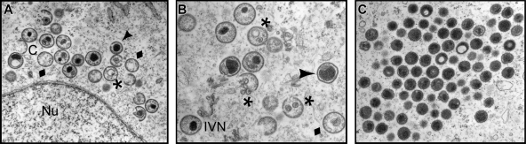
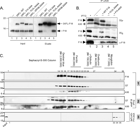
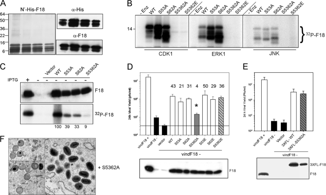
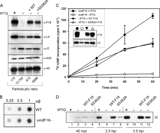
Poxvirus virions, whose outer membrane surrounds two lateral bodies and a core, contain at least 70 different proteins. The F18 phosphoprotein is one of the most abundant core components and is essential for the assembly of mature virions. We report here the results of a structure/function analysis in which the role of conserved cysteine residues, clusters of charged amino acids and clusters of hydrophobic/aromatic amino acids have been assessed. Taking advantage of a recombinant virus in which F18 expression is IPTG (isopropyl-beta-d-thiogalactopyranoside) dependent, we developed a transient complementation assay to evaluate the ability of mutant alleles of F18 to support virion morphogenesis and/or to restore the production of infectious virus. We have also examined protein-protein interactions, comparing the ability of mutant and WT F18 proteins to interact with WT F18 and to interact with the viral A30 protein, another essential core component. We show that F18 associates with an A30-containing multiprotein complex in vivo in a manner that depends upon clusters of hydrophobic/aromatic residues in the N' terminus of the F18 protein but that it is not required for the assembly of this complex. Finally, we confirmed that two PSSP motifs within F18 are the sites of phosphorylation by cellular proline-directed kinases in vitro and in vivo. Mutation of both of these phosphorylation sites has no apparent impact on virion morphogenesis but leads to the assembly of virions with significantly reduced infectivity.


Figures

FIG. 1.
FIG. 1.
Electron microscopic examination reveals a clear morphogenesis defect in cells infected with vindF18-IPTG. BSC40 cells were infected with vindF18 (MOI of 2) in the absence of IPTG for 17 h and were then fixed in situ and processed for transmission electron microscopy. (A and B) Structures seen during these nonpermissive infections are indicated. Normal structures: C, crescents; IVN, immature virions with nucleoids. Abnormal structures: ⧫; empty immature virions; *, empty immature particles with unusual membranous inclusions; ▴, immature virions with aberrant nucleoids. Nu, nucleus. (C) Aberrant spherical particles that appear to be abnormal mature virions. Note the absence of MV as well as the absence of DNA crystalloids in the cytoplasm.
FIG. 2.
FIG. 2.
F18:F18 interactions in vivo and in vitro. (A) Interactions between F18 proteins can be detected in infected cell lysates. BSC40 cells were infected with vindF18 virus in the presence (+) (lanes 1, 3 to 5, and 6 to 9) or absence (−) (lanes 2) of IPTG and, at 3 hpi, transfected with empty vector (lanes 3) or plasmids encoding 3×FLAG-F18 (lanes 2 and 4 to 9). A portion of each clarified cytoplasmic lysate was removed prior to affinity purification (input); the remainder was used for affinity purification of 3×FLAG-F18 and any associated untagged F18 (eluate). For the samples shown in lanes 1 to 5, the beads were washed with buffer containing 150 mM NaCl prior to elution, whereas the beads for the samples shown in lanes 6, 7, and 8 were washed with buffer containing 100, 300, and 600 mM NaCl, respectively. For the sample shown in lane 9, the beads were treated with DNase prior to being washed with 150 mM NaCl. Input and eluates were resolved by SDS-PAGE and subjected to immunoblot analysis with α-F18 serum. (B) Purified, recombinant N′-His-F18 self-associates to form a high-molecular-weight complex. Recombinant N′-His-F18 protein purified from E. coli was resolved on a Sephacryl S-200 column; 1-ml fractions were analyzed by SDS-PAGE and visualized by silver staining. The fraction numbers are depicted at the top of the immunoblot. The arrowhead indicates the void volume as assessed by dextran blue elution. The elution of the other standards is illustrated above the relevant fractions. Fractions 15 to 17 represent the peak elution of F18.
FIG. 3.
FIG. 3.
Alignment of poxviral F18 homologs and identification of motifs and residues chosen for targeted mutagenesis. An alignment of the predicted amino acid sequence is shown for F18 homologs encoded by: vaccinia virus (VV, WR strain; GenBank accession no. YP_232938), variola virus (VAR; GenBank ABF23617) cowpox virus (CPV; GenBank NP_619853), monkeypox virus (MPX; GenBank NP_536476), myxoma virus (MYX; GenBank NP_051740), rabbit fibroma virus (RPX; GenBank NP_051915); swinepox virus (SPV; GenBank NP_570189), Yaba-like disease virus (YLDV; GenBank NP_073416), lumpy skin disease virus (LSDV; GenBank AAN02756), and molluscum contagiosum virus (MCV; GenBank NP_043981) (the sequences were obtained from www.poxvirus.org). Residues identical to the vaccinia virus F18 sequence are shaded in gray. The F18 mutants constructed for the present study are marked within the alignment and described in the legend below. Conserved cysteine residues evaluated for their possible participation in forming covalent F18-F18 dimers (blue and red circles, linked by horizontal lines) were changed to Ser individually and in combination. Several clusters of hydrophobic or charged residues that are highly conserved among the Poxviridae homologs were changed to Ala residues; these motifs are marked with colored lines and colored triangles, respectively. Red stop signs indicate new termination sites engineered after amino acids 70 and 82. The two PSSP motifs predicted to be recognition sites for cellular proline-directed cellular kinases are marked with curly brackets; these motifs were changed to PSAP and PSEP, both individually and in combination.
FIG. 4.
FIG. 4.
Establishment of a transient complementation assay for structure/function analysis of F18; conserved cysteine residues are not required for the biological activity of F18. Cells were infected with vindF18 in the presence (+) or absence (−) of IPTG and either left untransfected or transfected with empty vector or plasmids encoding mutant alleles in which Cys residues at positions 30, 37, 44, or 56 were changed to Ser, either individually or in pairs, as shown. At 24 hpi, the yield of infectious virus was determined by plaque assay. The data are shown graphically. Here and in later figures, the horizontal black line represents the baseline yield obtained from cells infected without IPTG and transfected with empty vector. The expression of each form of F18 was confirmed by immunoblot analysis, which is shown beneath the graph.
FIG. 5.
FIG. 5.
Assessing the contribution of conserved clusters of charged residues to the function of F18 in vivo. (A) Transient complementation assay. Alleles of F18 in which clusters of charged residues had been changed to Ala residues or in which premature stop codons had been introduced were tested in the transient-complementation assay. The viral yield was determined at 24 hpi (graph), and protein expression was confirmed by immunoblot assay (bottom panel). (B) The KEGR→AAGA form of F18 retains the ability to interact with endogenous F18. Cells were infected with vindF18+IPTG and transfected with empty vector or plasmids encoding the 3×FL-WT and 3×FL-KEGR→AAGA forms of F18. As described for Fig. 2, affinity pulldown assays were used to compare the ability of the two tagged proteins to interact with endogenous F18. (C) Transient expression of WT F18, but not the KEGR→AAGA or KAVKVCD→AAVACQ forms of F18, restores virion morphogenesis in the context of a vindF18 -IPTG infection. Cells were infected with vindF18-IPTG and transfected with empty vector (data not shown) or plasmids encoding WT F18 or the KEGR→AAGA or KAVKVCD→AAVACQ forms of F18. At 17 hpi, the cells were processed for conventional electron microscopy. Representative images are shown.
FIG. 6.
FIG. 6.
Assessing the contribution of conserved clusters of hydrophobic and/or aromatic residues to the function of F18 in vivo. (A) Transient complementation assay. Alleles of F18 in which clusters of hydrophobic and/or aromatic residues had been changed to Ala were assessed in the transient-complementation assay. The viral yield was determined at 24 hpi, and protein expression was confirmed by immunoblot assay (bottom panel). (B to D) F18 variants containing FYI→AAA, YLVL→AAAA or VPFM→AAAA substitutions cannot support virion morphogenesis. Cells were infected with vindF18-IPTG and transfected with plasmids encoding WT F18 (see Fig. 5) or the FYI→AAA, YLVL→AAAA, or VPFM→AAAA variants. At 17 hpi, cells were processed for conventional electron microscopy. Representative images are shown.
FIG. 7.
FIG. 7.
Hydrophobic/aromatic motifs within the F18 protein mediate its self-association and interaction with other proteins. (A) The FYI→AAA and YLVL→AAAA forms of F18 lack the ability to interact with endogenous F18. Cells were infected with vindF18+IPTG and transfected with empty vector (lanes 1) or plasmids encoding 3×FLAG-tagged WT or mutant forms of F18 (lanes 2 to 5). α-FLAG beads were used to retrieve the 3×FLAG-tagged F18 and any interacting proteins. Both the input and the eluate fractions were subjected to immunoblot analysis with α-F18. (B) Mutations in hydrophobic clusters prevent the interaction of F18 and A30. Cells were infected with vindF18+IPTG or vindF18-IPTG as shown and left untransfected or transfected with plasmids encoding WT F18 or the YLVL→AAAA or FYI→AAAA forms of F18. Cells were metabolically labeled with 32PPi or [35S]Met from 6 to 24 hpi prior to being harvested and analyzed by immunoprecipitation with α-A30 serum. The A30 protein (⧫) and any coprecipitating F18 protein (▸) were visualized by autoradiography. The bottom panel represents an immunoblot analysis of the lysates, confirming the expression of F18 proteins (▸). (C) Utilization of gel filtration chromatography to assess the native molecular weight of protein complexes containing the A30, G7, or F18 proteins. Cells were infected with WT virus (group 1) in the presence of RIF. Lysates were prepared at 24 hpi and resolved on a Sephacryl S-300 column. A similar protocol was used to monitor the native molecular weight of G7 and A30 during vindF18-IPTG infections (also performed +RIF) (group 2). A third experiment was performed to monitor the elution profile of the 3×FL-FYI→AAA of F18 that was expressed in cells infected with vindF18-IPTG (+RIF); this protein was detected by immunoblot analysis with an α-FLAG antibody (group 3). The elution of molecular-weight standards and relevant fraction numbers are shown above the immunoblots.
FIG. 8.
FIG. 8.
Characterization of the role of the PSSP motifs in the phosphorylation of F18 in vitro and the phosphorylation and biological activity of F18 in vivo. (A) Purification of recombinant F18. Recombinant N′-His-F18 was expressed in E. coli and purified by metal ion chromatography; the peak of purified protein was examined by silver staining (left panel) and immunoblot analysis with α-His and α-F18 probes (right panels). (B) The phosphorylation of F18 in vitro by proline-directed kinases depends upon the two PSSP motifs. N′-His-tagged preparation of WT F18 or the S53A, S62A, S5362A, and S5362E mutants were used as substrates for in vitro kinase assays performed with CDK1, ERK1, or JNK in the presence of [γ-32P]ATP. Phosphorylation was assessed by autoradiography; only the relevant portion of the film is shown. (C) The phosphorylation of F18 in vivo depends on the PSSP motifs. Cells were infected with vindF18+IPTG (lane 1) or vindF18-IPTG (lanes 2 to 7) and left untransfected (lanes 1 and 2) or transfected with empty vector (lane 3) or plasmids encoding WT F18 (lane 4) or the S53A, S62A, and S5362A variants of F18 (lanes 5 to 7, respectively). Cells were metabolically labeled with 32PPi. The expression of each form of F18 was confirmed by immunoblot analysis (top panel), and the F18 was retrieved by immunoprecipitation and visualized by autoradiography (bottom panel). 32P incorporation was normalized to that seen for WT F18; the values are shown beneath the lanes. (D) The S5362A allele of F18 shows a loss of biological activity. Plasmids encoding WT F18 or the S53A, S62A, S5362A, S53E, S62E, and S5362E variants of F18 were assessed for their transient-complementation activity. The fold increase in viral yield relative to what was seen after transfection with empty vector is shown above the relevant bar. For the S5362A variant, the value obtained for viral yield (*, 4-fold) was statistically different from that seen after expression of the other forms of F18. The protein was confirmed by immunoblot (bottom panel). (E) Incorporation of a 3×FLAG epitope restores the biological activity of the S5362A variant of F18. The same transient-complementation protocol was used to compare the biological activity of 3×FL-WT F18 and 3×FL-S5362A F18. Expression of the proteins was confirmed by immunoblot (bottom panel). (F) The nonphosphorylatable S5362A variant of F18 can support virion morphogenesis. Cells were infected with vindF18-IPTG and transfected with plasmids encoding WT F18 (see Fig. 5) or the S5362A variant. At 17 hpi, cells were processed for conventional electron microscopy; representative images are shown.
FIG. 9.
FIG. 9.
Characterization of the protein content, DNA content, and transcriptional competence of virions that lack F18 or encapsidate WT or phosphorylation-deficient F18. (A) Protein content of purified virions. Cells were infected with vindF18 virus in the presence or absence of IPTG and left untransfected or transfected with plasmids encoding WT F18 or S5362A F18. At 24 hpi, the cells were harvested, and virions were purified by sucrose gradient centrifugation and quantitated by measuring the optical density at 260 nm. Then, 1-μg portions of the different preparations of purified virions were resolved electrophoretically and subjected to immunoblot analysis with sera directed against the F18, L4, I1, A30, or A5 core proteins. The infectivity of the preparations was also quantitated by plaque assay; the particle/PFU ratio of each preparation is shown at the bottom of the appropriate lane. (B) Purified vindF18-IPTG virions contain viral DNA. Different amounts (0.25, 0.5, and 1 μg) of purified wt and vindF18-IPTG particles were subjected to Southern dot blot analysis to quantitate the levels of encapsidated viral DNA. The levels of hybridized probe were visualized and quantitated by autoradiography. (C) In situ transcription assays of purified virions. Portions (4 μg) of purified virions were permeabilized and incubated with [32P]UTP in order to activate the in situ transcription of early genes. The incorporation of [32P]UTP into RNA was quantitated for aliquots removed after 0, 20, 40, and 60 min of incubation. The immunoblot shown in the inset confirms that the virion preparations contain comparable levels of the L4 protein and do or do not contain F18 as expected. (D) Virions containing only the S5362A form of F18 are delayed and diminished in their ability to induce early gene expression in vivo. Cells were inoculated on ice with equivalent numbers of virions (300/cell) purified from cells infected with vindF18+IPTG (lanes 1, 5, and 9), vindF18-IPTG (lanes 2, 6, and 10), or vindF18-IPTG and transfected with plasmids encoding WT F18 (lanes 3, 7, and 11) or the S5362A form of F18 (lanes 4, 8, and 12). After 1 h, the inocula were removed, and the cultures were shifted to 37°C. At 40 min postinfection, 2.5 hpi, and 3.5 hpi, the cells were harvested, and the expression of the early protein I3 was monitored by immunoblot analysis.

Similar articles

Cited by

References

    1. Cassetti, M. C., M. Merchlinsky, E. J. Wolffe, A. S. Weisberg, and B. Moss. 1998. DNA packaging mutant: repression of the vaccinia virus A32 gene results in noninfectious, DNA-deficient, spherical, enveloped particles. J. Virol. 72:5769-5780. - PMC - PubMed
    1. Chakrabarti, S., J. R. Sisler, and B. Moss. 1997. Compact, synthetic, vaccinia virus early/late promoter for protein expression. Biotechniques 23:1094-1097. - PubMed
    1. Chung, C. S., C. H. Chen, M. Y. Ho, C. Y. Huang, C. L. Liao, and W. Chang. 2006. Vaccinia virus proteome: identification of proteins in vaccinia virus intracellular mature virion particles. J. Virol. 80:2127-2140. - PMC - PubMed
    1. Condit, R. C., N. Moussatche, and P. Traktman. 2006. In a nutshell: structure and assembly of the vaccinia virion. Adv. Virus Res. 66:31-124. - PubMed
    1. Dyster, L. M., and E. G. Niles. 1991. Genetic and biochemical characterization of vaccinia virus genes D2L and D3R which encode virion structural proteins. Virology 182:455-467. - PubMed

Publication types

MeSH terms

LinkOut - more resources