. 2010 Jun;24(6):1187-202.
doi: 10.1210/me.2009-0480. Epub 2010 Apr 14.

Cooperative activation of cyclin D1 and progesterone receptor gene expression by the SRC-3 coactivator and SMRT corepressor

Affiliations

Cooperative activation of cyclin D1 and progesterone receptor gene expression by the SRC-3 coactivator and SMRT corepressor

Sudipan Karmakar et al. Mol Endocrinol. 2010 Jun.

Abstract

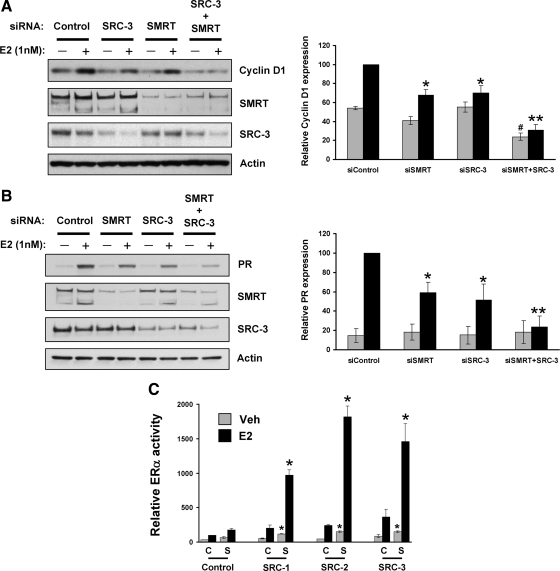
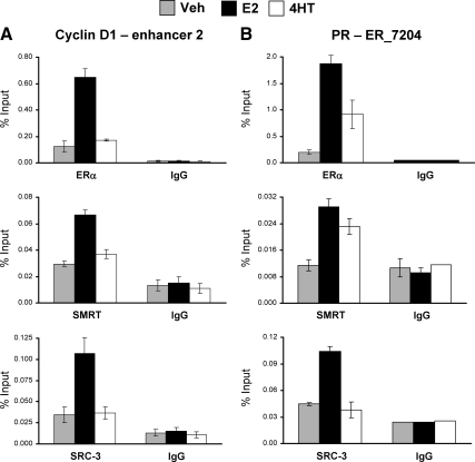
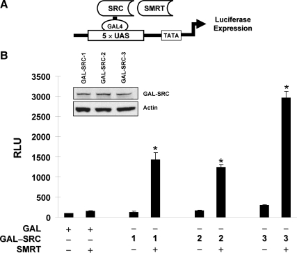
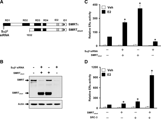
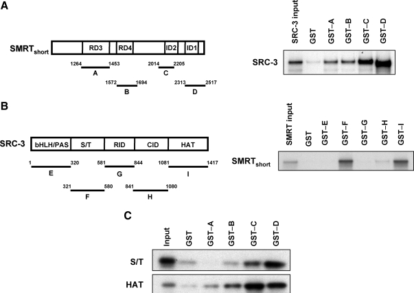
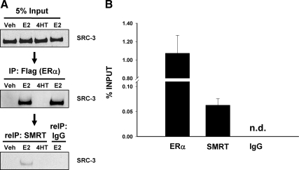
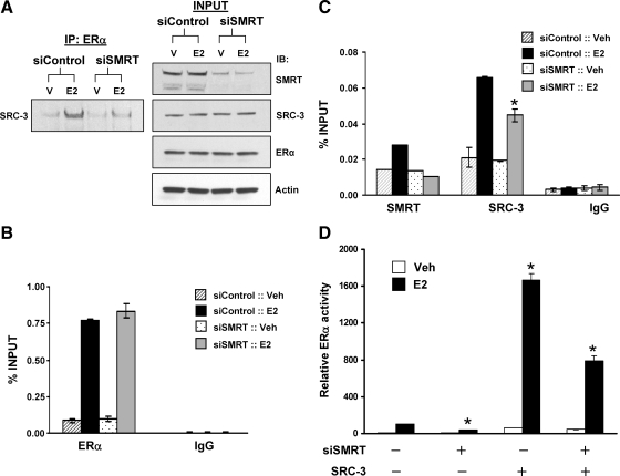
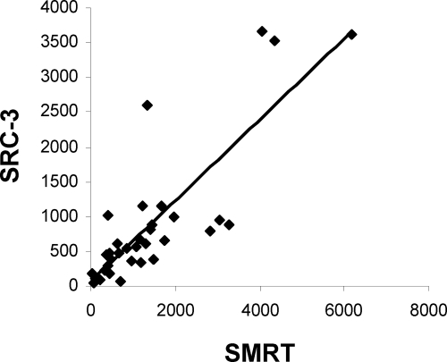
Although the ability of coactivators to enhance the expression of estrogen receptor-alpha (ERalpha) target genes is well established, the role of corepressors in regulating 17beta-estradiol (E2)-induced gene expression is poorly understood. Previous studies revealed that the silencing mediator of retinoic acid and thyroid hormone receptor (SMRT) corepressor is required for full ERalpha transcriptional activity in MCF-7 breast cancer cells, and we report herein the E2-dependent recruitment of SMRT to the regulatory regions of the progesterone receptor (PR) and cyclin D1 genes. Individual depletion of SMRT or steroid receptor coactivator (SRC)-3 modestly decreased E2-induced PR and cyclin D1 expression; however, simultaneous depletion revealed a cooperative effect of this coactivator and corepressor on the expression of these genes. SMRT and SRC-3 bind directly in an ERalpha-independent manner, and this interaction promotes E2-dependent SRC-3 binding to ERalpha measured by co-IP and SRC-3 recruitment to the cyclin D1 gene as measured by chromatin IP assays. Moreover, SMRT stimulates the intrinsic transcriptional activity of all of the SRC family (p160) coactivators. Our data link the SMRT corepressor directly with SRC family coactivators in positive regulation of ERalpha-dependent gene expression and, taken with the positive correlation found for SMRT and SRC-3 in human breast tumors, suggest that SMRT can promote ERalpha- and SRC-3-dependent gene expression in breast cancer.


Figures

Figure 1
Figure 1
Stimulatory effect of SMRT and SRC coactivators on ERα transcriptional activity and endogenous target gene expression. MCF-7 cells were transfected with either 6 pmol control siRNA or siRNA against SRC-3 (1 pmol) or SMRT (5 pmol) alone or in combination and then treated with vehicle (0.1% ethanol) or 1 nm E2 for 3 h (for cyclin D1) or 24 h (for PR). Subsequently, expression of cyclin D1 (panel A) and PR (panel B) was assessed by Western blotting. Expression of SMRT, SRC-3, and actin also was measured for each set of samples. The experiment was repeated three times, and representative blots are shown. Blots obtained from three independent experiments were quantitated by densitometry, and values normalized to actin are expressed as mean ± sem. Statistical analyses were done by one-way ANOVA. *, Significance at P < 0.05; **, P < 0.001 vs. E2-treated siControl cells; #, significance at P < 0.05 vs. vehicle-treated siControl cells. Panel C, HeLa cells were transfected with 10 ng ERα expression vector and 1 μg ERE-E1b-Luc reporter gene along with the indicated plasmids for empty pCR3.1 (C, control), SMRTτ (S, SMRT) or SRC-1, -2, or -3. Cells were subsequently treated with vehicle or 1 nm E2 for 24 h followed by measurement of luciferase activity. Values represent the average ± sem of three independent experiments. *, P < 0.01 in comparison with the respective vehicle (Veh)- or E2-treated SMRT-transfected cells.
Figure 2
Figure 2
Interaction of ERα, SRC-3, and SMRT with enhancer regions of the cyclin D1 and PR genes. Chromatin was prepared from MCF-7 cells treated with vehicle (Veh), 10 nm E2, or 100 nm 4HT for 45 min and then subjected to ChIP assay using antibodies for ERα (top), SMRT (middle), or SRC-3 (bottom) in parallel to the appropriate IgG negative control. Immunoprecipitated DNA was quantitated by qPCR using primers to amplify enhancer-2 of the cyclin D1 gene (A) or the ER_7204 binding site in the PR gene (B). Data represent an average ± sem of three independent experiments.
Figure 3
Figure 3
In vivo interaction of ERα, SMRT, and SRC-3 in MCF-7 cells. MCF-7 cells were grown 24 h in phenol red-free DMEM supplemented with 10% sFBS and then treated with vehicle (Veh), 1 nm E2, or 100 nm 4HT for 60 min. Thereafter, cells were harvested, lysed, and immunoprecipitated with antibodies to SMRT (A) or ERα (B) and subsequently Western blotted for SMRT, SRC-3, and ERα (left panels). IPs performed with rabbit IgG were incorporated into each experiment as negative controls, and 2.5% of the cell lysates (INPUT) was assessed for SMRT, SRC-3, and ERα by Western blot. IB, Immunoblot.
Figure 4
Figure 4
SMRT enhancement of intrinsic SRC-3 transcriptional activity is ERα independent. A, Schematic diagram illustrating a modified mammalian one-hybrid assay for evaluating functional interaction of GAL4-SRC fusion proteins with SMRT. B, HeLa cells were transfected with 5×UAS-Luc reporter plasmid, along with expression vectors for the GAL4 DNA-binding domain fused to full-length SRC-1 (1), SRC-2 (2), or SRC-3 (3) in the presence of the pCR3.1 control vector (−) or a SMRTτ expression plasmid (+). Twenty-four hours after transfection, cells were harvested for measurement of luciferase activity. Values represent the average ± sem from three independent experiments. *, P < 0.01 in comparison with the respective control group. Equal expression of GAL-SRC fusion proteins were confirmed by Western blot analysis using an antibody specific for the GAL4 DNA-binding domain. A representative blot of three independent experiments is shown (inset).
Figure 5
Figure 5
The N terminus of SMRT is dispensable for synergistic coactivation of ERα by SRC-3 and SMRT. A, Schematic representation of SMRTτ and SMRTshort illustrating repression domains (black box) and CoRNR box motifs (striped box). The 47-amino-acid splice deletion in the C-terminal region of both SMRTτ and SMRTshort adjacent to ID1 is represented as a single horizontal line. The arrow indicates the location of the sequence targeted by this SMRT siRNA (Sαβ2). B, Representative Western blot analysis of SMRT (top) or actin (bottom) expression in cells obtained from HeLa cells transfected in parallel. C, HeLa cells were transfected with negative control (−) siRNA or the Sαβ2 (+) siRNA directed against SMRT before transfection with vectors for ERα, ERE-E1b-Luc, and either 500 ng SMRTshort or control (pCR3.1) expression vector followed by treatment with vehicle or 1 nm E2 for 24 h. The Mr of SMRTshort is approximately 175 kDa. Values represent the average ± sem of three independent experiments. *, P < 0.01 in comparison with control E2 values. D, HeLa cells were transfected with 250 ng expression vectors for control, SMRTshort, or SRC-3 alone or in combination in addition to ERα and ERE-E1b-Luc. Cells were subsequently treated with vehicle or E2 for 24 h. Values represent the average ± sem of four independent experiments. *, P < 0.01 in comparison with control E2 values.
Figure 6
Figure 6
SMRT binds directly to SRC-3 in vitro. Panel A, Schematic diagram showing SMRTshort and the GST-tagged fragments of SMRT (A–D) used in the experiment (left). GST pull-down assay demonstrating the interaction of full-length in vitro-transcribed and -translated SRC-3 with GST-SMRT fusion proteins or GST control (right). Panel B, Schematic representation of full-length SRC-3 and the GST-tagged SRC-3 fragments used in this study (left). Interaction of in vitro-transcribed and -translated SMRTshort with GST-SRC-3 fusion proteins or GST control (right). Panel C, Interaction of in vitro-translated S/T or HAT domains of SRC-3 with GST-SMRT fusion proteins or GST control. In each assay, bound 35S-labeled proteins were resolved by SDS-PAGE and detected by autoradiography. Input lanes represent 10% of 35S-labeled full-length SRC-3 or fragments (S/T or HAT) of SRC-3 or 0.5% of SMRTshort proteins used in pull-down assays. Results are representative of three independent experiments.
Figure 7
Figure 7
ERα, SMRT, and SRC-3 form a trimeric complex in vivo. A, HeLa cells were transfected with expression vector for FLAG-tagged ERα and SMRTshort and 24 h thereafter were treated with vehicle (Veh), 1 nm E2, or 100 nm 4HT for 60 min. Five percent of the cell lysate (top panel) was set aside, and remaining lysates were immunoprecipitated with Sigma's EZview Red anti-FLAG M2 affinity gel. Immune complexes were eluted with 3X FLAG peptide, and a portion of this eluant was retained (middle panel), whereas the rest was subjected to a second IP [re-immunoprecipitated (reIP)] with either SMRT antibody or rabbit IgG (bottom panel). Material from the first and second IPs as well as input samples were analyzed by SDS-PAGE and Western blotted with SRC-3 antibody. B, MCF-7 cells were treated with 10 nm E2 for 45 min and first subjected to ChIP with SRC-3 antibody, followed by re-IP with antibodies to ERα, SMRT, or IgG. Chromatin recovered from the second IP was quantitated by qPCR using primers to an amplicon in the enhancer-2 region of the cyclin D1 gene. Data represent an average ± sem of three independent experiments. Signal was not detected (n.d.) in the IgG control.
Figure 8
Figure 8
Depletion of SMRT inhibits SRC-3 and ERα interaction, recruitment of SRC-3 to cyclin D1 enhancer and ERα transcriptional activity. A, MCF-7 cells were transfected with 20 nm control or SMRT siRNA and 48 h thereafter were treated with either vehicle (V) or 10 nm E2 for 60 min, followed by cell lysis and IP with ERα antibody. Levels of SRC-3 immunoprecipitated with ERα were assessed by Western blot (left). Five percent of the total cell lysates was reserved (INPUT), and Western blots were employed to assess SMRT, SRC-3, ERα, and actin expression (right). The experiment was performed three times, and a representative blot is shown. B and C, MCF-7 cells were transfected with 20 nm control or SMRT siRNA, and 48 h later, cells were treated with either vehicle (Veh) or 10 nm E2 for 45 min and subjected to ChIP assay using antibodies for ERα, SMRT, SRC-3, or IgG. Immunoprecipitated chromatin was quantitated by qPCR using primers for the cyclin D1 enhancer 2. Data represent an average ± sem of two to three independent experiments. D, HeLa cells were transfected with control siRNA or Sαβ2 siRNA before transfection with an ERE-E1b-Luc reporter gene and expression vectors for ERα, SRC-3, or vector control (−; pCR3.1) and treated with vehicle (Veh) or 1 nm E2. Values represent the average ± sem of three independent experiments. *, P < 0.01 in comparison with the respective E2-treated control values.
Figure 9
Figure 9
SMRT and SRC-3 protein expression are positively correlated in breast cancer. Total protein from tumor lysates of 34 breast cancer patients was analyzed by Western blot. Expression of SMRT, SRC-3, and actin protein was quantitated by densitometry using ImageJ software (http://rsbweb.nih.gov/ij/), and the relative expression of actin-normalized SRC-3 vs. actin normalized SMRT was plotted.

Similar articles

Cited by

References

    1. Katzenellenbogen BS 1996 Estrogen receptors: bioactivities and interactions with cell signaling pathways. Biol Reprod 54:287–293 - PubMed
    1. Sarrel PM, Lufkin EG, Oursler MJ, Keefe D 1994 Estrogen actions in arteries, bone and brain. Sci Am Sci Med 1:44–53
    1. Henderson BE, Ross R, Bernstein L 1988 Estrogens as a cause of human cancer: the Richard and Hinda Rosenthal Foundation Award Lecture. Cancer Res 48:246–253 - PubMed
    1. Glass CK 1994 Differential recognition of target genes by nuclear receptor monomers, dimers, and heterodimers. Endocr Rev 15: 391–407 - PubMed
    1. Oñate SA, Tsai SY, Tsai MJ, O'Malley BW 1995 Sequence and characterization of a coactivator for the steroid hormone receptor superfamily. Science 270:1354–1357 - PubMed

Publication types

MeSH terms