. 2010 Apr 14;30(15):5167-75.
doi: 10.1523/JNEUROSCI.5591-09.2010.

Kv1.1 potassium channel deficiency reveals brain-driven cardiac dysfunction as a candidate mechanism for sudden unexplained death in epilepsy

Affiliations

Kv1.1 potassium channel deficiency reveals brain-driven cardiac dysfunction as a candidate mechanism for sudden unexplained death in epilepsy

Edward Glasscock et al. J Neurosci. .

Abstract

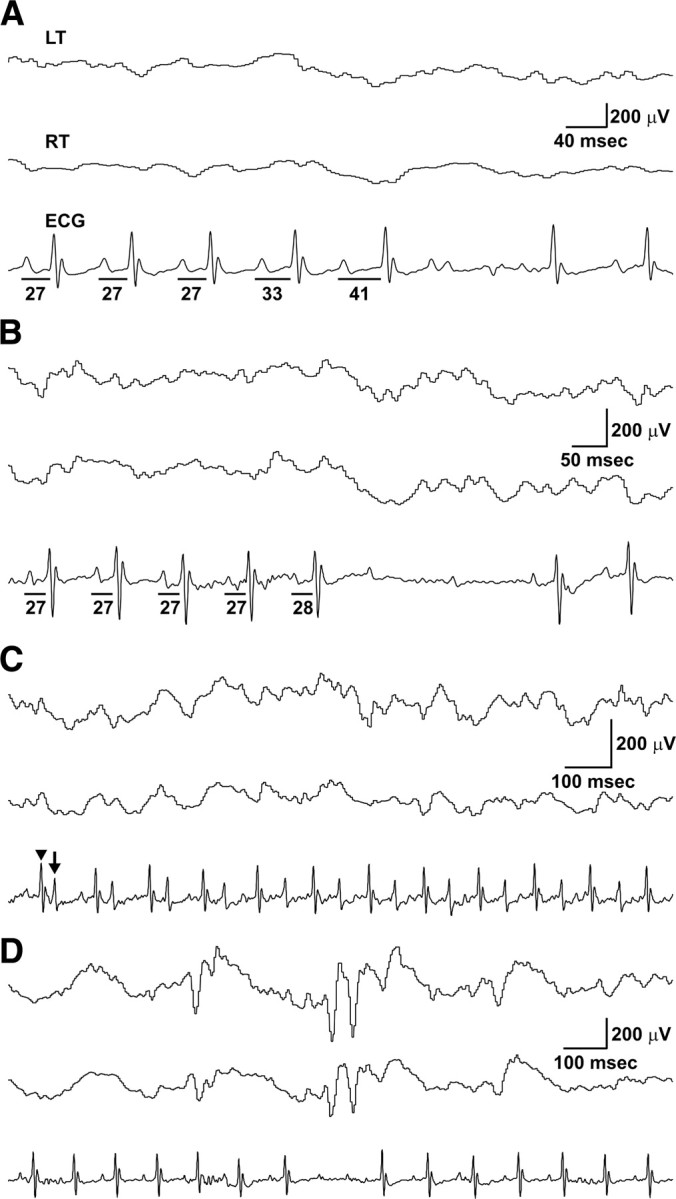
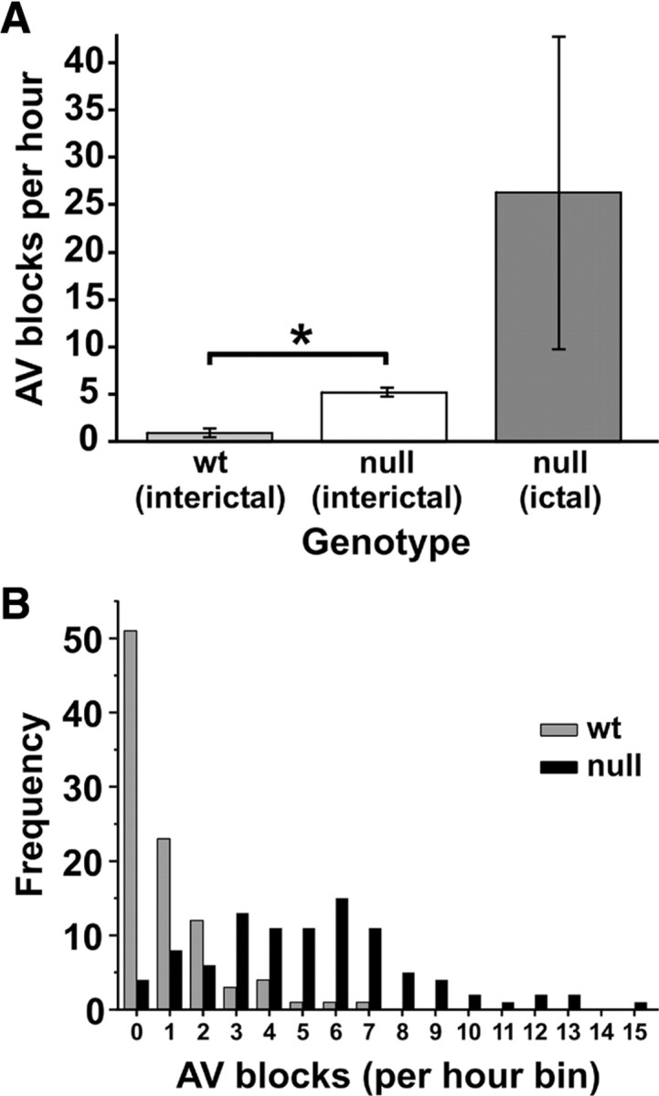
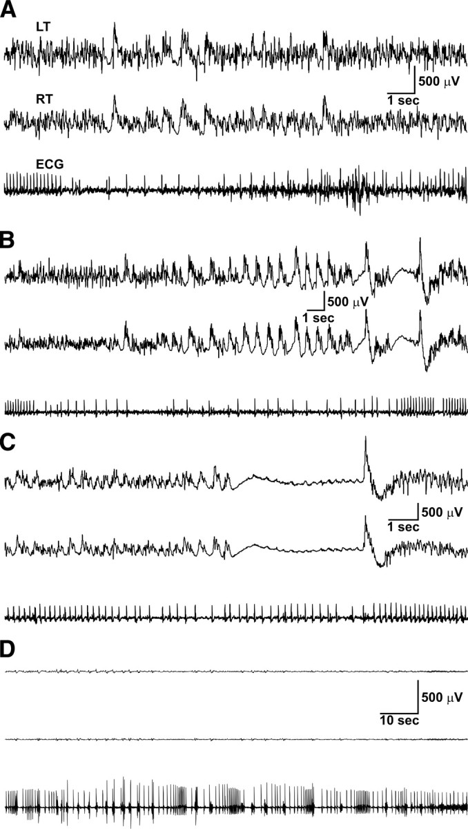
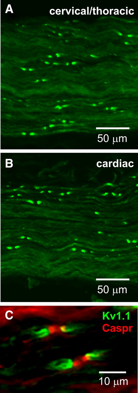
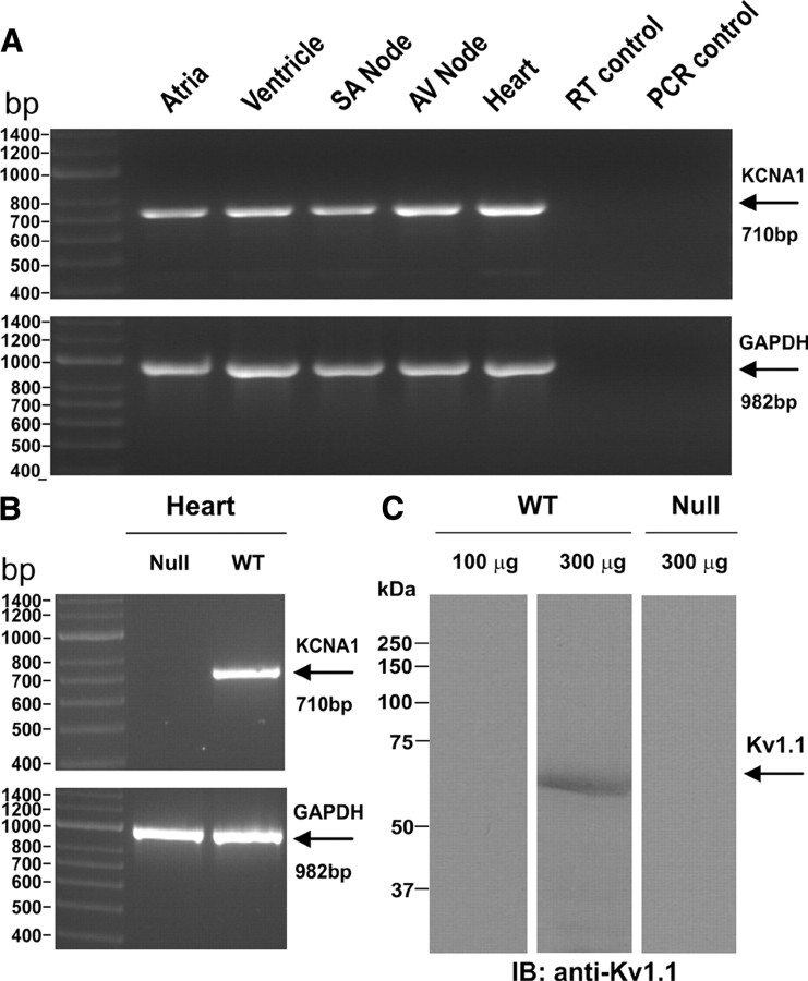
Mice lacking Kv1.1 Shaker-like potassium channels encoded by the Kcna1 gene exhibit severe seizures and die prematurely. The channel is widely expressed in brain but only minimally, if at all, in mouse myocardium. To test whether Kv1.1-potassium deficiency could underlie primary neurogenic cardiac dysfunction, we performed simultaneous video EEG-ECG recordings and found that Kcna1-null mice display potentially malignant interictal cardiac abnormalities, including a fivefold increase in atrioventricular (AV) conduction blocks, as well as bradycardia and premature ventricular contractions. During seizures the occurrence of AV conduction blocks increased, predisposing Kv1.1-deficient mice to sudden unexplained death in epilepsy (SUDEP), which we recorded fortuitously in one animal. To determine whether the interictal AV conduction blocks were of cardiac or neural origin, we examined their response to selective pharmacological blockade of the autonomic nervous system. Simultaneous administration of atropine and propranolol to block parasympathetic and sympathetic branches, respectively, eliminated conduction blocks. When administered separately, only atropine ameliorated AV conduction blocks, indicating that excessive parasympathetic tone contributes to the neurocardiac defect. We found no changes in Kv1.1-deficient cardiac structure, but extensive Kv1.1 expression in juxtaparanodes of the wild-type vagus nerve, the primary source of parasympathetic input to the heart, suggesting a novel site of action leading to Kv1.1-associated cardiac bradyarrhythmias. Together, our data suggest that Kv1.1 deficiency leads to impaired neural control of cardiac rhythmicity due in part to aberrant parasympathetic neurotransmission, making Kcna1 a strong candidate gene for human SUDEP.


Figures

Figure 1.
Figure 1.
Simultaneous EEG–ECG reveals interictal cardiac abnormalities, including AV blocks and premature ventricular contractions, in Kcna1 −/− mice. A, Example of a type 1, second-degree AV block (Wenckebach) in a Kcna1 −/− mouse demonstrating the characteristic progressive lengthening of the PR interval (denoted by lines with time interval in milliseconds underneath) until the QRS complex is dropped. B, Example of a type 2, second-degree AV block in a Kcna1 −/− mouse characterized by a skipped beat (in this case 2) preceded by normal length PR intervals. C, In at least two null mice, PVCs appeared in a bigeminy pattern composed of normal sinus rhythm (sharper, higher amplitude waves; arrowhead) alternating with abnormal PVCs (broader, lower amplitude waves; arrow). D, Occasionally, AV blocks corresponded with interictal cortical discharges in Kcna1-null mice, but these instances were rare. LT, Left temporal EEG; RT, right temporal EEG.
Figure 2.
Figure 2.
Kcna1 −/− mice exhibit a fivefold increase in basal AV blocks, a difference that is exacerbated by seizure activity. A, Comparison of mean number of AV blocks per hour during interictal periods in wild-type and null mice (n = 4 per genotype) showed a fivefold increase in mutants. During seizures in null mice, the frequency of AV blocks increased another fivefold to 25 times the wild-type rate. B, Histogram distribution of interictal AV blocks per hour in wild-type and null mice during 24 h recording sessions (n = 4 mice per genotype) shows that wild-type mice usually exhibited only 0–2 conduction blocks per hour compared to null mice, which exhibited conduction blocks with much higher frequency, up to 15 or more per hour. At least one AV block was observed in 96% of recording hours in null mice, whereas less than half of the recording hours showed a conduction block in controls. *p < 0.001; two-tailed t test.
Figure 3.
Figure 3.
SUDEP in a Kcna1 −/− mouse was preceded by ictal cardiac abnormalities including severe bradycardia and asystole. A–C, Simultaneous EEG–ECG recordings showing cortical and cardiac activity during the second, third, and fourth seizures, respectively, in the series of five severe seizures culminating with death. Note the regular cardiac rhythm in A and B, with an abrupt transition to profound bradycardia (∼100 bpm) interspersed with episodes of asystole lasting up to 20 s before resuming normal rhythm. The postictal heart rate was progressively depressed by each successive seizure as shown in Table 2. D, Simultaneous EEG–ECG recording showing electrocerebral silence coupled with severe bradycardia, asystole, and arrhythmias shortly preceding death. LT, Left temporal EEG; RT, right temporal EEG.
Figure 4.
Figure 4.
Selective pharmacological blockade of the autonomic nervous system suggests parasympathetic exacerbation of AV blocks in Kcna1 −/− mice. The first three panels are scatter plots of raw data showing the number of interictal AV blocks per hour before (Pre) and after (Post) drug administration. First panel, The elimination of interictal AV blocks in Kv1.1-deficient mice following administration of atropine and propranolol together (Atr + Pro) implicates autonomic transmission in arrhythmogenesis (n = 6). Second panel, Interictal AV blocks disappeared with atropine alone, suggesting predominant effect of parasympathetic tone (n = 4). Third panel, In contrast, injection of propranolol alone to inhibit sympathetic activity did not result in a significant change in the rate of AV conduction blocks (n = 6). Fourth panel, Summary of the percentage change in interictal AV blocks (symmetrized; mean ± SEM) following each drug treatment. Both complete autonomic blockade (Atr + Pro) and selective parasympathetic blockade (Atr) significantly decreased the average number of AV blocks per hour, but sympathetic blockade (Pro) had no significant effect. *p < 0.01; one-sample t test with μ = 0. NS, Not statistically significant.
Figure 5.
Figure 5.
Kv1.1 channels localize to juxtaparanodes of vagus nerve axons. Both the cervical/thoracic portion of the vagus nerve (A) and the cardiac branch (B) exhibited strong Kv1.1 immunoreactivity in a juxtaparanodal staining pattern. C, Double labeling with antibodies to Kv1.1 subunits (green) and to the paranodal protein Caspr (red) confirms that the Kv1.1 staining pattern in vagus nerve corresponds to juxtaparanodes.
Figure 6.
Figure 6.
Kv1.1 transcript and protein expression in mouse heart. A, RT-PCR using wild-type tissue reveals Kv1.1 transcripts (710 bp) in atria, ventricle, SA node, AV node, and whole heart tissue. RT-PCR of GAPDH mRNA (982 bp) as a positive control showed adequate tissue and transcript levels of the preparations. Omitting template during either the reverse transcriptase (RT control) or PCR (PCR control) steps to control for DNA contamination resulted in no unwanted amplification. B, As expected, RT-PCR of cardiac ventricular tissue from a Kcna1 −/− mouse as a negative control failed to amplify any Kv1.1 mRNA. C, A Kv1.1-immunoreactive band with an apparent molecular weight of ∼58 kDa was detected in 300 μg but not 100 μg of wild-type heart protein lysate following SDS-PAGE and immunoblotting using anti-Kv1.1 polyclonal antibody. As expected, the immunoreactive band was not detected in 300 μg of heart protein lysate from a Kcna1 −/− mouse.

Comment in

References

    1. Adelman JP, Bond CT, Pessia M, Maylie J. Episodic ataxia results from voltage-dependent potassium channels with altered functions. Neuron. 1995;15:1449–1454. - PubMed
    1. Altenmüller DM, Zehender M, Schulze-Bonhage A. High-grade atrioventricular block triggered by spontaneous and stimulation-induced epileptic activity in the left temporal lobe. Epilepsia. 2004;45:1640–1644. - PubMed
    1. Berry D, Ayers G. Symmetrized percent change for treatment comparisons. Am Stat. 2006;60:27–31.
    1. Berthoud HR, Neuhuber WL. Functional and chemical anatomy of the afferent vagal system. Auton Neurosci. 2000;85:1–17. - PubMed
    1. Bird JM, Dembny KAT, Sandeman D, Butler S. Sudden unexplained death in epilepsy: an intracranially monitored case. Epilepsia. 1997;38(Suppl 11):S52–S56.

Publication types

MeSH terms