. 2010 Apr 14;30(15):5334-45.
doi: 10.1523/JNEUROSCI.5963-09.2010.

Dlx5 and Dlx6 regulate the development of parvalbumin-expressing cortical interneurons

Affiliations

Dlx5 and Dlx6 regulate the development of parvalbumin-expressing cortical interneurons

Yanling Wang et al. J Neurosci. .

Abstract

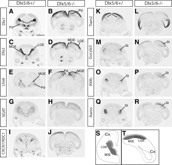
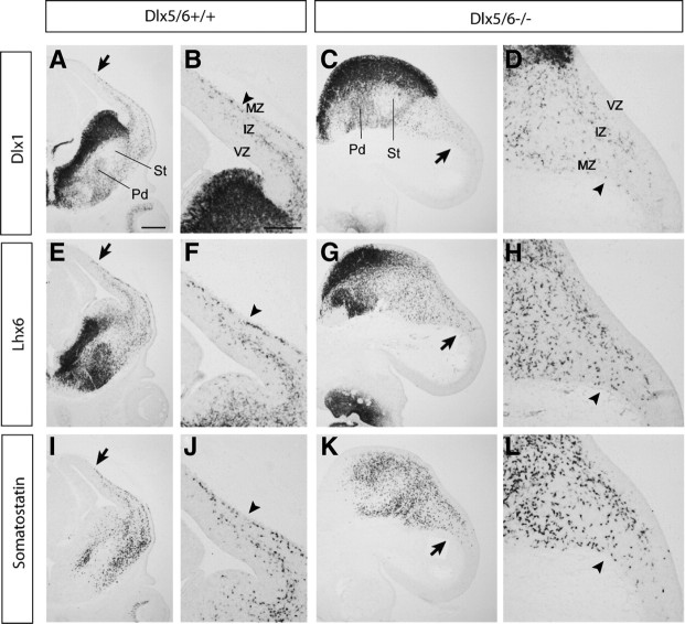
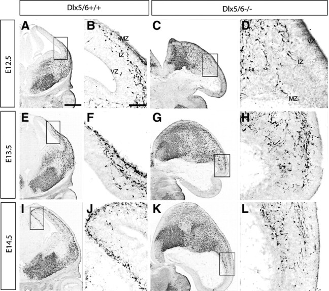
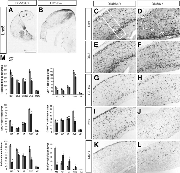
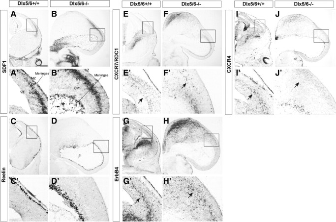
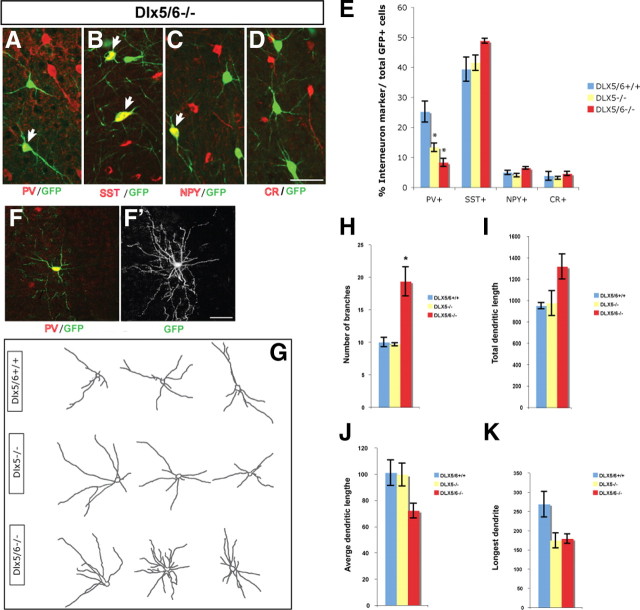
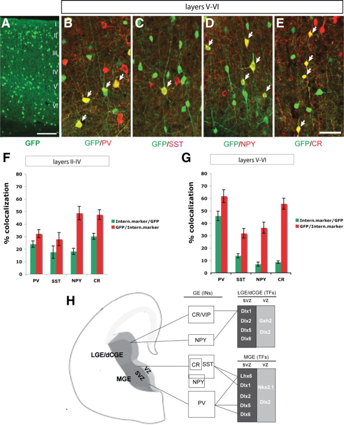
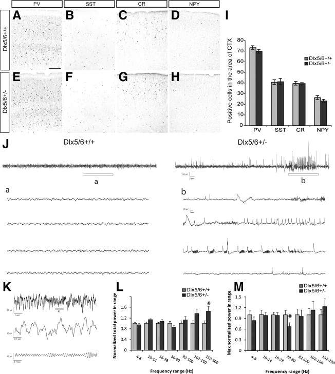
Dlx5 and Dlx6 homeobox genes are expressed in developing and mature cortical interneurons. Simultaneous deletion of Dlx5 and 6 results in exencephaly of the anterior brain; despite this defect, prenatal basal ganglia differentiation appeared largely intact, while tangential migration of Lhx6(+) and Mafb(+) interneurons to the cortex was reduced and disordered. The migration deficits were associated with reduced CXCR4 expression. Transplantation of mutant immature interneurons into a wild-type brain demonstrated that loss of either Dlx5 or Dlx5&6 preferentially reduced the number of mature parvalbumin(+) interneurons; those parvalbumin(+) interneurons that were present had increased dendritic branching. Dlx5/6(+/-) mice, which appear normal histologically, show spontaneous electrographic seizures and reduced power of gamma oscillations. Thus, Dlx5&6 appeared to be required for development and function of somal innervating (parvalbumin(+)) neocortical interneurons. This contrasts with Dlx1, whose function is required for dendrite innervating (calretinin(+), somatostatin(+), and neuropeptide Y(+)) interneurons (Cobos et al., 2005).


Figures

Figure 1.
Figure 1.
A–T, Preservation of telencephalic regional patterning and features of subpallial differentiation in E15.5 Dlx5/6−/− mutants. The Dlx5/6−/− mutants are exencephalic; a schematic representation of Dlx5/6+/ (S) and Dlx5/6−/− (T) is presented to help the reader orient the regions of the exencephalic telencephalon. Coronal sections from Dlx5/6+/ and Dlx5/6−/− were labeled by in situ hybridization with markers of LGE and MGE progenitor and mantle zones; the Dlx5/6−/− mutants do not show major gene expression defects, although the morphologies of the telencephalic regions are abnormal (A–R). Cx, Cortex; Pd, pallidum (including globus pallidus); St, striatum. Scale bar, 1.5 mm.
Figure 2.
Figure 2.
A–L, Reduced tangential migration in Dlx5/6−/− mutants at E13.5. Coronal sections from Dlx5/6+/+ and Dlx5/6−/− were labeled by in situ hybridization against Dlx1 (A–D), Lhx6 (E–H) and Somatostatin (I–L). The arrows point to the leading migrating cells. Note superficial migratory stream (arrowheads) in MZ is poorly formed in Dlx5/6−/− mutants. Scale bars: (in A) A, C, E, G, I, K, 300 μm; (in B) B, D, F, H, J, L, 200 μm.
Figure 3.
Figure 3.
A–L, Reduced tangential migration and accumulation of Lhx6-GFP-positive cells in the SVZ of the LGE of Dlx5/6−/− mutants at E12.5, E13.5, and E14.5. Immunohistochemistry for GFP was performed on coronal sections from E12.5 (A–D), E13.5 (E–H), and E14.5 (I–L) Dlx5/6+/+ and Dlx5/6−/− mutants. Boxed areas shown in high magnification are the migrating cells at the front of migration. Dlx5/6−/− mutants show reduced tangential migration and an accumulation of Lhx6-GFP cells in the SVZ of the LGE. Scale bars: (in A) A, C, E, G, I, K, 500 μm; (in B) B, D, F, H, J, L, 200 μm.
Figure 4.
Figure 4.
A–M, Reduced number of Dlx1+, Dlx2+, GAD67+, and Lhx6+ cells in the lateral cortex of Dlx5/6−/− mutants at E16.5. in situ hybridization with probes for Lhx6 (A, B, I, J), Dlx1 (C, D), Dlx2 (E, F), GAD67 (G, H), and MafB (K, L) was performed on coronal sections of Dlx5/6+/ and Dlx5/6−/− mutants. The boxes in A and B show the region that is shown at higher magnification in C-L. The box in C shows the size of the region used for cell counting; the total number of cells expressing these genes within 125,000 μm2 of the lateral neocortex and the number of positive cells in each of the cortical layers are presented (M). The reduction is most severe in MZ and SVZ, particularly for Lhx6 and MafB within the MZ. Scale bars: (in A) A, B, 1 mm; (in C) C–L, 200 μm.
Figure 5.
Figure 5.
A–J′, Loss of CXCR4 expression in deep migratory interneurons of Dlx5/6−/− mutants at E13.5. Coronal sections from Dlx5/6+/+ (A, C, E, G, I) and Dlx5/6−/− (B, D, F, H, J) were labeled by in situ hybridization against SDF1, Reelin, CXCR7 (RDC1), ErbB4 and CXCR4.Boxed areas are shown below in high magnification. The expression of SDF1 and reelin was maintained in Dlx5/6−/− mutants (B, D). The cavity formed by the everted exencephalic cortex contained scattered SDF1+ cells (* in B'). Although the expression of CXCR7 and ErbB4 was intact, CXCR4 expression was not detected in deep migratory interneurons (arrows in E′, F′, G′, H′, I′, J′). Scale bar: (in A) A–J′, 300 μm.
Figure 6.
Figure 6.
Cell-autonomous role for Dlx5/6 in controlling differentiation of PV+ cortical interneurons. E13.5 control (+/+), Dlx5−/−, or Dlx5/6−/− mutant MGE cells were transplanted into a wild-type postnatal d 0 (P0) cortex; the Lhx6-BAC GFP transgene was a reporter to follow the fate of MGE-derived cells several weeks after transplantation. A–D, Neocortical interneurons differentiated from transplanted Lhx6-GFP-expressing Dlx5/6−/− precursors, as shown by double immunofluorescence with anti-PV, anti-SST, anti-NPY or anti-CR antibodies. E, Percentage of double-labeled cells in the neocortex of 2-month-old mice grafted with control (blue), Dlx5−/− (yellow), and Dlx5/6−/− (red) cells. The percentage of GFP+/PV+ double-positive cells is reduced in mice grafted with either the Dlx5−/− or Dlx5/6−/− MGE cells. F, F′, To analyze the morphology of GFP+/PV+ interneurons, Z-stack confocal image for dendrite analysis was used; a representative GFP+/PV+ grafted cell from Dlx5/6−/− mutant is shown (F); the same cell was captured with Z-stack confocal image for dendrite analysis (F′). G, Representative images of grafted PV+ neocortical interneurons from control, Dlx5−/−, and Dlx5/6−/− mutants. H–K, Quantification of dendrite branching of PV+ interneurons from control (blue), Dlx5−/− (yellow), and Dlx5/6−/− (red) mutants. Note that the number of branches is increased in Dlx5/6−/− grafted PV+ interneurons. Scale bar: (in F′) A–F′, 100 μm.
Figure 7.
Figure 7.
Characterization of GFP expression in adult neocortical interneurons from the Dlx5 BAC transgenic mouse. A, GFP immunofluorescence in coronal sections through somatosensory cortex at 2 months of age. B–E, Double immunofluorescence confocal images with anti-GFP and anti-PV (B), anti-SST (C), anti-NPY (D), or anti-CR (E) antibodies. F, G, Quantification of the percentage of GFP+ cells that express each of the different interneuron markers (green bar) and the percentage of PV, SST, NPY, or CR cells that express GFP (red bar) in layer II–IV (F) and layer V–VI (G). H, Model of transcription factors that control the development of cortical and hippocampal interneurons. LGE/dCGE (dorsal CGE) are proposed to generate CR/VIP+ and a subset of NPY+ (late born) interneurons, which express Dlx1 and require it for their survival. The dorsal MGE generates SST+ (including SST/CR+ and SST/NPY+) interneurons that express Dlx1 and Lhx6, and require them for their survival and differentiation, respectively. The ventral MGE produces PV+ interneurons that express Dlx5 and Lhx6, and require Dlx5, Dlx6, and Lhx6 for their differentiation. Scale bar: (in E) A–E, 100 μm.
Figure 8.
Figure 8.
Dlx5/6+/− mice show spontaneous electrographic seizures and reduced maximum gamma power in the absence of gross histological abnormalities. A–H, Expression of PV, SST, CR, and NPY in somatosensory cortex of Dlx5/6+/+ and Dlx5/6+/− littermates. Scale bar: (in A) A–H, 200 μm. I, Quantification of PV+, SST+, CR+, NPY+ cells within a region of 380,000 μm2 of the somatosensory cortex. J, Sample EEG traces obtained during daytime recordings from freely moving adult Dlx5/6+/+ and Dlx5/6+/− mice. Top, Sixty-second-long EEG recordings. Bottom, Enlargement of region indicated by a or b in the top trace. Note the presence of an abnormal epileptiform-like electrographic discharge in b. Scale bars (in b) a, b, top, 20 μV, 2 s; bottom, 20 μV, 1 s. K, Sample EEG traces used for power analysis. Top, Ten-second-long EEG recording from a Dlx5/6+/+ mouse. Middle, Enlargement of region indicated by an asterisk in the top trace. Bottom, Same as middle, but filtered between 30 and 80 Hz. L, Total power as a function of frequency band for Dlx5/6+/+ or Dlx5/6+/− mice. For each frequency band, total power was normalized by the mean total power in Dlx5/6+/+ mice. Each bar represents an average over four mice from each group, five 60 s epochs from each mouse, and two EEG recording sites (n = 40 per group). M, Maximum power within each frequency band for Dlx5/6+/+ or Dlx5/6+/− mice. For each mouse, we selected the 60-s epoch with the most power in each frequency band. Each bar represents an average over four mice from each group, and two EEG recording sites in each mouse (n = 8 per group). *p < 0.05 by one-way ANOVA.

References

    1. Anderson SA, Qiu M, Bulfone A, Eisenstat DD, Meneses J, Pedersen R, Rubenstein JL. Mutations of the homeobox genes Dlx-1 and Dlx-2 disrupt the striatal subventricular zone and differentiation of late born striatal neurons. Neuron. 1997a;19:27–37. - PubMed
    1. Anderson SA, Eisenstat DD, Shi L, Rubenstein JL. Interneuron migration from basal forebrain to neocortex: dependence on Dlx genes. Science. 1997b;278:474–476. - PubMed
    1. Baraban SC, Southwell DG, Estrada RC, Jones DL, Sebe JY, Alfaro-Cervello C, Garcia-Verdugo JM, Rubenstein JL, Alvarez-Buylla A. Reduction of seizures by transplantation of cortical GABAergic interneuron precursors into Kv1.1 mutant mice. Proc Natl Acad Sci U S A. 2009;106:15472–15477. - PMC - PubMed
    1. Bielle F, Griveau A, Narboux-Neme N, Vigneau S, Sigrist M, Arber S, Wassef M, Pierani A. Multiple origins of Cajal-Retzius cells at the borders of the developing pallium. Nat Neurosci. 2005;8:1002–1012. - PubMed
    1. Borrell V, Marin O. Meninges control tangential migration of hem-derived Cajal-Retzius cells via CXCL12/CXCR4 signaling. Nat Neurosci. 2006;9:1284–1293. - PubMed

Publication types

MeSH terms

LinkOut - more resources