. 2010 Apr 14;30(15):5394-403.
doi: 10.1523/JNEUROSCI.5831-09.2010.

Neuron dysfunction is induced by prion protein with an insertional mutation via a Fyn kinase and reversed by sirtuin activation in Caenorhabditis elegans

Affiliations

Neuron dysfunction is induced by prion protein with an insertional mutation via a Fyn kinase and reversed by sirtuin activation in Caenorhabditis elegans

Nicolas Bizat et al. J Neurosci. .

Abstract

Although prion propagation is well understood, the signaling pathways activated by neurotoxic forms of prion protein (PrP) and those able to mitigate pathological phenotypes remain largely unknown. Here, we identify src-2, a Fyn-related kinase, as a gene required for human PrP with an insertional mutation to be neurotoxic in Caenorhabditis elegans, and the longevity modulator sir-2.1/SIRT1, a sirtuin deacetylase, as a modifier of prion neurotoxicity. The expression of octarepeat-expanded PrP in C. elegans mechanosensory neurons led to a progressive loss of response to touch without causing cell death, whereas wild-type PrP expression did not alter behavior. Transgenic PrP molecules showed expression at the plasma membrane, with protein clusters, partial resistance to proteinase K (PK), and protein insolubility detected for mutant PrP. Loss of function (LOF) of src-2 greatly reduced mutant PrP neurotoxicity without reducing PK-resistant PrP levels. Increased sir-2.1 dosage reversed mutant PrP neurotoxicity, whereas sir-2.1 LOF showed aggravation, and these effects did not alter PK-resistant PrP. Resveratrol, a polyphenol known to act through sirtuins for neuroprotection, reversed mutant PrP neurotoxicity in a sir-2.1-dependent manner. Additionally, resveratrol reversed cell death caused by mutant PrP in cerebellar granule neurons from prnp-null mice. These results suggest that Fyn mediates mutant PrP neurotoxicity in addition to its role in cellular PrP signaling and reveal that sirtuin activation mitigates these neurotoxic effects. Sirtuin activators may thus have therapeutic potential to protect from prion neurotoxicity and its effects on intracellular signaling.


Figures

Figure 1.
Figure 1.
Human mutant PrP produces a progressive neuronal dysfunction in C. elegans transgenics. A, Progressive loss of response to touch at the tail caused by mutant (PG13-PrP) PrP expression. Four lines of each genotype (Wt-PrP or PG13-PrP), all of them expressing similar levels of transgene (see supplemental Fig. 1, available at www.jneurosci.org as supplemental material, and Fig. 4A), were tested at different developmental stages (L3 larvae, L4 larvae, and young adults). Each bar corresponds to the mean ± SEM for touch response at the tail. ***p < 0.0001 versus control (mean value for the four Wt-PrP lines). B, Compared to a population of Wt-PrP animals, PG13-PrP animals show a shift toward insensitivity in the distribution of responsiveness/animal. The responses to light touch at the tail were recorded for every animal and expressed as mean percentage of responsiveness/animal ± SEM as shown on the x-axis. C, PrP is expressed in Pmec-7 target cells. Two lines expressing either Wt-PrP or PG13-PrP were crossed with animals stably expressing GFP under the control of Pmec-7. Shown here is a representative immunohistochemical analysis of PrP localization in PLM cells from young adults, with Nomarski analysis shown in the left panel. Magnification is ×63 and scale bar is 5 μm. Posterior is to the bottom in all panels. D, PrP is expressed at the cell body membrane, with accumulation detected in the cell body and axons for PG13-PrP. PrP expression was assessed using confocal analysis of PLM neurons in animals coexpressing GFP and PrP (2 lines/genotype) upon PrP immunostaining. Shown here are representative images of the most frequent phenotypes observed in PLM cells. Wt-PrP primarily shows diffuse expression at the cell body membrane, with few signals detected in the axon in some occasions. PG13-PrP expression shows clustering in the cell body and along the axonal process (white arrows). Magnification is ×100 and scale bar is 2 μm. E, Quantification of PrP clusters in PLM cells. Neurons expressing PG13-PrP exhibit a higher level of PrP clustering (mean ± SEM) in the cell body and axon. ***p < 0.001 compared to Wt-PrP. F, Representative confocal images of the most frequent morphology of PLM neurons in day-4 adults coexpressing GFP and PrP. R and L refer to the right and left PLM cells. Magnification is ×63 and scale bar is 5 μm. Posterior is to the left in all panels. G, Quantification of PLM neurons showing a normal morphology in young (day 1) and old (day 4) adults. Two lines/genotype were scored, and 80–90% of normal PLM cells were detected in mutant PrP and control animals at the old adult stage. H, Image of PLM cell degeneration in old adults. A weak level of cell body loss or axonal degeneration (white arrows) was observed in all the genetic background tested, thus corresponding to normal cellular senescence. Magnification is ×63 and scale bar is 5 μm. Posterior is to the left in all panels.
Figure 2.
Figure 2.
Biochemical properties of human prion protein expressed in C. elegans neurons. A, Similar expression levels for transgenic PrP proteins as shown by Western blotting in the four lines expressing Wt-PrP and four lines expressing PG13-PrP. A molecular weight shift observed for PG13-PrP as expected. Human brain homogenate and protein extracts from the N2 nematode strain were used as controls. B, Calibration of resistance to PK digestion using PK at 1000 to 0.1 μg/ml. PG13-PrP is partially resistant to PK digestion in a PK-concentration-dependent manner, whereas Wt-PrP is not resistant to PK at all the enzyme concentrations tested. C, The resistance of PG13-PrP to PK digestion is weaker than that of sporadic CJD PrPSc. PG13-PrP is resistant to PK in a dose-dependent manner (10 μg/ml and 0.1 μg/ml) at concentrations that fully degrade PrP from healthy human brain (human CTL). At these concentrations, PrP from human brain CJD (Hum CJD) is PK resistant, indicating that PG13-PrP show partial and weaker PK resistance compared to PrP from human CJD. D, Resistance to PK digestion in lines expressing PG13-PrP. Prion protein in the PG13-PrP lines are partially protected from PK digestion, whereas Wt-PrP lines do not show PK resistance. Healthy (CTL) and sporadic disease (CJD) human brain homogenates were used as digestion controls. E, Western blot signature of PK-resistant PrP associated with the PG13 mutation. T1, Control sample with PK-resistant PrP type 1 from patient with sporadic CJD. T2A, Control sample with PK-resistant PrP type 2A from a patient with sporadic CJD. FC, Frontal cortex from a patient with the PG13 mutation. Tha, Thalamus from a patient with the PG13 mutation. WT, C. elegans line expressing wild-type PrP. PG13, C. elegans line expressing PG13-PrP. Samples from a patient with the PG13 mutation and from PG13-PrP-expressing animals showed a comparable pattern of PK-resistant PrPres with, compared to PrPres type from sporadic CJD, additional bands at 17–18 and 7–8 kDa. F, PG13-PrP is insoluble. Upon fractionation, Wt-PrP is primarily found in the soluble fraction, whereas mutant PrP is primarily found in the sedimented fraction.
Figure 3.
Figure 3.
Quinacrine protects C. elegans neurons from the effects of PG13-PrP expression. A, Quinacrine reversed neuronal dysfunction (mean ± SEM) in PG13-PrP animals with no effect detected in Wt-PrP animals. Three independent lines were tested per genotype. *p < 0.05 and ***p < 0.0001 versus untreated PG13-PrP animals. B, Quinacrine does not alter PG13-PrP transgene expression as detected by RT-PCR. C, Representative Western blot showing that quinacrine does not alter the expression level of the mutated prion protein and restores sensitivity to PK digestion.
Figure 4.
Figure 4.
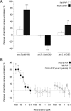
Modulation of PG13-PrP cytotoxicity in C. elegans neurons by scr-2/FRK, sir-2.1/SIRT1, and resveratrol. A, The LOF src-2 (ok819) greatly reduced neuronal dysfunction in PG13-PrP animals with no effect seen in Wt-PrP animals. This effect was not due to a change in the resistance to PK digestion (see supplemental Fig. S4A, available at www.jneurosci.org as supplemental material), nor to a change in PG-13-PrP expression levels (supplemental Fig. S4B, available at www.jneurosci.org as supplemental material). Increased sir-2.1 dosage specifically rescues neuronal dysfunction in PG13-PrP animals, whereas a sir-2.1 deletion allele (ok434) enhanced neuronal dysfunction. These effects were not due to a change in the resistance to PK digestion (see supplemental Fig. S4C,E, available at www.jneurosci.org as supplemental material), nor to a change in PG-13-PrP expression levels (supplemental Fig. S4D,F, available at www.jneurosci.org as supplemental material). Results are given as the percentage of rescue (mean ± SEM), a negative value means aggravation, and the maximal achievable rescue is 100% (see Materials and Methods). Three independent lines were tested per genotype. ***p < 0.0001; **p < 0.001 compared to PrP transgenics. B, Resveratrol rescues neuronal dysfunction in PG13-PrP animals with no effect seen in Wt-PrP animals. This effect was not due to a change in PG13-PrP expression levels (supplemental Fig. S2G, available at www.jneurosci.org as supplemental material). Rescue by resveratrol is lost in PG13-PrP transgenics harboring sir-2.1 LOF. Three independent lines were tested per genotype. **p < 0.001; *p < 0.05 compared to animals treated with vehicle.
Figure 5.
Figure 5.
Resveratrol protects mouse neurons from cell death induced by human mutant (PG14-PrP) prion protein expression. A, Representative images of cerebellar granule neurons transfected with Wt-PrP or PG14-PrP constructs. To detect intracellular aggregated form of PG14, cells were permeabilized and immunostained after 4 d of culture (green, dendritic MAP2; red, PrP; blue, DAPI). Wt-PrP and PG14-PrP are expressed in cell bodies and neuronal processes (note that intracellular Wt-PrP staining is fainter than membrane-bound PrP signal in C). Accumulation and aggregation in neuronal processes (white arrows) is observed for PG14-PrP. Magnification is ×400 and scale bar is 10 μm. B, Quantification of PG14-PrP-induced neuronal death and neuroprotection by resveratrol. Data are expressed as the mean ratio ± SEM of surviving cells at day 6 compared to day 3 after transfection. A ratio equal to 1 means no cell death. PG14-PrP expression strongly alters neuronal survival (***p < 0.0001), whereas Wt-PrP expression has no effect. Resveratrol (duration of treatment: 72 h starting at day 3 after transfection) prevents neuronal death induced by PG14-PrP expression. ***p < 0.0001. C, Representative fields of cerebellar granule neurons transfected with Wt-PrP or PG14-PrP and treated for 72 h with either vehicle or resveratrol. Membrane-bound (no permeabilization) forms of PrP are immunostained after 6 d in culture (green, membrane-bound PrP; blue, cell nuclei, DAPI). Note the drastic reduction in PrP-expressing cell number caused by PG14-PrP expression in untreated neurons. Magnification is ×200 and scale bar is 20 μm. Inset [red, membrane-bound PrP; yellow, nuclear DAPI (pseudocolor)] shows a healthy Wt-PrP-expressing neuron, whereas remaining PG14-expressing neurons shows condensed chromatin (white arrow) with dystrophic neurites.

Similar articles

Cited by

References

    1. Aguzzi A, Sigurdson C, Heikenwaelder M. Molecular mechanisms of prion pathogenesis. Annu Rev Pathol. 2008a;3:11–40. - PubMed
    1. Aguzzi A, Baumann F, Bremer J. The prion's elusive reason for being. Annu Rev Neurosci. 2008b;31:439–477. - PubMed
    1. Araki T, Sasaki Y, Milbrandt J. Increased nuclear NAD biosynthesis and SIRT1 activation prevent axonal degeneration. Science. 2004;305:1010–1013. - PubMed
    1. Baur JA Advance online publication. Biochemical effects of SIRT1 activators. Biochim Biophys Acta. 2009 doi: 10.1016/j.bbapap.2009.10.025. - DOI - PMC - PubMed
    1. Bolton DC, McKinley MP, Prusiner SB. Molecular characteristics of the major scrapie prion protein. Biochemistry. 1984;23:5898–5906. - PubMed

Publication types

MeSH terms