

Dissociable neural responses to hands and non-hand body parts in human left extrastriate visual cortex
- PMID: 20393066
- PMCID: PMC2888254
- DOI: 10.1152/jn.00215.2010
Dissociable neural responses to hands and non-hand body parts in human left extrastriate visual cortex
Abstract
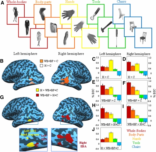
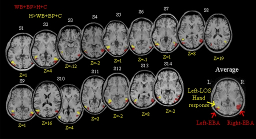
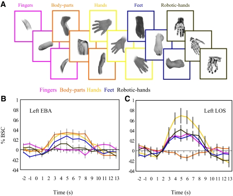
Accumulating evidence points to a map of visual regions encoding specific categories of objects. For example, a region in the human extrastriate visual cortex, the extrastriate body area (EBA), has been implicated in the visual processing of bodies and body parts. Although in the monkey, neurons selective for hands have been reported, in humans it is unclear whether areas selective for individual body parts such as the hand exist. Here, we conducted two functional MRI experiments to test for hand-preferring responses in the human extrastriate visual cortex. We found evidence for a hand-preferring region in left lateral occipitotemporal cortex in all 14 participants. This region, located in the lateral occipital sulcus, partially overlapped with left EBA, but could be functionally and anatomically dissociated from it. In experiment 2, we further investigated the functional profile of hand- and body-preferring regions by measuring responses to hands, fingers, feet, assorted body parts (arms, legs, torsos), and non-biological handlike stimuli such as robotic hands. The hand-preferring region responded most strongly to hands, followed by robotic hands, fingers, and feet, whereas its response to assorted body parts did not significantly differ from baseline. By contrast, EBA responded most strongly to body parts, followed by hands and feet, and did not significantly respond to robotic hands or fingers. Together, these results provide evidence for a representation of the hand in extrastriate visual cortex that is distinct from the representation of other body parts.
Figures
References
-
- Amedi A, Malach R, Hendler T, Peled S, Zohary E. Visuo-haptic object-related activation in the ventral visual pathway. Nat Neurosci 4: 324–330, 2001 - PubMed
-
- Astafiev SV, Stanley CM, Shulman GL, Corbetta M. Extrastriate body area in human occipital cortex responds to the performance of motor actions. Nat Neurosci 7: 542–548, 2004 - PubMed
-
- Barton JJ. Disorders of face perception and recognition. Neurol Clin 21: 521–548, 2003 - PubMed
-
- Beauchamp MS, Lee KE, Haxby JV, Martin A. Parallel visual motion processing streams for manipulable objects and human movements. Neuron 34: 149–159, 2002 - PubMed
-
- Beauchamp MS, Lee KE, Haxby JV, Martin A. FMRI responses to video and point-light displays of moving humans and manipulable objects. J Cogn Neurosci 15: 991–1001, 2003 - PubMed
Publication types
MeSH terms
Substances
Grants and funding
LinkOut - more resources
Full Text Sources
Other Literature Sources