. 2010 Oct;51(10):5002-11.
doi: 10.1167/iovs.09-5110. Epub 2010 Apr 14.

TGF-β-stimulated CTGF production enhanced by collagen and associated with biogenesis of a novel 31-kDa CTGF form in human corneal fibroblasts

Affiliations

TGF-β-stimulated CTGF production enhanced by collagen and associated with biogenesis of a novel 31-kDa CTGF form in human corneal fibroblasts

Edward G Tall et al. Invest Ophthalmol Vis Sci. 2010 Oct.

Abstract

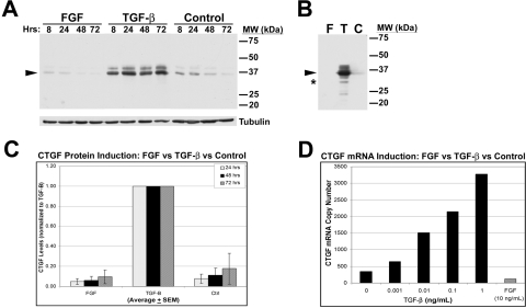
Purpose: Connective tissue growth factor (CTGF) is induced by transforming growth factor-beta (TGF-β) after corneal wounding. This study addressed the role of the extracellular matrix in the induction of CTGF by TGF-β.

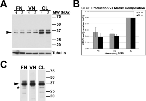
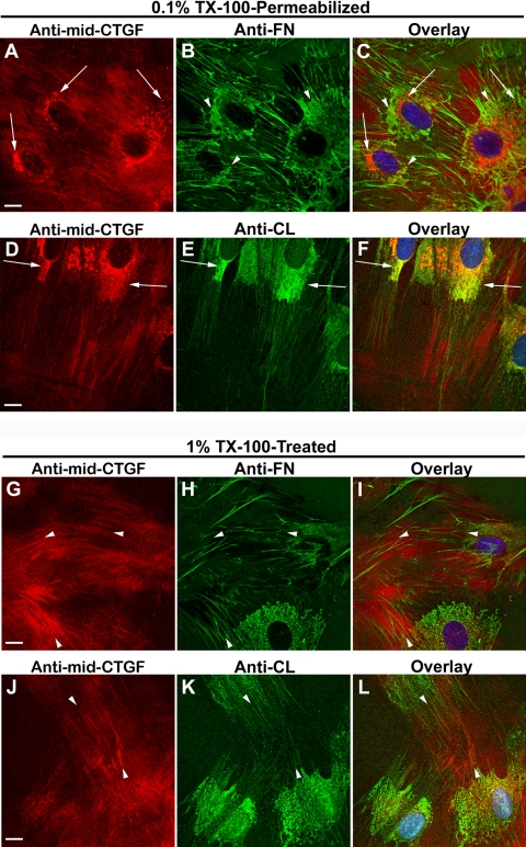
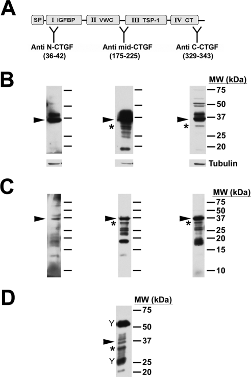
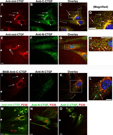
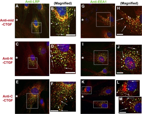
Methods: Human corneal fibroblasts (HCFs) were grown on fibronectin (FN), vitronectin (VN), or collagen (CL) in supplemented serum-free media alone or with TGF-β1 or fibroblast growth factor plus heparin. CTGF mRNA was analyzed by qPCR and protein expression by Western blot analysis of Triton X-100 (TX-100)-soluble and TX-100-insoluble cell lysates using antibodies to N-terminal, mid, and C-terminal CTGF regions. Immunocytochemistry was performed on nonconfluent or scrape-wounded confluent HCFs.

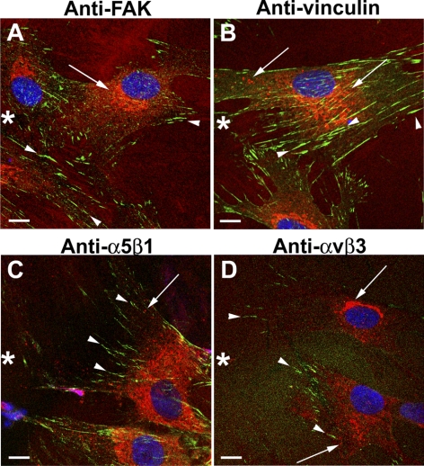
Results: TGF-β-treated HCFs grown on CL produced five times more 38-kDa CTGF than untreated controls (72 hours). TGF-β-treated HCFs on CL secreted twofold more CTGF than those on FN or VN. Furthermore, a 31-kDa CTGF form, lacking the N-terminal domain, was detected in Triton X-100 insoluble fractions in Western blot analysis. Immunodetectable extracellular CTGF formed linear arrays parallel to, but not colocalized with, CL or FN. It also did not colocalize with FAK, vinculin, or integrins α(v)β(3) and α(5)β(1). Intracellular CTGF was detected in the Golgi apparatus and vesicles, including endosomes.

Conclusions: Enhanced CTGF secretion induced by TGF-β in CL-grown cells may contribute to positive feedback in which CL is overexpressed in CTGF-induced fibrosis. N-terminal CTGF fragments in the plasma of patients with severe fibrotic disease may be a product of CTGF proteolysis that also produces the newly identified 31-kDa CTGF that remains cell associated and may have its impact by non-integrin signaling pathways.


Figures

Figure 1.
Figure 1.
CTGF production by HCFs is stimulated by TGF-β. HCFs were plated on collagen in SSFM only or with FGF or TGF-β for 8, 24, 48, and 72 hours. (A) Anti–mid-CTGF antibody detected a doublet of 36- to 38-kDa CTGF (38 kDa) in TX-100–soluble lysates in Western blot analysis (arrowhead). Tubulin controls confirm equal loading. (B) Triton X-100–insoluble fractions from HCFs at 24 hours were solubilized in 2% SDS buffer followed by sonication and SDS-PAGE and Western blot analysis. As with the soluble lysates, increased levels of CTGF were detected in the TGF-β–treated cells and not from those treated with FGF or with no growth factor. In addition to the 38-kDa CTGF (arrowhead), a previously undescribed band at 31 kDa (asterisk) was strongly immunodetected in this fraction with anti–mid-CTGF antibody, as were other lower MWt bands (e.g., 24 kDa). (C) Quantification by densitometry of TGF-β–induced production of 38-kDa CTGF at 24, 48, and 72 hours in the TX-100–soluble fraction (n = 20). TGF-β–treated HCF cultures had a fivefold increase in immunodetected CTGF production compared with FGF-treated or nontreated control cultures. (D) qPCR analysis showed a dose-response increase of CTGF mRNA in HCFs treated for 24 hours with TGF-β (black bars). In contrast, 24-hour FGF (10 ng/mL) treatment (gray bar) decreased CTGF mRNA levels to below the level of control cultures grown without addition of growth factors (“0” TGF-β).
Figure 2.
Figure 2.
TGF-β–induced CTGF was greater in HCFs growing on CL than on FN or VN. HCFs were plated on FN, VN, or CL in duplicate experiments (lanes 1 and 2) in the presence of TGF-β for 24 hours. (A) Anti–mid-CTGF Ab was used to detect 38-kDa CTGF in TX-100 soluble lysates by Western blot. An increasing amount of CTGF (arrowhead) was detected in cells plated on CL compared with cells plated on FN and VN. Tubulin controls confirm equal loading. (B) Quantification of a 38-kDa CTGF doublet in HCFs treated with TGF-β for 24 or 72 hours on the three different matrices. Bars represent averages + SEM normalized to CTGF levels of samples plated on CL. (C) In TX-100–insoluble fractions of lysates of HCFs grown on FN, VN, or CL, anti–mid-CTGF immunodetected 38-kDa CTGF (arrowhead). Novel 31-kDa form of immunodetectable CTGF was enriched in the TX-100–insoluble fraction in cells grown on all three matrices (asterisk). Because we loaded these lanes based on equal sample volume rather than protein concentration, quantitative comparisons cannot be made. Previously described 24-kDa and 18- to 20-kDa CTGF bands were also detected.
Figure 3.
Figure 3.
Organization of CTGF with fibronectin or collagen. HCFs grown on FN-coated or CL-coated coverslips in SSFM plus TGF-β for 24 hours were fixed and then permeabilized with 0.1% TX-100 for 1.5 minutes in a standard immunocytochemical protocol (A–F). To “visualize” the TX-100–insoluble fraction, the coverslips were permeabilized with 1% TX-100 on ice for 20 minutes before fixation (G–L). (A–C, arrows) CTGF (red) detected in the ER/Golgi that does not colocalize with intracellular regions of FN (green) enrichment (arrowheads). (D–F) Juxtanuclear ER/Golgi (arrows) in which detectable CTGF (red) and CL (green) overlap (yellow). (G–L) CTGF (red) was detected in linear arrangement outside of cells (G, J, arrowheads) and aligned with fibrillar FN (H, I, arrowheads) or fibrillar CL but did not colocalize with either (K, L, arrowheads). Vesicular and Golgi-localized CTGF staining (seen in A–F) is absent after the prolonged and stronger TX-100 permeabilization (G–J). Scale bars, 10 μm.
Figure 4.
Figure 4.
CTGF did not colocalize at focal adhesions with FAK, vinculin, or integrin α5β1 or αvβ3. HCFs growing on CL-coated coverslips were scrape-wounded and incubated with TGF-β for 10 hours and then fixed, permeabilized, and immunodetected for CTGF (red) and FAK, vinculin, and integrin α5β1 or αvβ3 (green). In the cells migrating into the wound (asterisk), CTGF was detected in intracellular vesicles and the Golgi (arrows). Focal adhesions enriched in FAK, vinculin, α5β1, or αvβ3 (arrowheads) did not colocalize with intracellular or extracellular CTGF (A–D). Scale bars, 10 μm.
Figure 5.
Figure 5.
31-kDa CTGF was not detected by the anti–N-CTGF antibody. (A) Diagram of CTGF showing its modular architecture and the domains to which the specific antibodies were raised, indicated at “Y”: signal peptide (SP), insulin-like growth factor binding protein (IGFBP), von Willebrand factor type C repeat (VWC), thrombospondin type 1 (TSP-1), and C-terminal (CT). (B) HCFs were treated with TGF-β for 24 hours before lysis and detection with CTGF antibodies on Western blot. Full-length CTGF (arrowheads) was detected by all three antibodies. 31-kDa CTGF (asterisk) was detected in the blots probed with anti–mid-CTGF or anti–C-CTGF antibodies. In contrast, anti–N-CTGF did not detect the 31-kDa form. Tubulin was used as a loading control (lower panels). (C) TX-100–insoluble fractions were probed with the same CTGF antibodies and yielded similar results. (D) Immunoprecipitation by anti–mid-CTGF antibody of conditioned media of HCFs cultured on CL with TGF-β for 72 hours yielded multiple forms of CTGF. Full-length CTGF (arrowhead) and the 31-kDa form (asterisk), as well as other molecular weight forms, including 20 and 26 to 28 kDa, were immunodetected. (“Y” symbols indicate bands from heavy and light IgG chains of the immunoprecipitation antibody.)
Figure 6.
Figure 6.
Immunocytochemical localizations using anti–mid-CTGF, anti–N-CTGF, anti–C-CTGF, and anti–p230 Golgi antibodies in HCFs. HCFs growing on CL-coated coverslips were scrape-wounded and incubated with TGF-β before fixation and immunodetection with antibodies used for the Western blot analysis in Figure 5. Although the images are from cells adjacent to the wound (asterisk), they are representative of CTGF domain staining seen in cells not adjacent to a scrape wound. (A–D) Antibody detection of CTGF central domain (A, red) and C-terminal domain (B, green) colocalized to the Golgi apparatus (A, B, arrowheads; C, yellow) and some adjacent vesicles (D, arrowheads). In addition, the C-terminal CTGF domain was detected in vesicles that were distal from the Golgi apparatus (arrow and brackets in C [an overlay of A and B]) and in (D [magnification of boxed region in C]). (E–H) Antibody to the N terminus of CTGF was seen in vesicles adjacent to the Golgi and throughout the cell rather than in the Golgi lamellae (F, J, arrows). Anti–mid-CTGF and anti–N-CTGF colocalized in vesicles in the Golgi region (G; magnified in H, yellow vesicles, arrowheads). However, many vesicles stained only for either N-CTGF (green, bracket in H) or mid-CTGF (red, arrows in H). (I–L) Most vesicles had either immunodetectable C-CTGF or immunodetectable N-CTGF, but not both. Using an mAb, anti–C-CTGF was immunodetected in the Golgi lamellae and vesicles (I, red) but was usually not colocalized with anti–N-CTGF (K, L), suggesting that each terminal fragment may be taken up into separate vesicles. Arrows: vesicles that stain only with C-terminal domain (red) or with N-terminal domain (green). As in (G), antibody to N-CTGF did not colocalize with Golgi lamellae (K). Colocalization with antibodies to trans-Golgi protein p230 (M–O, red) confirms that anti–mid-CTGF (M, green) and anti–C-CTGF (O, green) detect CTGF in the Golgi apparatus. However, anti–N-CTGF (N, green) does not colocalize with antibodies to Golgi (N, red). Scale bars, 10 μm.
Figure 7.
Figure 7.
Immunodetection of the scavenger receptor LRP and the endosome marker EEA1 in vesicles: colocalization with CTGF and its N- or C-terminal domain. HCFs on CL-coated coverslips were scrape-wounded (asterisk) and incubated with TGF-β for 8 to 10 hours and then fixed, permeabilized, and immunostained with antibodies, as indicated. (A–F) CTGF central (A) or N-terminal (C) or C-terminal (E) domains (all red) were detected in vesicles, including those proximal to the Golgi apparatus. LRP (green) was similarly localized to vesicles (arrows) distributed throughout the cell. In these overlay images, there were three patterns of vesicular localization of CTGF antibody and LRP antibody (better seen in B, D, F, which are magnifications of the boxed regions in A, C, E). Many CTGF-stained vesicles were also stained with LRP antibody. The two other vesicular localization patterns were vesicles that were either green or red (arrows) and that had a “traffic light pattern” of adjacent vesicles (arrowheads), one stained with CTGF antibody (red) and the other stained with LRP antibody (green), sometimes with regions of apparent colocalization between them (yellow). The traffic light vesicles were not the result of the nonalignment of images because the orientation of the red to the green varies within a small region as seen in (B, D, F). Similar patterns of CTGF and LRP staining were seen in cells not adjacent to a scrape wound (data not shown). Scale bars, 10 μm. (G–M) Anti–EEA1-identified endosomal vesicles (green). Some endosomes were colocalized by antibodies to CTGF central (G) or N-terminal (I) or C-terminal (K) domains (all red), better seen in magnified regions (H, J, L, M). The same three patterns of overlap/nonoverlap of EEA1 and CTGF domains are seen, as was described for LRP and CTGF. Scale bars, 10 μm.

Similar articles

Cited by

References

    1. Blalock TD, Duncan MR, Varela JC, et al. Connective tissue growth factor expression and action in human corneal fibroblast cultures and rat corneas after photorefractive keratectomy. Invest Ophthalmol Vis Sci. 2003;44:1879–1887 - PubMed
    1. Secker GA, Shortt AJ, Sampson E, Schwarz QP, Schultz GS, Daniels JT. TGFβ stimulated re-epithelialisation is regulated by CTGF and Ras/MEK/ERK signalling. Exp Cell Res. 2008;314:131–142 - PubMed
    1. Grotendorst GR. Connective tissue growth factor: a mediator of TGF-β action on fibroblasts. Cytokine Growth Factor Rev. 1997;8:171–179 - PubMed
    1. Brigstock DR. The connective tissue growth factor/cysteine-rich 61/nephroblastoma overexpressed (CCN) family. Endocr Rev. 1999;20:189–206 - PubMed
    1. Gao R, Brigstock DR. Connective tissue growth factor (CCN2) induces adhesion of rat activated hepatic stellate cells by binding of its C-terminal domain to integrin alpha (v) beta(3) and heparan sulfate proteoglycan. J Biol Chem 2004;279:8848–8855 - PubMed

Publication types

MeSH terms