. 2010 May 4;107(18):8153-8.
doi: 10.1073/pnas.0912509107. Epub 2010 Apr 14.

Structure of the histone chaperone CIA/ASF1-double bromodomain complex linking histone modifications and site-specific histone eviction

Affiliations

Structure of the histone chaperone CIA/ASF1-double bromodomain complex linking histone modifications and site-specific histone eviction

Yusuke Akai et al. Proc Natl Acad Sci U S A. .

Abstract

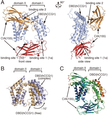
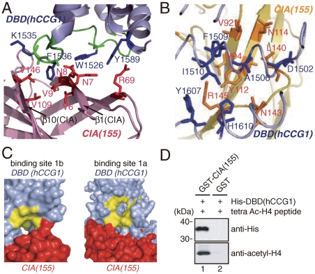
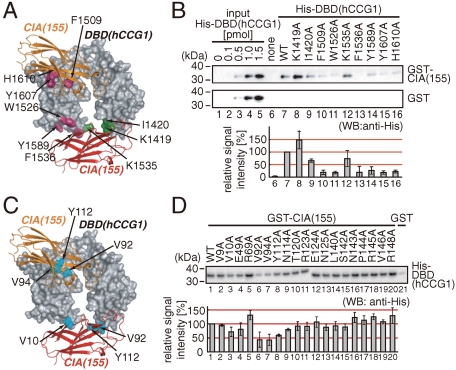
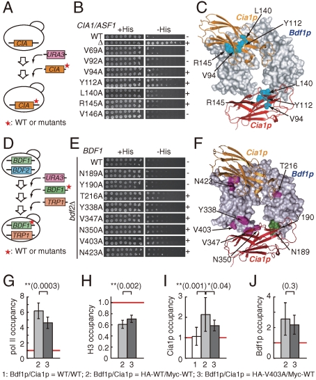
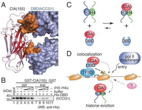
Nucleosomes around the promoter region are disassembled for transcription in response to various signals, such as acetylation and methylation of histones. Although the interactions between histone-acetylation-recognizing bromodomains and factors involved in nucleosome disassembly have been reported, no structural basis connecting histone modifications and nucleosome disassembly has been obtained. Here, we determined at 3.3 A resolution the crystal structure of histone chaperone cell cycle gene 1 (CCG1) interacting factor A/antisilencing function 1 (CIA/ASF1) in complex with the double bromodomain in the CCG1/TAF1/TAF(II)250 subunit of transcription factor IID. Structural, biochemical, and biological studies suggested that interaction between double bromodomain and CIA/ASF1 is required for their colocalization, histone eviction, and pol II entry at active promoter regions. Furthermore, the present crystal structure has characteristics that can connect histone acetylation and CIA/ASF1-mediated histone eviction. These findings suggest that the molecular complex between CIA/ASF1 and the double bromodomain plays a key role in site-specific histone eviction at active promoter regions. The model we propose here is the initial structure-based model of the biological signaling from histone modifications to structural change of the nucleosome (hi-MOST model).


Conflict of interest statement

The authors declare no conflict of interest.

Figures

Fig. 1.
Fig. 1.
Physical and functional interactions between CIA/ASF1 and DBD(CCG1). (A) Schematic drawing of human CIA/ASF1, the CCG1 subunit containing DBD(hCCG1), and their yeast homologs, yCia1p/Asf1p, yTAFII145, and yBdf1p. The evolutionarily conserved N-terminal region (colored in gray) of CIA/ASF1 can bind the histone H3–H4 dimer (28, 29). BD stands for bromodomain. (B) Physical interaction between human GST-CIA/ASF1(155) and His-DBD(hCCG1). CIA/ASF1(155) and the full-length CIA/ASF1 [CIA/ASF1(204)] have nearly the same binding activities for DBD(hCCG1). (C) Physical interaction between yeast GST-Cia1p/Asf1p(155) and His-DBD(Bdf1p). (DG) Relative occupancies of pol II (D), histone H3 (E), Cia1p/Asf1p (F), and Bdf1p (G) on the promoter regions of the PMA1, ACT1, PHO11, GAL10, GAL1, and HO genes as well as one ORF-free region. The occupancy of each protein on the PHO11 promoter region (indicated by a red line in each graph) was utilized as a reference.
Fig. 2.
Fig. 2.
Crystal structure of the CIA/ASF1(155)–DBD(hCCG1) complex. (A) Crystal structure of the CIA/ASF1(155)–DBD(hCCG1) complex (front and side views). CIA/ASF1(155) at binding sites 1 and 2, and DBD(hCCG1) are shown in red, orange, and light blue, respectively. All molecular graphics were prepared by PyMOL (http://www.pymol.org). (B) Conformational change of DBD(hCCG1) induced by the CIA/ASF1(155) binding. The Cα atoms in domain II of DBD(hCCG1) were superposed by the least-square fittings. The DBD(hCCG1) of CIA/ASF1-free form (PDB ID code 1EQF) (33) and that in the present complex are shown in brown and light blue, respectively. (C) The result of the TLS-tensor analysis of the CIA/ASF1(155)–DBD(hCCG1) complex. Temperature factors calculated from the TLS tensors are displayed in the crystal structure of the CIA/ASF1(155)–DBD(hCCG1) complex (blue, low; red, high).
Fig. 3.
Fig. 3.
Interacting surface among CIA/ASF1(155), DBD(hCCG1), and acetylated-histone N tail. (A) Interaction between CIA/ASF1(155) (purple) and DBD(hCCG1) (light blue) at binding site 1b. The residues of CIA/ASF1(155) and DBD(hCCG1) are shown in red and blue, respectively. The main chain of the ZA loop is indicated by green. (B) Interaction between CIA/ASF1(155) (yellow) and DBD(hCCG1) (light blue) at binding site 2. The residues of CIA/ASF1(155) and DBD(hCCG1) are shown in orange and blue, respectively. (C) Surface representation of the binding sites 1b (Left) and 1a (Right) of DBD(hCCG1) (light blue) with CIA/ASF1(155) (red). The predicted acetylated lysine-binding residues on DBD(CCG1) (40) shown in yellow are mostly exposed to the solvent. (D) In vitro binding assay between DBD(hCCG1) and CIA/ASF1(155) in the presence of tetraacetylated histone H4 N-tail peptide (Ac-Lys5/8/12/16) (lane 1). The upper and lower images show the bound His-DBD(hCCG1) and tetraacetylated histone H4 N-tail peptide as detected by Western blotting analysis, respectively.
Fig. 4.
Fig. 4.
In vitro physical interaction between CIA/ASF1(155) and DBD(hCCG1). (A) Positions of the mutations of DBD(hCCG1) used in the in vitro binding assay. Residues with and without significant effects from mutation are shown in violet and green, respectively. (B) Result of the GST pull-down assay using GST-CIA/ASF1(155) (WT: wild type) and His-DBD(hCCG1) (WT and mutants). Result of densitometric analysis showing means with error bars (SD) of triplicate experiments. (C) CIA/ASF1(155) residues showing significant effects from mutation are shown in cyan. (D) Result of the GST pull-down assay using GST-CIA/ASF1(155) (WT and mutants) and His-DBD(hCCG1) (WT). The result of densitometric analysis showing means with error bars (SD) of triplicate experiments.
Fig. 5.
Fig. 5.
In vivo functional interaction between CIA1/ASF1 and BDF1. (A) Construction of the cia1/asf1 point mutant strains for the Spt phenotype analysis. (B) The Spt phenotype analysis of cia1/asf1 point mutant strains. The mutants showing and not showing the Spt(-) phenotype are indicated by “+” and “-,” respectively. (C) Summary of the yeast genetic analyses of cia1/asf1 depicted on the corresponding residues of the CIA/ASF1—DBD(CCG1) complex (Table S5). Cia1p/Asf1p residues showing the Spt(-) phenotype by mutation are shown in cyan. (D) Construction of the bdf1 point mutant strains lacking BDF2 for the Spt phenotype analysis. (E) The Spt phenotype analysis of yeast bdf1 point mutant strains lacking BDF2 (bdf2Δ). (F) Summary of the yeast genetic analyses on Bdf1p depicted on the corresponding residues of the CIA/ASF1–DBD(CCG1) complex (Table S4). Bdf1p residues showing the Spt(-) phenotype and not showing the Spt(-) phenotype by mutation in the BDF2-deletion strain are shown in violet and green, respectively. (GJ) ChIP analysis of pol II, histone H3, Cia1p/Asf1p, and Bdf1p on the ACT1 promoter. Relative occupancies of pol II (G), histone H3 (H), Cia1p/Asf1p (I), and Bdf1p (J) at the promoter region of the ACT1 gene in the wild-type and mutant (Val403Ala) strains for Bdf1p. The results of the two-sided t test are shown above the graph.
Fig. 6.
Fig. 6.
The effect of the histone H3–H4 dimer on the CIA/ASF1(155)–DBD(hCCG1) interaction. (A) CIA/ASF1(155) residues that interact with the histone H3–H4 dimer are shown by a stick model (red). CIA/ASF1(155) (purple) and DBD(hCCG1) (light blue) are shown by ribbon and surface models, respectively. CIA/ASF1(155) in the CIA/ASF1—histone-H3–H4 complex was superposed onto CIA/ASF1(155) in the CIA/ASF1(155)–DBD(hCCG1) complex, and close contacts were analyzed. The DBD(hCCG1) residues (in binding site 1) that make close contacts with the histone H3–H4 dimer are shown in orange. (B) In vitro competition assay between the histone H3–H4 dimer and DBD(hCCG1) for CIA/ASF1(155). (C) Summary of the competition assay in (B). (D) The colocalization and histone eviction model of CIA/ASF1 and DBD(CCG1).

References

    1. Kornberg RD. Chromatin structure: A repeating unit of histones and DNA. Science. 1974;184:868–871. - PubMed
    1. Arents G, Burlingame RW, Wang BC, Love WE, Moudrianakis EN. The nucleosomal core histone octamer at 3.1 Å resolution: A tripartite protein assembly and a left-handed superhelix. Proc Natl Acad Sci USA. 1991;88:10148–10152. - PMC - PubMed
    1. Arents G, Moudrianakis EN. Topography of the histone octamer surface: Repeating structural motifs utilized in the docking of nucleosomal DNA. Proc Natl Acad Sci USA. 1993;90:10489–10493. - PMC - PubMed
    1. Luger K, Mäder AW, Richmond RK, Sargent DF, Richmond TJ. Crystal structure of the nucleosome core particle at 2.8 Å resolution. Nature. 1997;389:251–260. - PubMed
    1. Groth A, Rocha W, Verreault A, Almouzni G. Chromatin challenges during DNA replication and repair. Cell. 2007;128:721–733. - PubMed

Publication types

Associated data

LinkOut - more resources