Review
. 2010 Apr;90(2):419-63.
doi: 10.1152/physrev.00037.2009.

Plastic synaptic networks of the amygdala for the acquisition, expression, and extinction of conditioned fear

Affiliations
Review

Plastic synaptic networks of the amygdala for the acquisition, expression, and extinction of conditioned fear

Hans-Christian Pape et al. Physiol Rev. 2010 Apr.

Erratum in

  • Physiol Rev. 2010 Jul;90(3):1269

Abstract

The last 10 years have witnessed a surge of interest for the mechanisms underlying the acquisition and extinction of classically conditioned fear responses. In part, this results from the realization that abnormalities in fear learning mechanisms likely participate in the development and/or maintenance of human anxiety disorders. The simplicity and robustness of this learning paradigm, coupled with the fact that the underlying circuitry is evolutionarily well conserved, make it an ideal model to study the basic biology of memory and identify genetic factors and neuronal systems that regulate the normal and pathological expressions of learned fear. Critical advances have been made in determining how modified neuronal functions upon fear acquisition become stabilized during fear memory consolidation and how these processes are controlled in the course of fear memory extinction. With these advances came the realization that activity in remote neuronal networks must be coordinated for these events to take place. In this paper, we review these mechanisms of coordinated network activity and the molecular cascades leading to enduring fear memory, and allowing for their extinction. We will focus on Pavlovian fear conditioning as a model and the amygdala as a key component for the acquisition and extinction of fear responses.


Figures

Fig. 1
Fig. 1
Macroscopic organization of the rat amygdala. Coronal sections. (A) Cresyl violet stain. (B) Two adjacent sections processed to reveal immunoreactivity for GABA (B1) or μ opioid receptors (μOR; B2). Note spatial correspondence between zones expressing high levels of GABA and μOR immunoreactivity. Arrows point to ITC cell clusters. Asterisks indicate main ITC cluster. Cross indicates orientation of the sections where D, V, L, and M respectively stand for dorsal, ventral, lateral, and medial. Abbreviations: AB, accessory basal nucleus; BL, basolateral nucleus; CE, central nucleus; CO, cortical nucleus; LA, lateral nucleus; ME, medial nucleus; OT, optic tract.
Fig. 2
Fig. 2
Intrinsic connectivity and CS-US input pathways of the amygdala. (A) Scheme showing the directionally polarized connections that exist between different ITCm cell clusters in guinea pigs. These connections prevalently run from lateral to medial. Cross indicates orientation of the sections where D, V, L, and M respectively stand for dorsal, ventral, lateral, and medial. (B) Summary of main internuclear connections between the BLA, CE, and ITC cells. Note that BL and AB also contribute projections to CeL but these were omitted from the scheme for clarity. (C) Scheme illustrating the various routes that exist for the transfer of CS or US information to the amygdala. Note the contrasting termination patterns of PO vs. MGm-PIN in the amygdala. (D) Scheme illustrating the various indirect routes that exist between LA and CeM along with their expected impact on CeM neurons (right). Abbreviations: AB, accessory basal; BL, basolateral; CeL, central lateral; CeM, central medial; ITC, intercalated; LA, lateral; MGm, medial sector of the medial geniculate nucleus; OT, optic tract; PIN, posterior intralaminar nucleus; PO, posterior thalamic nucleus.
Fig. 3
Fig. 3
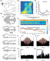
Theta oscillations in the BLA. (A) LA neuron recorded intracellularly in vivo. Near threshold membrane depolarization by intracellular current injection (numbers on right) elicits intrinsic membrane potential oscillations in the theta frequency range. (B) Principal BLA neuron exhibit rhythmic firing at the theta frequency during paradoxical sleep. (B1) Unit activity (top) and LFP (bottom) recorded by the same microelectrode and obtained by high vs. low-pass digital filtering, respectively. (C) Perirhinal (C1) and entorhinal (C2) neurons fire rhythmically at the theta frequency. Traces obtained as in B. (D) Synchronized theta activity in LA and CA1 during retrieval of conditioned fear. LFP recordings (D1) and their color-coded power spectra (D2) demonstrate theta activity in both LA and CA1 during CS+-evoked freezing. White bar in D1 denotes CS+ presentation; f, freezing; r, risk-assessment behavior. (E) LA-CA1 activity during retrieval of conditioned fear at short-term, long-term and remote stages, recorded at 2 hours, 24 hours and 30 days after fear training, respectively. (E1) Crosscorrelograms indicate synchronized theta during long-term (middle; obtained from recordings in D), but not short-term or remote stages. (E2) Significant increase in CS+-evoked freezing (black bars; compared to CS-, white bars) at short, long-term and remote stages is accompanied by synchronized theta in LA-CA1 (grey bars) only at long-term memory stages. * P<0.01, ** P<0.001, ***P<0.0001. Data in D, E modified from (335).
Fig. 4
Fig. 4
Coherent gamma oscillations in the BLA and its targets. (A) Simultaneous LFP recordings of gamma activity in the BLA and rhinal cortices. (A1) Scheme showing position of recording sites for activity depicted in A2. (A2) Top and bottom traces respectively show raw vs. digitally filtered (35-45 Hz) LFPs. (B) Correlated amygdalo-rhinal gamma activity. (B1) Power fluctuations: long periods of spontaneous field potential activity recorded during the waking state were segmented in one-second windows. Fast-Fourier Transforms were computed for each window and the power in each frequency was correlated with all others for BL and entorhinal (ER) recording sites. (B2) Gamma coherence. Coherence (y-axis) as a function of frequency (x-axis) for recording sites in the BLA and perirhinal cortex. (C) Inhibition of BLA activity by local muscimol infusions produces a selective reduction in striatal gamma activity. (C1) Striatal LFP power (color-coded) in different frequencies (y-axis) plotted as a function of time (x-axis) in experiments where muscimol was slowly infused in the BLA, over a period of 25 min. (C2) Gamma power (y-axis) ± s.e.m. (dashed lines) as a function of time (x-axis) when either saline (black) or muscimol (red) was infused in the BLA. The thick black lines indicate infusion periods. (D-E) Gamma-related unit activity in the BLA (D) and striatum (E). Peri-event histograms of unit activity computed around the positive peaks of high-amplitude gamma cycles recorded by the same electrode as used to record unit activity. (F) Gamma oscillations increase coupling between the activity of BLA and striatal neurons. (F1) Crosscorrelogram that included all spikes generated by a simultaneously recorded couple of BLA and striatal neurons. (F2) Crosscorrelogram of unit activity for the same cell couple after excluding striatal spikes occurring during periods of low amplitude gamma.
Fig. 5
Fig. 5
Molecular cascades of fear memory stabilization in the amygdala. A postsynaptic increase in intracellular Ca2+ concentration, mediated through Ca2+ influx via NMDA receptors and voltage-gated Ca2+ channels (VGCCs) and through release from intracellular stores upon activation of metabotropic glutamate receptors (mGluRs), triggers a plethora of signalling steps. Three major, mutually interconnected signalling routes involve Ca2+/calmodulin-dependent protein kinases II (CaMKII), the protein kinase (PK) family of enzymes, and tyrosine kinase (TK) pathways. Signalling cascades can reach the nucleus to induce macromolecular synthesis, and they can control translational processes. Consequently, they can act on cytoskeletal and adhesion molecules to re-organize and stabilize synaptic structures, or regulate AMPA receptor trafficking to the synapse. At intermediate steps, protein kinase signals converge on the mitogen-activated protein kinase (MAPK) signal transduction pathways, including the extracellular regulated kinases (ERK). RAS, RAF, and MEK kinases transduce intra- and extracellular signals, mediated for instance through tyrosin receptor kinases (Trk), to the MAPK/ERK pathway. Scaffolding proteins dictate specificity of activation as well as entry in the nucleus. MAPKs translocated into the nucleus phosphorylate transcription factors, such as cAMP response element binding protein (CREB). Actin rearrangement is under the control of RhoGTPases, whose activation from a GDP- to a GTP-bound form is controlled via Ca2+ or kinase pathways, including tyrosine kinases (TK) and SRC kinases. RhoGTPases control activity of Rho-associated kinases (ROCK), a key molecule for regulation of the cytosekeleton.
Fig. 6
Fig. 6
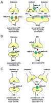
Long-term synaptic plasticity related to conditioned fear in the basolateral amygdaloid complex. A. Long-term potentiation (LTP) in projection neurons (PN). At thalamic inputs, LTP is homosynaptic upon stimulation of postsynaptic NMDA receptors and/or voltage-gated Ca2+ channels. At cortical inputs, LTP is heterosynaptic upon stimulation of presynaptic NMDA receptors through concurrent activation of thalamic inputs. B. Long-term depression (LTD) in PN can be mediated via stimulation of postsynaptic metabotropic glutamate receptors (mGluRs) at thalamic inputs, or via presynaptic mGluRs at LA-BLA synaptic connections. C. LTP in local GABAergic interneurons (IN) at thalamic and cortical inputs can be homosynaptic upon stimulation of Ca2+ permeable AMPA receptors, or heterosynaptic upon stimulation of NMDA receptors.
Fig. 7
Fig. 7
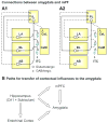
Connections between the amygdala, mPFC, and hippocampus. (A) Reciprocal connections of the infralimbic (A1) and prelimbic (A2) components of the mPFC with the amygdala. Solid lines indicate major projections whereas dashed lines indicate weaker ones. (B) Multiples direct and indirect paths for the transfer of contextual influences to the amygdala.
Fig. 8
Fig. 8
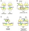
Synaptic plasticity related to fear extinction. A. Activation of NMDA receptors occurs in the basolateral amygdaloid complex (BLA) and the prefrontal cortex (PFC) during within-session and consolidation of extinction, respectively, most likely inducing long-term potentiation (LTP). B. Postsynaptic release of endocannabinoids (eCB) mediates long-term depression of GABAergic transmission (LTDi) via activation of CB1 receptors on cholecystokinin-positive interneurons (CCK-IN). Release of eCB can be stimulated via metabotropic glutamate receptors (mGluRs). C. Increase in glutamatergic transmission to GABAergic mITC neurons mediated through NPS receptors in presynaptic LA principal neurons. D. Both NMDA receptor-dependent LTP and LTD exist at BLA inputs to mITC, which can be induced homo- and heterosynaptically, and which keep the overall synaptic strength in balance.
Fig. 9
Fig. 9. Molecular mechanisms of unlearning and new learning related to early and late stages of fear extinction
Reversal of conditioned fear (unlearning) involves activation of the phosphatase calcineurin and regulated AMPA receptor endocytosis. Extinction learning and consolidation (new learning) involve activation of NMDA receptors (in particular the NR2B subtype), kinase pathways (for instance the mitogen-activated protein kinase (MAPK), extracellular regulated kinase (ERK) pathway), transcriptional regulation (via transcription factors, such as cAMP response element binding protein (CREB)), and structural organization (involving cytoskeletal proteins such as actin).

Similar articles

Cited by

References

    1. Abraham WC. Metaplasticity: tuning synapses and networks for plasticity. Nat Rev Neurosci. 2008;9:387. - PubMed
    1. Aggleton JP, Saunders RC. The amygdala - What's happened in the last decade? In: Aggleton JP, editor. The Amygdala. Oxford, UK: Oxford University Press; 2000. pp. 1–30.
    1. Aitkin LM, Irvine DR, Nelson JE, Merzenich MM, Clarey JC. Frequency representation in the auditory midbrain and forebrain of a marsupial, the northern native cat (Dasyurus hallucatus) Brain Behav Evol. 1986;29:17–28. - PubMed
    1. Albrecht A, Bergado J, Pape HC, Stork O. Role of NCAM in amygdalo-hippocampal interactions and stress modulation of context fear memory in revision. 2009.
    1. Alexander JC, McDermott CM, Tunur T, Rands V, Stelly C, Karhson D, Bowlby MR, An WF, Sweatt JD, Schrader LA. The role of calsenilin/DREAM/KChIP3 in contextual fear conditioning. Learn Mem. 2009;16:167–177. - PMC - PubMed

Publication types