

Ethanol blocks long-term potentiation of GABAergic synapses in the ventral tegmental area involving mu-opioid receptors
- PMID: 20393452
- PMCID: PMC2904870
- DOI: 10.1038/npp.2010.51
Ethanol blocks long-term potentiation of GABAergic synapses in the ventral tegmental area involving mu-opioid receptors
Abstract
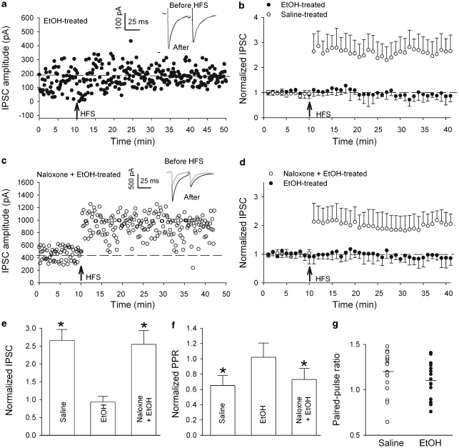
It is well documented that ethanol exposure alters GABA (gamma-aminobutyric acid)-releasing synapses, and ethanol addiction is associated with endogenous opioid system. Emerging evidence indicates that opioids block long-term potentiation in the fast inhibitory GABA(A) receptor synapses (LTP(GABA)) onto dopamine-containing neurons in the ventral tegmental area (VTA), a brain region essential for reward-seeking behavior. However, how ethanol affects LTP(GABA) is not known. We report here that in acute midbrain slices from rats, clinically relevant concentrations of ethanol applied both in vitro and in vivo prevents LTP(GABA), which is reversed, respectively, by in vitro and in vivo administration of naloxone, a mu-opioid receptor (MOR) antagonist. Furthermore, the blockade of LTP(GABA) induced by a brief in vitro ethanol treatment is mimicked by DAMGO ([D-Ala(2), N-MePhe(4), Gly-ol]-enkephalin), a MOR agonist. Paired-pulse ratios are similar in slices, 24 h after in vivo injection with either saline or ethanol. Sp-cAMPS, a stable cAMP analog, and pCPT-cGMP, a cGMP analog, potentiates GABA(A)-mediated inhibitory postsynaptic currents in slices from ethanol-treated rats, indicating that a single in vivo ethanol exposure does not maximally increase GABA release, instead, ethanol produces a long-lasting inability to generate LTP(GABA). These neuroadaptations to ethanol might contribute to early stage of addiction.
Figures


Similar articles
-
Ethanol dually modulates GABAergic synaptic transmission onto dopaminergic neurons in ventral tegmental area: role of mu-opioid receptors.Neuroscience. 2008 Apr 22;153(1):240-8. doi: 10.1016/j.neuroscience.2008.01.040. Epub 2008 Feb 6. Neuroscience. 2008. PMID: 18343590 Free PMC article.
-
PKG and PKA signaling in LTP at GABAergic synapses.Neuropsychopharmacology. 2009 Jun;34(7):1829-42. doi: 10.1038/npp.2009.5. Epub 2009 Feb 4. Neuropsychopharmacology. 2009. PMID: 19194373 Free PMC article.
-
Opioids block long-term potentiation of inhibitory synapses.Nature. 2007 Apr 26;446(7139):1086-90. doi: 10.1038/nature05726. Nature. 2007. PMID: 17460674
-
LTP of GABAergic synapses in the ventral tegmental area and beyond.J Physiol. 2008 Mar 15;586(6):1487-93. doi: 10.1113/jphysiol.2007.148098. Epub 2007 Dec 13. J Physiol. 2008. PMID: 18079157 Free PMC article. Review.
-
Progress in opioid reward research: From a canonical two-neuron hypothesis to two neural circuits.Pharmacol Biochem Behav. 2021 Jan;200:173072. doi: 10.1016/j.pbb.2020.173072. Epub 2020 Nov 20. Pharmacol Biochem Behav. 2021. PMID: 33227308 Free PMC article. Review.
Cited by
-
Synaptic Plasticity at Inhibitory Synapses in the Ventral Tegmental Area Depends upon Stimulation Site.eNeuro. 2019 Nov 15;6(6):ENEURO.0137-19.2019. doi: 10.1523/ENEURO.0137-19.2019. Print 2019 Nov/Dec. eNeuro. 2019. PMID: 31619451 Free PMC article.
-
Complex NADASE Infusions Improve Clinical Outcome in Substance Use Disorder: Descriptive Annotation in Fifty Cases.J Addict Psychiatry. 2024 Aug 23;8(1):95-157. J Addict Psychiatry. 2024. PMID: 39949994 Free PMC article.
-
Lithium prevents long-term neural and behavioral pathology induced by early alcohol exposure.Neuroscience. 2012 Mar 29;206:122-35. doi: 10.1016/j.neuroscience.2011.12.059. Epub 2012 Jan 8. Neuroscience. 2012. PMID: 22266347 Free PMC article.
-
Shared Mechanisms of GABAergic and Opioidergic Transmission Regulate Corticolimbic Reward Systems and Cognitive Aspects of Motivational Behaviors.Brain Sci. 2023 May 17;13(5):815. doi: 10.3390/brainsci13050815. Brain Sci. 2023. PMID: 37239287 Free PMC article. Review.
-
Stressed and wired: The effects of stress on the VTA circuits underlying motivated behavior.Curr Opin Endocr Metab Res. 2022 Oct;26:100388. doi: 10.1016/j.coemr.2022.100388. Epub 2022 Aug 7. Curr Opin Endocr Metab Res. 2022. PMID: 36406203 Free PMC article.
References
-
- Bergevin A, Girardot D, Bourque MJ, Trudeau LE. Presynaptic mu-opioid receptors regulate a late step of the secretory process in rat ventral tegmental area GABAergic neurons. Neuropharmacology. 2002;42:1065–1078. - PubMed
Publication types
MeSH terms
Substances
Grants and funding
LinkOut - more resources
Full Text Sources
Research Materials
