. 2010 Jun;24(6):1171-8.
doi: 10.1038/leu.2010.50. Epub 2010 Apr 15.

Ectopic expression of wild-type FGFR3 cooperates with MYC to accelerate development of B-cell lineage neoplasms

Affiliations

Ectopic expression of wild-type FGFR3 cooperates with MYC to accelerate development of B-cell lineage neoplasms

A Zingone et al. Leukemia. 2010 Jun.

Abstract

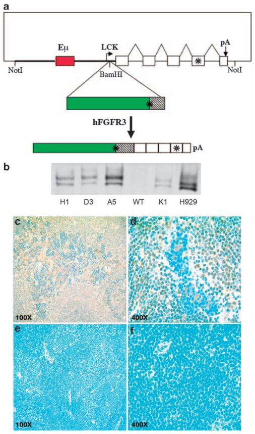
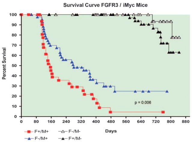
The t(4;14) translocation in multiple myeloma (MM) simultaneously dysregulates two apparent oncogenes: fibroblast growth factor receptor 3 (FGFR3) controlled by the 3' immunoglobulin heavy chain enhancer on der(14) and MMSET controlled by the intronic Emu enhancer on der(4). Although all MM tumors and cell lines with a t(4;14) translocation have dysregulated MMSET, about 25% do not express FGFR3. Therefore, the function of dysregulated wild-type (WT) FGFR3 in the pathogenesis of MM remains unclear. We developed a murine transgenic (TG) model in which WT FGFR3 is overexpressed in B lymphoid cells. Although high levels of FGFR3 resulted in lymphoid hyperplasia in about one-third of older mice, no increase in tumorigenesis was observed. However, double TG FGFR3/Myc mice develop mature B lymphoma tumors that occur with a higher penetrance and shorter latency than in single TG Myc mice (P=0.006). We conclude that expression of high levels of WT FGFR3 can be oncogenic and cooperate with MYC to generate B lymphoid tumors. This suggests that dysregulated FGFR3 expression is likely to be essential at least for the early stages of pathogenesis of MM tumors that have a t(4;14) translocation.


Conflict of interest statement

Conflict of interest

The authors declare no conflict of interest.

Figures

Figure 1
Figure 1
Expression of hFGFR3 in TG mice. (a) Schematic diagram of p1026X-hFGFR3 TG vector. The vector includes a 9.7 kb NotI fragment that contains 3.2 kb of Lck proximal promoter sequences (thick line), a 1 kb Eμ enhancer fragment (red box) inserted into these sequences, a 3.4 kb human FGFR3 cDNA (green box is coding) cloned into a BamHI site located 37 bp from the lck promoter, and a 2.1 kb human growth hormone (hGH) genomic fragment with five exons (boxes), and a polyadenylation site. FGFR3 and the fourth exon of hGH contain stop codons, as indicated by the asterisks. The spliced hFGFR3-hGH transcript is 4.4 kb. (b) Immunoblot of hFGFR3 in plasma cells. Plasma cells were generated in vitro by 5 days LPS stimulation of spleen cells from four independent F1 TG mice (founders H1, D3, A5, K1) and a non-TG littermate (WT). Proteins from total cell lysates were immunoprecipitated with a polyclonal anti-hFGFR3, fractionated by electrophoresis on a 7.5% polyacrylamide gel in SDS, transferred to nitrocellulose filters, and reacted with a monoclonal anti-hFGFR3 antibody. H929 MM cell line expresses FGFR3 as a result of a t(4;14) translocation. The FGFR3 doublet, which is present in the H929 myeloma cell line and TG splenocytes, probably reflects differential glycosylation as described for other tyrosine kinase receptors. (c–f) Expression of hFGFR3 in spleen cells by immunohistochemistry. Sections of normal spleens from hFGFR3 TG mice and normal littermates were immunostained with anti-hFGFR3 antibody (brown) and counterstained with Meyer’s hematoxylin (Ventana) (blue); hFGFR3 TG at × 100 (c) and × 400 magnification (d); non-TG littermate at × 100 (e) and × 400 magnification (f).
Figure 2
Figure 2
Survival curves for FGFR3 (F) and iMyc (M) TG mice. Tumor incidence is similarly low in F+/M− and F−/M− mice, but is higher in F+/M+ mice versus F−/M+ mice (P=0.006).
Figure 3
Figure 3
Southern blots of representative B lymphoma tumors from FGFR3/iMyc TG mice. (a) Tumor DNA was digested with EcoRI, subjected to electrophoresis, blotted, and hybridized with a 1.2 kb JH probe. Size markers are to the left, arrows to right indicate the 6.4 kb normal germline locus, and the 5.5 kb locus containing the knocked-in c-myc gene. Rearranged fragments are indicated by an R. (b) Tumor DNA was digested with BamHI and EcoRI, subjected to electrophoresis, blotted, and hybridized with a 1 kB Ck probe. The normal germline locus is indicated by an arrow.
Figure 4
Figure 4
RT–PCR and qRT–PCR to detect hFGFR3 expression in primary tumors and cell lines. (a) Detection of expression of hFGFR3 transgene in tumors derived from F+/M+ mice by RT–PCR. (b) qRT–PCR in F+/M+ tumors (white bar), and in the corresponding cell lines (CL, gray bar) derived from primary tumors #228 and #229 and cultured continuously with FGF. (c) qRT–PCR in three F+/M+ cell lines cultured in the presence (F1, gray bar) or in the absence (F0, white bar) of FGF. hFGFR3 expression is −ΔΔCt, normalized to USF2 and to the H929 human myeloma cell line.
Figure 5
Figure 5
Expression profiling by microarrays of FGFR3 + iMyc TG mice and iMyc TG mice. Two-dimensional hierarchical clustering of gene expression profiles, using distance matrix of Pearson’s correlation, for lymphomas from F+/M+ mice (labeled FM with case number) and F−/M+ mice (labeled M plus case number). Scale bar shown at the top is in log2 scale.

Similar articles

Cited by

References

    1. Jemal A, Siegel R, Ward E, Murray T, Xu J, Thun MJ. Cancer statistics, 2007. CA Cancer J Clin. 2007;57:43–66. - PubMed
    1. Bergsagel PL, Chesi M, Nardini E, Brents LA, Kirby SL, Kuehl WM. Promiscuous translocations into immunoglobulin heavy chain switch regions in multiple myeloma. Proc Natl Acad Sci USA. 1996;93:13931–13936. - PMC - PubMed
    1. Kuehl WM, Bergsagel PL. Multiple myeloma: evolving genetic events and host interactions. Nat Rev Cancer. 2002;2:175–187. - PubMed
    1. Bergsagel PL, Kuehl WM. Molecular pathogenesis and a consequent classification of multiple myeloma. J Clin Oncol. 2005;23:6333–6338. - PubMed
    1. Fonseca R, Blood E, Rue M, Harrington D, Oken MM, Kyle RA, et al. Clinical and biologic implications of recurrent genomic aberrations in myeloma. Blood. 2003;101:4569–4575. - PubMed

Publication types

MeSH terms