

Increased perfusion and angiogenesis in a hindlimb ischemia model with plasmid FGF-2 delivered by noninvasive electroporation
- PMID: 20393507
- PMCID: PMC3138216
- DOI: 10.1038/gt.2010.43
Increased perfusion and angiogenesis in a hindlimb ischemia model with plasmid FGF-2 delivered by noninvasive electroporation
Abstract
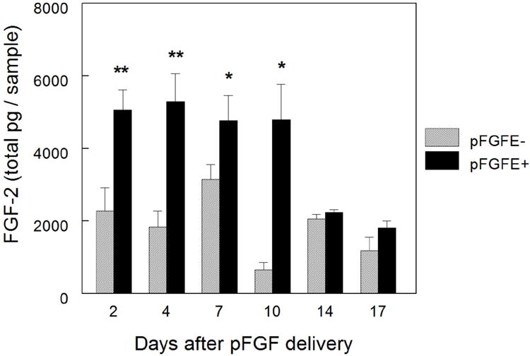
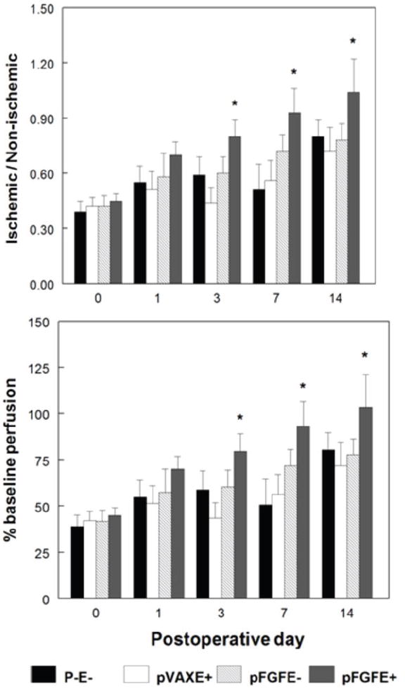
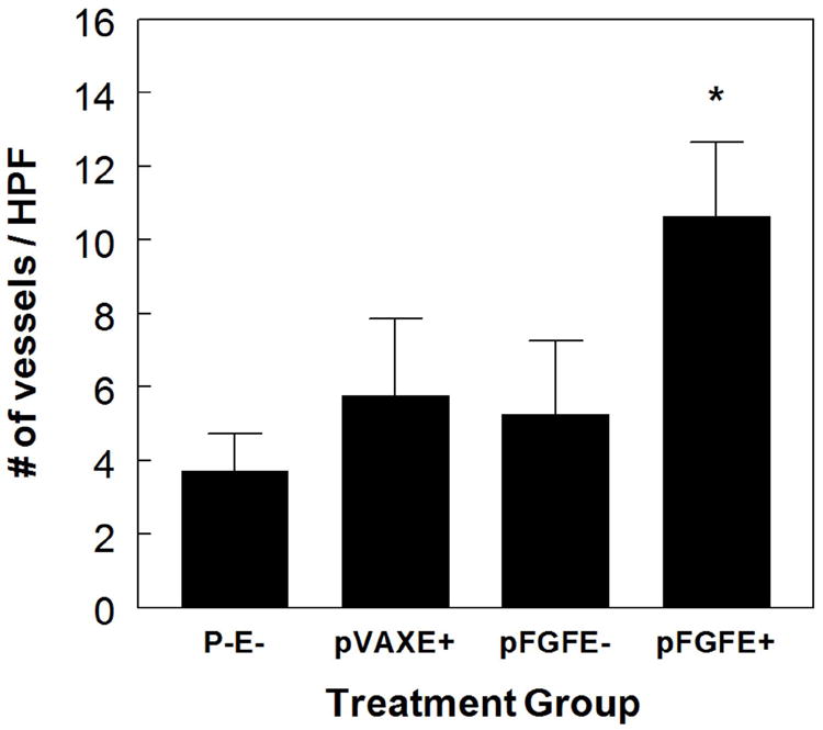
Gene therapy approaches delivering fibroblast growth factor-2 (FGF-2) have shown promise as a potential treatment for increasing blood flow to ischemic limbs. Currently, effective noninvasive techniques to deliver plasmids encoding genes of therapeutic interest, such as FGF-2, are limited. We sought to determine if intradermal injection of plasmid DNA encoding FGF-2 (pFGF) followed by noninvasive cutaneous electroporation (pFGFE+) could increase blood flow and angiogenesis in a rat model of hindlimb ischemia. pFGFE+ or control treatments were administered on postoperative day 0. Compared to injection of pFGF alone (pFGFE-), delivery of pFGFE+ significantly increased FGF-2 expression for 10 days. Further, the increase in FGF-2 expression with pFGFE+ was sufficient to significantly increase ischemic limb blood flow, measured by laser Doppler perfusion imaging, beginning on postoperative day 3. Ischemic limb blood flow in the pFGFE+ treatment group remained significantly higher than all control groups through the end point of the study, postoperative day 14. Immunohistochemical staining of gastrocnemius cross sections determined there was a twofold increase in capillary density in the pFGFE+ treatment group. Our results suggest that pFGFE+ is a potential noninvasive, nonviral therapeutic approach to increase perfusion and angiogenesis for the treatment of limb ischemia.
Conflict of interest statement
Figures


Comment in
-
DNA electrotransfer to the skin: a highly translatable approach to treat peripheral artery disease.Gene Ther. 2010 Jun;17(6):691. doi: 10.1038/gt.2010.68. Epub 2010 May 13. Gene Ther. 2010. PMID: 20463758 No abstract available.
Similar articles
-
In vivo electroporation of constitutively expressed HIF-1α plasmid DNA improves neovascularization in a mouse model of limb ischemia.J Vasc Surg. 2014 Mar;59(3):786-93. doi: 10.1016/j.jvs.2013.04.043. Epub 2013 Jul 11. J Vasc Surg. 2014. PMID: 23850058 Free PMC article.
-
In vivo electroporation enhances plasmid-based gene transfer of basic fibroblast growth factor for the treatment of ischemic limb.J Surg Res. 2004 Jul;120(1):37-46. doi: 10.1016/j.jss.2003.12.016. J Surg Res. 2004. PMID: 15172188
-
Therapeutic angiogenesis induced by human hepatocyte growth factor gene in rat and rabbit hindlimb ischemia models: preclinical study for treatment of peripheral arterial disease.Gene Ther. 2001 Feb;8(3):181-9. doi: 10.1038/sj.gt.3301379. Gene Ther. 2001. PMID: 11313789
-
Synergistic effects of FGF-2 and PDGF-BB on angiogenesis and muscle regeneration in rabbit hindlimb ischemia model.Microvasc Res. 2010 Jul;80(1):10-7. doi: 10.1016/j.mvr.2009.12.002. Epub 2010 Jan 4. Microvasc Res. 2010. PMID: 20045007
-
CD151 promotes neovascularization and improves blood perfusion in a rat hind-limb ischemia model.J Endovasc Ther. 2005 Aug;12(4):469-78. doi: 10.1583/04-1478R.1. J Endovasc Ther. 2005. PMID: 16048379
Cited by
-
Targets and delivery methods for therapeutic angiogenesis in peripheral artery disease.Vasc Med. 2012 Jun;17(3):174-92. doi: 10.1177/1358863X12438270. Epub 2012 Apr 11. Vasc Med. 2012. PMID: 22496126 Free PMC article. Review.
-
Rescue of murine hind limb ischemia via angiogenesis and lymphangiogenesis promoted by cellular communication network factor 2.Sci Rep. 2023 Nov 16;13(1):20029. doi: 10.1038/s41598-023-47485-y. Sci Rep. 2023. PMID: 37973852 Free PMC article.
-
A Thermoresponsive Chitosan/β-Glycerophosphate Hydrogel for Minimally Invasive Treatment of Critical Limb Ischaemia.Polymers (Basel). 2021 Oct 16;13(20):3568. doi: 10.3390/polym13203568. Polymers (Basel). 2021. PMID: 34685327 Free PMC article.
-
Moderate Heat-Assisted Gene Electrotransfer as a Potential Delivery Approach for Protein Replacement Therapy through the Skin.Pharmaceutics. 2021 Nov 11;13(11):1908. doi: 10.3390/pharmaceutics13111908. Pharmaceutics. 2021. PMID: 34834323 Free PMC article.
-
Evaluation of delivery conditions for cutaneous plasmid electrotransfer using a multielectrode array.Gene Ther. 2011 May;18(5):496-500. doi: 10.1038/gt.2010.171. Epub 2010 Dec 23. Gene Ther. 2011. PMID: 21179175 Free PMC article.
References
-
- Gardner AW, Poehlman ET. Exercise rehabilitation programs for the treatment of claudication pain. A meta-analysis. JAMA. 1995;274:975–980. - PubMed
-
- Robeer G, Brandsma J, van den Heuvel S, Smit B, Oostendorp R, Wittens C. Exercise therapy for intermittent claudication: a review of the quality of randomized clinical trials and evaluation of predictive factors. Eur J Vasc Endovasc. 1998;15:36–43. - PubMed
-
- Dormandy J, Rutherford R. Management of Peripheral Arterial Disease (PAD) J Vasc Surg. 2000;31:s3. - PubMed
-
- Rissanen TT, Vajanto I, Yla-Herttuala S. Gene therapy for therapeutic angiogenesis in critically ischaemic lower limb - on the way to the clinic. European Journal of Clinical Investigation. 2001;31:651–666. - PubMed
-
- Norgren L, Hiatt WR, Dormandy JA, Nehler MR, Harris KA, Fowkes FGR. Inter-Society Consensus for the Management of Peripheral Arterial Disease (TASC II) European Journal of Vascular and Endovascular Surgery. 2007;33:S1–S75. - PubMed
Publication types
MeSH terms
Substances
Grants and funding
LinkOut - more resources
Full Text Sources
Other Literature Sources
Medical
