. 2010 Apr 15;464(7291):999-1005.
doi: 10.1038/nature08989.

Genome remodelling in a basal-like breast cancer metastasis and xenograft

Affiliations

Genome remodelling in a basal-like breast cancer metastasis and xenograft

Li Ding et al. Nature. .

Abstract

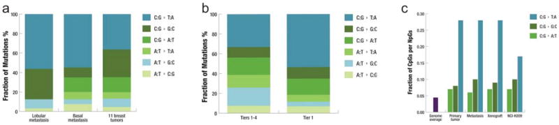
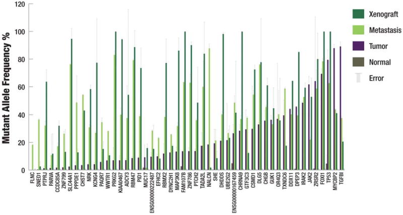
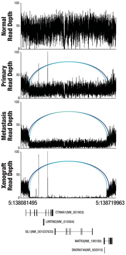
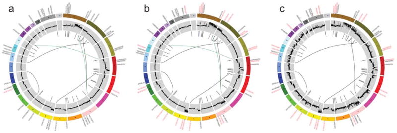
Massively parallel DNA sequencing technologies provide an unprecedented ability to screen entire genomes for genetic changes associated with tumour progression. Here we describe the genomic analyses of four DNA samples from an African-American patient with basal-like breast cancer: peripheral blood, the primary tumour, a brain metastasis and a xenograft derived from the primary tumour. The metastasis contained two de novo mutations and a large deletion not present in the primary tumour, and was significantly enriched for 20 shared mutations. The xenograft retained all primary tumour mutations and displayed a mutation enrichment pattern that resembled the metastasis. Two overlapping large deletions, encompassing CTNNA1, were present in all three tumour samples. The differential mutation frequencies and structural variation patterns in metastasis and xenograft compared with the primary tumour indicate that secondary tumours may arise from a minority of cells within the primary tumour.


Figures

Figure 1
Figure 1. Mutational signatures in the basal breast tumor
a, Fraction of mutations in each of the transition and transversion categories in the metastasis of a lobular breast tumor, the metastasis of the basal breast tumor under study, and the 11 breast tumors reported by Wood et al. from which 1,104 coding mutations identified in the discovery set were used in the analysis. b, Fraction of mutations in each of the transition and transversion categories in 43 tier1 mutations and 3,204 tier 1-4 mutations in the metastasis under study. c, Fraction of guanine mutations at CpGs in primary tumor, metastasis, xenograft, and NCI-H209 as reported by Pleasance et al..
Figure 2
Figure 2. Mutant allele frequency from deep read count data
The mutant allele frequency of each somatic mutation is shown. Mutations were validated using both 454 and Illumina sequencing. Each bar represents the average of the frequency yielded by the two technologies for a single primer pair and the error bars represent the standard deviation. Data were considered only if there were at least 200 reads from Illumina sequencing and at least 20 reads from 454 sequencing. If no error bar exists, then data were only available from a single sequencing platform.
Figure 3
Figure 3. Two overlapping CTNNA1 deletions on chromosome 5 in three tumors
A graph of sequence depths, read pairs, and genes in a 638,468 bp region containing two overlapping deletions. The top four panels display the read depths at each base and the reads within the region whose mates mapped at an abnormal distance are displayed as blue bars, with matched pairs connected by arcs. Two different shades of blue indicate the two separate allelic deletion events (538,467 bp and 515,465 bp in length). The bottom panel displays genes annotated in this genomic region.
Figure 4
Figure 4. Circos plots for the primary tumor, metastasis, and xenograft genomes
Circos plots display the validated tier 1 somatic mutations, DNA copy number, and validated structural rearrangements in the primary tumor (a), metastasis (b), and xenograft (c). Mutations enriched in the primary tumor (a) are labeled in red and mutations enriched in the metastasis or xenograft are in red (b and c). Mutations and the large deletion unique to the metastasis are in blue (b). Translocations only present in primary tumor and metastasis are in green. All shared events are in black. The copy number difference between the tumor and normal is shown (scale: -4 to 4). No purity-based copy number corrections were used for plotting.

Comment in

Similar articles

Cited by

References

    1. Carey LA, et al. Race, breast cancer subtypes, and survival in the Carolina Breast Cancer Study. JAMA. 2006;295(21):2492–2502. - PubMed
    1. Citron ML, et al. Randomized trial of dose-dense versus conventionally scheduled and sequential versus concurrent combination chemotherapy as postoperative adjuvant treatment of node-positive primary breast cancer: first report of Intergroup Trial C9741/Cancer and Leukemia Group B Trial 9741. J Clin Oncol. 2003;21(8):1431–1439. - PubMed
    1. Kuperwasser C, et al. Reconstruction of functionally normal and malignant human breast tissues in mice. Proc Natl Acad Sci U S A. 2004;101(14):4966–4971. - PMC - PubMed
    1. Parker JS, et al. Supervised risk predictor of breast cancer based on intrinsic subtypes. J Clin Oncol. 2009;27(8):1160–1167. - PMC - PubMed
    1. Li H, Ruan J, Durbin R. Mapping short DNA sequencing reads and calling variants using mapping quality scores. Genome Res. 2008;18(11):1851–1858. - PMC - PubMed

Publication types