

Functional genomic screen for modulators of ciliogenesis and cilium length
- PMID: 20393563
- PMCID: PMC2929961
- DOI: 10.1038/nature08895
Functional genomic screen for modulators of ciliogenesis and cilium length
Abstract
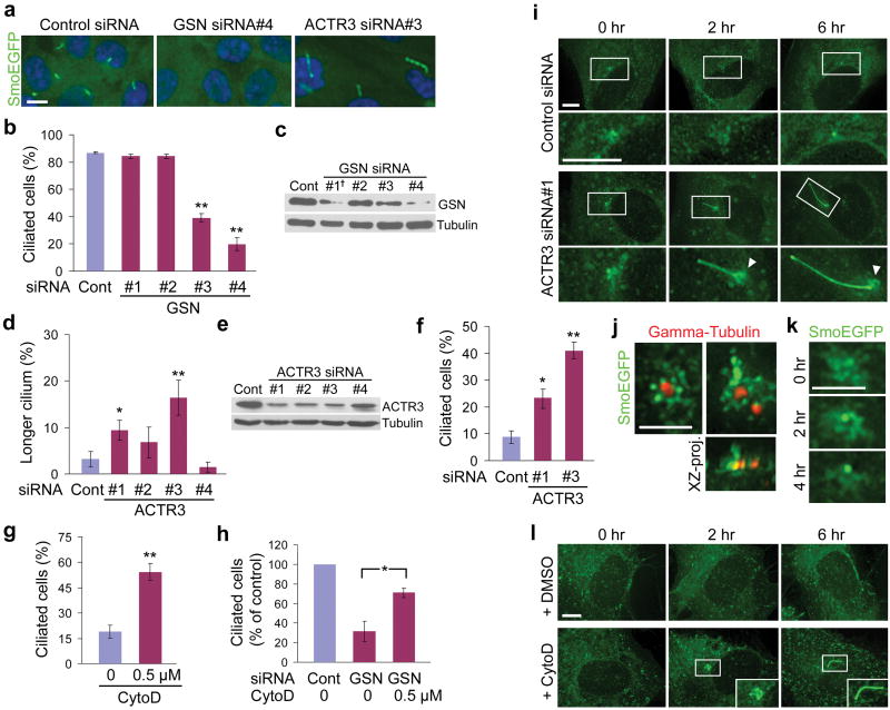
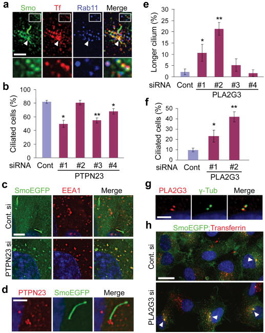
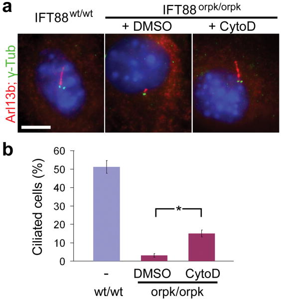
Primary cilia are evolutionarily conserved cellular organelles that organize diverse signalling pathways. Defects in the formation or function of primary cilia are associated with a spectrum of human diseases and developmental abnormalities. Genetic screens in model organisms have discovered core machineries of cilium assembly and maintenance. However, regulatory molecules that coordinate the biogenesis of primary cilia with other cellular processes, including cytoskeletal organization, vesicle trafficking and cell-cell adhesion, remain to be identified. Here we report the results of a functional genomic screen using RNA interference (RNAi) to identify human genes involved in ciliogenesis control. The screen identified 36 positive and 13 negative ciliogenesis modulators, which include molecules involved in actin dynamics and vesicle trafficking. Further investigation demonstrated that blocking actin assembly facilitates ciliogenesis by stabilizing the pericentrosomal preciliary compartment (PPC), a previously uncharacterized compact vesiculotubular structure storing transmembrane proteins destined for cilia during the early phase of ciliogenesis. The PPC was labelled by recycling endosome markers. Moreover, knockdown of modulators that are involved in the endocytic recycling pathway affected the formation of the PPC as well as ciliogenesis. Our results uncover a critical regulatory step that couples actin dynamics and endocytic recycling with ciliogenesis, and also provides potential target molecules for future study.
Conflict of interest statement
The authors declare competing financial interests (patent application).
Figures
References
-
- Fliegauf M, Benzing T, Omran H. When cilia go bad: cilia defects and ciliopathies. Nat Rev Mol Cell Biol. 2007;8:880–893. - PubMed
-
- Rosenbaum JL, Witman GB. Intraflagellar transport. Nature Rev Mol Cell Biol. 2002;3:813–825. - PubMed
-
- Christensen ST, et al. The primary cilium coordinates signaling pathways in cell cycle control and migration during development and tissue repair. Curr Top Dev Biol. 2008;85:261–301. - PubMed
Publication types
MeSH terms
Substances
Grants and funding
LinkOut - more resources
Full Text Sources
Other Literature Sources
Molecular Biology Databases
