. 2010 Aug;6(8):1411-8.
doi: 10.1039/c000237b. Epub 2010 Apr 14.

Characterization of mice lacking candidate N-acyl ethanolamine biosynthetic enzymes provides evidence for multiple pathways that contribute to endocannabinoid production in vivo

Affiliations

Characterization of mice lacking candidate N-acyl ethanolamine biosynthetic enzymes provides evidence for multiple pathways that contribute to endocannabinoid production in vivo

Gabriel M Simon et al. Mol Biosyst. 2010 Aug.

Abstract

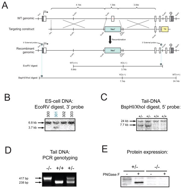
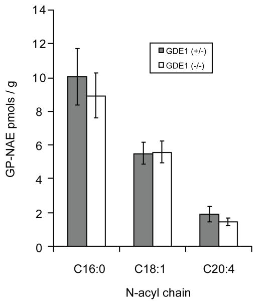
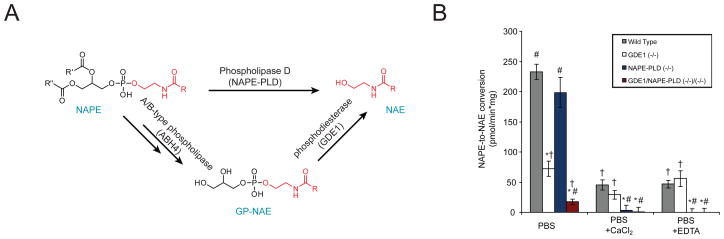
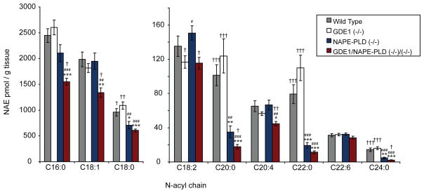
The biosynthesis of the endocannabinoid anandamide (AEA) and related N-acyl ethanolamine (NAE) lipids is complex and appears to involve multiple pathways, including: (1) direct release of NAEs from N-acyl phosphatidyl ethanolamine (NAPE) precursors by the phosphodiesterase NAPE-PLD, and (2) double O-deacylation of NAPEs followed by phosphodiester bond hydrolysis of the resulting glycero-phospho (GP)-NAEs. We recently identified GDE1 as a GP-NAE phosphodiesterase that may be involved in the second pathway. Here, we report the generation and characterization of GDE1(-/-) mice, which are viable and overtly normal in their cage behavior. Brain homogenates from GDE1(-/-) mice exhibit a near-complete loss of detectable GP-NAE phosphodiesterase activity; however, bulk brain levels of AEA and other NAEs were unaltered in these animals. To address the possibility of compensatory pathways, we generated GDE1(-/-)/NAPE-PLD(-/-) mice. Conversion of NAPE to NAE was virtually undetectable in brain homogenates from these animals as measured under standard assay conditions, but again, bulk changes in brain NAEs were not observed. Interestingly, significant reductions in the accumulation of brain NAEs, including anandamide, were detected in GDE1(-/-)/NAPE-PLD(-/-) mice treated with a fatty acid amide hydrolase (FAAH) inhibitor that blocks NAE degradation. Finally, we determined that primary neurons from GDE1(-/-)/NAPE-PLD(-/-) mice can convert NAPEs to NAEs by a pathway that is not preserved following cell homogenization. In summary, combined inactivation of GDE1 and NAPE-PLD results in partial disruption of NAE biosynthesis, while also pointing to the existence of an additional enzymatic pathway(s) that converts NAPEs to NAEs. Characterization of this pathway should provide clarity on the multifaceted nature of NAE biosynthesis.


Figures

Figure 1
Figure 1. Generation of GDE1(−/−) mice
(A) The genomic structure of the GDE1 gene locus on chromosome 7 is shown, along with the targeting construct used to delete exon 2 (which contains several residues essential for catalysis, Glu97, Asp99 and His112) and the final recombined locus. Only relevant restriction sites are designated. The locations of DNA probes used for Southern blotting 5’ and 3’ to the targeting construct are also shown. (B) Southern blot of EcoRV-digested ES cell DNA showing the heterozygous (targeted) clone #301. (C) Sothern blot of BspHI/XhoI-digested tail-DNA from heterozygous and wild-type mice demonstrating germ-line transmission. (D) PCR strategy in which the wild-type locus is identified by a 238 bp band and the targeted locus is identified by a 417 bp band. (E) Western blot of the particulate fraction from brain tissue confirming loss of GDE1 protein. GDE1 is a PNGaseF-sensitive glycosylated protein.
Figure 2
Figure 2. GP-NAE phosphodiesterase activity is lost in GDE1(−/−) tissue
(A) Near-complete loss of conversion of 14C-GP-NAE to 14C-NAE is evident from a representative thin layer radiochromatogram (left) and quantified data from replicate experiments (right). (B) Loss of conversion of 14C-lysoNAPE to 14C-NAE is evident from a representative thin layer radiochromatogram (left) and quantified data from replicate experiments (right). Note also the accumulation of the GP-NAE intermediate in the assay with GDE1(−/−) tissue. All assays were performed in PBS buffer with tissue homogenates from brains of GDE1(+/+) and (−/−) mice. (C) Ex vivo assay demonstrating that EDTA induces GP-NAE accumulation in brain homogenates due to disruption of GDE1 activity. Brain tissue from GDE1(+/−) and (−/−) mice was homogenized in the presence or absence of 10 mM EDTA (which inhibits GDE1) and incubated at room temperature for 4 hours. The addition of EDTA leads to a rapid accumulation of GP-NAEs in wild-type brain, as previously reported. GP-NAE accumulation was observed in brain tissue from GDE1(−/−) mice with- or without EDTA, demonstrating that GDE1 is the EDTA-sensitive GP-NAE phosphodiesterase in wild-type tissue. n = 4 mice per group. Results are presented as means ± standard error. Asterisks designate p < 0.05, p < 0.01, p < 0.005 (student’s t-test) for *, **, and ***, respectively. Asterisks represent difference from wild-type in panels (A) and (B), and difference from EDTA-treated GDE1(+/−) tissue in panel (C).
Figure 3
Figure 3. GP-NAE levels are unchanged in GDE1(−/−) brains
Endogenous GP-NAEs were measured in brain extracts from GDE1(+/−) and (−/−) mice via LC-MS/MS. No significant differences were observed. n = 8 mice per group. Results are presented as means ± standard error.
Figure 4
Figure 4. Bulk NAE levels in brains from mice lacking GDE1 and/or NAPE-PLD
Brains from wild-type, GDE1(−/−), NAPE-PLD(−/−) or GDE1(−/−)/NAPE-PLD(−/−) mice were subjected to organic extraction and endogenous NAE levels were measured by LC-MS/MS. No significant differences were observed between wild-type and GDE1(−/−) or between NAPE-PLD(−/−) and GDE1(−/−)/NAPE-PLD(−/−) brains.
Figure 5
Figure 5. NAPE conversion to NAE in brain lysates from mice lacking GDE1 and/or NAPE-PLD
(A) Pathway diagram showing potential routes for NAPE conversion to NAE. Note that both NAPE-PLD and GDE1 can liberate NAE from its esterified precursors (NAPE and GP-NAE, respectively). (B) Brain tissue from wild-type, GDE1(−/−), NAPE-PLD(−/−), or GDE1(−/−)/NAPE-PLD(−/−) mice was homogenized and assayed for the ability to convert NAPE to NAE in PBS buffer in the presence or absence of 10 mM CaCl2 or EDTA via thin layer radiochromatography. In the presence of calcium or EDTA, tissue lacking NAPE-PLD shows complete loss of NAPE-to-NAE conversion. However, assays performed without these additives show no impairment from loss of NAPE-PLD and a large reduction in activity in tissue lacking GDE1. Under all conditions, tissue from the GDE1(−/−)/NAPE-PLD(−/−) displayed negligible activity. The results are presented as means ± standard error. n = 4 mice/group. The symbols above the bars designate p < 0.05 (student’s t-test) with * for different from wild-type, # for different from GDE1(−/−) samples and † for different from NAPE-PLD(−/−) samples.
Figure 6
Figure 6. Impaired NAE accumulation is observed following FAAH inhibition in brains from mice lacking GDE1 and NAPE-PLD
Mice were treated with the selective FAAH inhibitor PF-3845 (10 mg/kg, i.p.) and, after 3 hours, sacrificed and brain NAE levels measured. Significant reductions were observed for several NAEs, including the endocannabinoid anandamide (C20:4), in brains from GDE1(−/−)/NAPE-PLD(−/−) mice. The results are presented as means ± standard error. n = 4 mice/group. The symbols above the bars designate p < 0.05, p < 0.01, p < 0.005 (student’s t-test) with *, **, *** for different from wild-type, #, ##, ### for different from GDE1(−/−) samples and †, ††, ††† for different from NAPE-PLD(−/−) samples, respectively.
Figure 7
Figure 7. In situ NAE release from NAPE in primary neuron cultures from mice lacking GDE1 and NAPE-PLD
(A) Time-dependence of NAE release in the presence or absence of the FAAH inhibitor PF-3845. NAE release in the absence of neurons was negligible on this timescale (not shown). (B) Assays conducted in primary neurons for 6 hours in the presence of PF-3845. Comparison of wild-type neurons to neurons from GDE1(−/−) or NAPE-PLD(−/−) mice reveals no impairment in the ability to release NAE from NAPE in situ. (C) Comparison of NAPE-PLD(−/−) neurons to GDE1(−/−)/NAPE-PLD(−/−) neurons reveals wild-type levels of NAE release in GDE1(−/−)/NAPE-PLD(−/−) neurons. (D) NAE release from 14C-NAPE or an ether-linked (non-hydrolyzable) NAPE analogue in neurons from GDE1(−/−)/NAPE-PLD(−/−) mice reveals the necessity of de-acylation prior to NAE release. These data represent single experiments performed in triplicate, representative of multiple experiments. *, p < 0.05.

Similar articles

Cited by

References

    1. Pacher P, Bátkai S, Kunos G. Pharmacol Rev. 2006;58:389–462. - PMC - PubMed
    1. Howlett AC, Breivogel CS, Childers SR, Deadwyler SA, Hampson RE, Porrino LJ. Neuropharmacology. 2004;47:345–358. - PubMed
    1. Walter L, Stella N. Br J Pharmacol. 2004;141:775–785. - PMC - PubMed
    1. Fowler CJ, Holt S, Nilsson O, Jonsson KO, Tiger G, Jacobsson SO. Pharmacol Biochem Behav. 2005;81:248–262. - PubMed
    1. Ahn K, McKinney MK, Cravatt BF. Chem Rev. 2008;108:1687–1707. - PMC - PubMed

Substances