

Evaluation of MR imaging findings differentiating cavernous haemangiomas from schwannomas in the orbit
- PMID: 20393718
- PMCID: PMC2914262
- DOI: 10.1007/s00330-010-1774-y
Evaluation of MR imaging findings differentiating cavernous haemangiomas from schwannomas in the orbit
Abstract
Objective: It is important to distinguish between orbital cavernous haemangioma and schwannoma because the treatments of choice for the two tumours are different. The aim was to evaluate MR imaging findings distinguishing the two tumours.
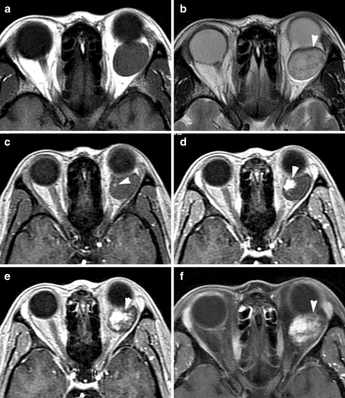
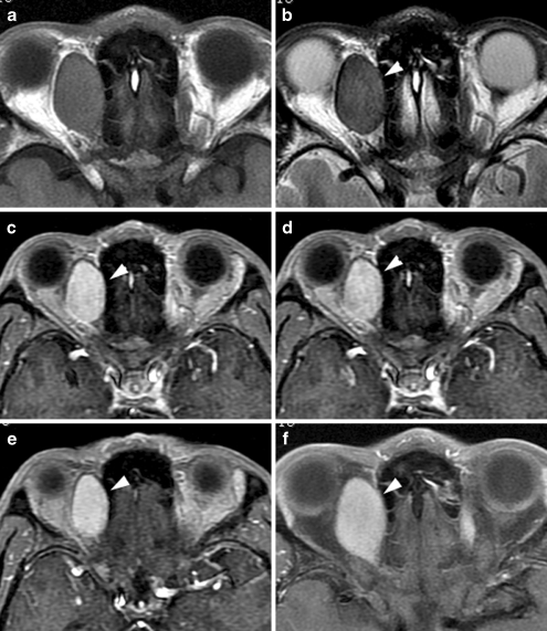
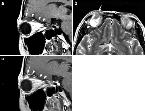
Methods: Magnetic resonance imaging including T1- and T2-weighted imaging and contrast-enhanced MR imaging was performed in 43 patients with cavernous haemangiomas and 16 patients with schwannomas confirmed by pathology. Location, configuration, margins, signal intensity, homogeneity and enhancement pattern of the tumour were retrospectively evaluated.
Results: There was a significant difference between cavernous haemangiomas and schwannomas regarding the location, configuration and margins of the mass, signal intensity and homogeneity on T1- and T2-weighted imaging, the spread pattern of contrast enhancement, the enhancement pattern and the type of time-intensity curve (P<0.05). Markedly homogeneous hyperintensity signal on T2-weighted imaging and the spread pattern of the contrast enhancement favoured cavernous haemangioma rather than schwannoma (P<0.01).
Conclusion: Cavernous haemangiomas and schwannomas have different MR imaging features that could be helpful in the differentiation between the tumours. The spread pattern of the contrast enhancement on dynamic contrast-enhanced MR imaging is the most reliable finding distinguishing cavernous haemangiomas from schwannomas.
Figures
References
-
- Shields JA, Bakewell B, Augsburger JJ, Flanagan JC. Classification and incidence of space-occupying lesions of the orbit. A survey of 645 biopsies. Arch Ophthalmol. 1984;102:1606–1611. - PubMed
-
- Moss HM. Expanding lesions of the orbit. A clinical study of 230 consecutive cases. Am J Ophthalmol. 1962;54:761–770. - PubMed
-
- Ruchman MC, Flanagan J. Cavernous hemangiomas of the orbit. Ophthalmology. 1983;90:1328–1336. - PubMed
-
- Bilaniuk LT. Orbital vascular lesions. Role of imaging. Radiol Clin North Am. 1999;37:169–183. - PubMed
Publication types
MeSH terms
LinkOut - more resources
Full Text Sources
Medical
