. 2010 Jun;460(1):69-76.
doi: 10.1007/s00424-010-0835-z.

TRPM5 regulates glucose-stimulated insulin secretion

Affiliations

TRPM5 regulates glucose-stimulated insulin secretion

Lili R Brixel et al. Pflugers Arch. 2010 Jun.

Abstract

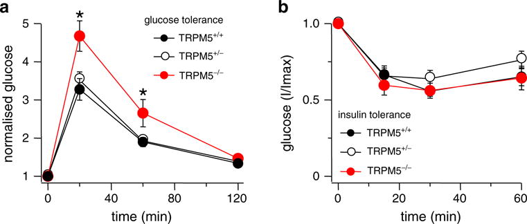
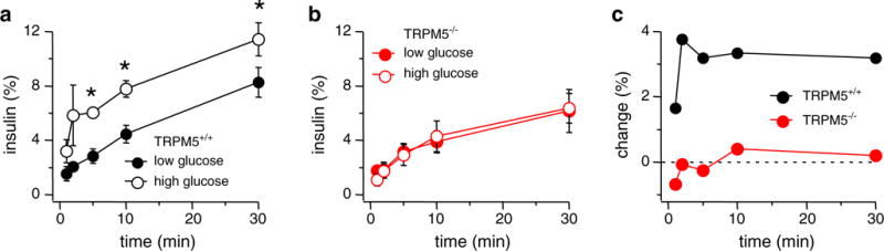
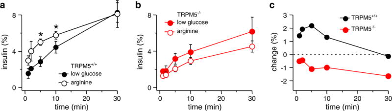
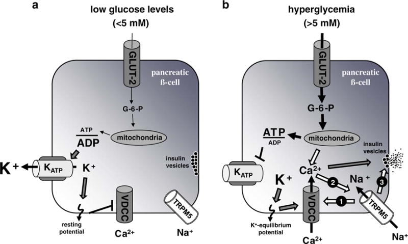
Insulin secretion in beta-pancreatic cells due to glucose stimulation requires the coordinated alteration of cellular ion concentrations and a substantial membrane depolarization to enable insulin vesicle fusion with the cellular membrane. The cornerstones of this cascade are well characterized, yet current knowledge argues for the involvement of additional ion channels in this process. TRPM5 is a cation channel expressed in beta-cells and proposed to be involved in coupling intracellular Ca(2+) release to electrical activity and cellular responses. Here, we report that TRPM5 acts as an indispensable regulator of insulin secretion. In vivo glucose tolerance tests showed that Trpm5 (-/-) -mice maintain elevated blood glucose levels for over an hour compared to wild-type littermates, while insulin sensitivity is normal in Trpm5 (-/-) -mice. In pancreatic islets isolated from Trpm5 (-/-) -mice, hyperglycemia as well as arginine-induced insulin secretion was diminished. The presented results describe a major role for TRPM5 in glucose-induced insulin secretion beyond membrane depolarization. Dysfunction of the TRPM5 protein could therefore be an important factor in the etiology of some forms of type 2 diabetes, where disruption of the normal pattern of secretion is observed.


Figures

Fig. 1
Fig. 1
Trpm5/ mice maintain prolonged elevated glucose levels. Time course of blood glucose levels before and after intraperitoneal glucose or insulin application in Trpm5+/+-, Trpm5+/-, and Trpm5−/−-mice (ten mice per genotype (n=10), see “Materials and Methods”). Error bars are SEM. Stars indicate statistical relevance. a Trpm5/-mice show an impaired glucose tolerance performance with statistical significance according to paired Student’s t test with P<0.01. b Insulin tolerance was unaffected in all three genotypes as depicted
Fig. 2
Fig. 2
Glucose-induced insulin secretion is impaired in pancreatic Trpm5/-islets. Pooled data from 20 Trpm5+/+-islets and 40 Trpm5/-islets are given. Islets were stimulated for 30 min in the presence of 2.8 or 16.8 mM glucose. a Insulin secretion measured in Trpm5+/+-islets stimulated with either 2.8 mM glucose (closed circles) or 16.8 mM glucose (open circles). Error bars are SEM. Stars indicate statistical relevance with P<0.003 (n=4; 5 min P=0.001, 10 min P=0.002, 30 min P=0.0002). b Insulin secretion measured in Trpm5/-islets stimulated with either 2.8 mM glucose (red closed circles) or 16.8 mM glucose (open circles). Error bars are SEM (n=8). c Change of insulin release assessed by subtracting low-glucose data from high-glucose data for Trpm5+/+- (upper graph) and Trpm5/- (lower graph) islets. Data sets were taken from panels a and b, respectively
Fig. 3
Fig. 3
Insulin secretion in islets of Trpm5/-mice cannot be stimulated by arginine-mediated depolarization. Pooled data from 20 Trpm5+/+-islets and 40 Trpm5/-islets are given. Islets were stimulated for 30 min in the presence of 20 mM arginine and compared to the respective low-glucose datasets shown in Fig. 2. a Insulin secretion measured in Trpm5+/+-islets stimulated with 2.8 mM glucose (closed circles, same data as in Fig. 2a) and 20 mM arginine (open circles). Error bars represent SEM. Star indicates statistical relevance with P<0.02 (n=4; 5 min P=0.0085, 10 min P=0.016). b Insulin secretion measured in Trpm5/-islets stimulated with 2.8 mM glucose (closed circles, same data set as in Fig. 2b) and 20 mM arginine (open circles). Error bars represent SEM (n=8). c Relative change of insulin release assessed by subtracting low-glucose data from 20 mM arginine data for both Trpm5+/+- (upper graph) and Trpm5/- (lower graph) islets. Data sets taken from a and b, respectively
Fig. 4
Fig. 4
Schematic representation of pathways involved in the control of insulin secretion and possible involvement of TRPM5. a During low levels of glucose (<5 mM), few glucose molecules are metabolized. This results in a low ATP/ADP ratio within the pancreatic β-cell. The (KATP) channel is active and transports K+ ions out of the cell contributing to the resting membrane potential causing VDCCs to remain closed and insulin-filled vesicles to pause within the cytoplasm beneath the outer cell membrane. b At elevated glucose levels (>5 mM), the glucose uptake increases and consequently leads to enhanced ATP production. The resulting high ATP/ADP ratio within the cell closes the (KATP) channel, and the membrane potential is shifted to the K+ equilibrium potential. This depolarization needs further amplification, e.g., via activation of TRPM5 resulting in Na+-influx (arrow 1), to reach the VDCC activation potential of around 0 mV with a consequential influx of Ca2+ ions that trigger insulin secretion (triggering pathway). The processing of glucose does not only lead to ATP production that closes (KATP) channels but also results in production of cADPR and NAADP that both can stimulate the release of stored Ca2+ from intracellular stores and thus are able to activate TRPM5. Thus, TRPM5 could also be part of the metabolic amplifying pathway of glucose-induced insulin secretion (arrow 2). Besides a possible function in the described triggering and/or amplifying pathway, the presented indispensability of TRPM5 for proper insulin secretion even with VDCC activation by arginine stimulation could also go along with a role of TRPM5 in the direct secretion process, e.g., as part of the process of vesicle–membrane fusion (arrow 3)

Comment in

References

    1. Ashcroft FM, Harrison DE, Ashcroft SJ. Glucose induces closure of single potassium channels in isolated rat pancreatic beta-cells. Nature. 1984;312:446–448. - PubMed
    1. Barg S, Eliasson L, Renstrom E, Rorsman P. A subset of 50 secretory granules in close contact with L-type Ca2+ channels accounts for first-phase insulin secretion in mouse beta-cells. Diabetes. 2002;51(Suppl 1):S74–S82. - PubMed
    1. Berggren PO, Yang SN, Murakami M, Efanov AM, Uhles S, Kohler M, Moede T, Fernstrom A, Appelskog IB, Aspinwall CA, Zaitsev SV, Larsson O, de Vargas LM, Fecher-Trost C, Weissgerber P, Ludwig A, Leibiger B, Juntti-Berggren L, Barker CJ, Gromada J, Freichel M, Leibiger IB, Flockerzi V. Removal of Ca2+ channel beta3 subunit enhances Ca2+ oscillation frequency and insulin exocytosis. Cell. 2004;119:273–284. - PubMed
    1. Cheng H, Beck A, Launay P, Gross SA, Stokes AJ, Kinet JP, Fleig A, Penner R. TRPM4 controls insulin secretion in pancreatic beta-cells. Cell Calcium. 2007;41:51–61. - PMC - PubMed
    1. Colsoul B, Schraenen A, Lemaire K, Quintens R, Van Lommel L, Segal A, Owsianik G, Talavera K, Voets T, Margolskee RF, Kokrashvili Z, Gilon P, Nilius B, Schuit FC, Vennekens R. Loss of high-frequency glucose-induced Ca2+ oscillations in pancreatic islets correlates with impaired glucose tolerance in Trpm5−/− mice. Proc Natl Acad Sci USA. 2010 doi: 10.1073/pnas.0913107107. - DOI - PMC - PubMed

Publication types

MeSH terms