. 2010 Jun 1;518(11):1925-45.
doi: 10.1002/cne.22311.

PDF receptor expression reveals direct interactions between circadian oscillators in Drosophila

Affiliations

PDF receptor expression reveals direct interactions between circadian oscillators in Drosophila

Seol Hee Im et al. J Comp Neurol. .

Abstract

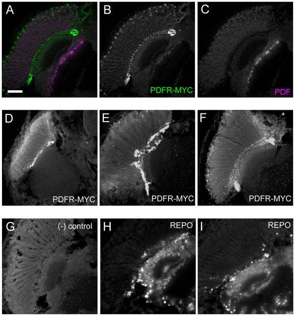
Daily rhythms of behavior are controlled by a circuit of circadian pacemaking neurons. In Drosophila, 150 pacemakers participate in this network, and recent observations suggest that the network is divisible into M and E oscillators, which normally interact and synchronize. Sixteen oscillator neurons (the small and large lateral neurons [LNvs]) express a neuropeptide called pigment-dispersing factor (PDF) whose signaling is often equated with M oscillator output. Given the significance of PDF signaling to numerous aspects of behavioral and molecular rhythms, determining precisely where and how signaling via the PDF receptor (PDFR) occurs is now a central question in the field. Here we show that GAL4-mediated rescue of pdfr phenotypes using a UAS-PDFR transgene is insufficient to provide complete behavioral rescue. In contrast, we describe a approximately 70-kB PDF receptor (pdfr) transgene that does rescue the entire pdfr circadian behavioral phenotype. The transgene is widely but heterogeneously expressed among pacemakers, and also among a limited number of non-pacemakers. Our results support an important hypothesis: the small LNv cells directly target a subset of the other crucial pacemaker neurons cells. Furthermore, expression of the transgene confirms an autocrine feedback signaling by PDF back to PDF-expressing cells. Finally, the results present an unexpected PDF receptor site: the large LNv cells appear to target a population of non-neuronal cells that resides at the base of the eye.


Figures

Figure 1
Figure 1. pdfr-GAL4 lines diagram and summary of expression patterns
Five different pdfr-GAL4 constructs were created, all of which contain a portion of the promoter, upstream of the first exon. (A) Schematic drawing of captured promoter region in each GAL4 construct. (B) Summary table of expression patterns of each GAL4 construct. The first row lists brain regions examined for β–GALACTOSIDASE expression driven by GAL4 lines, whereas the first column lists five pdfr-GAL4 lines and number of independent transgenic fly lines examined for each construct.
Figure 2
Figure 2. pdfr(B)-GAL4-2 expression pattern
pdfr(B)-GAL4-2;UAS-lacZ were reared at 29 °C and stained with anti-β-GAL (green), anti-PDF (Magenta), and anti-PER (blue). (A–D) A Z-series projection of the entire brain. (A) Merged image of (B) (β-GAL, green), (C) (PDF, magenta), and (D) (PER, blue). Scale bar, 100 μm. (E–H) LNvs express pdfr(B)-GAL4-2. (E) Merged image of (F) (β-GAL, green), (G) (PER, blue), and (H) (PDF, magenta). Among the LNvs, four PDF-expressing s-LNvs strongly express β-GAL, the 5th s-LNv (arrow) and two of four l-LNvs (arrowheads) weakly express it. Scale bar, 10 μm. (I–K) LNds express pdfr(B)-GAL4-2. (I) Merged image of (J) (β-GAL, green) and (K) (PER, blue). Three LNds out of six show very strong pdfr(B)-GAL4-2 expression (white arrowheads), whereas the others show none (magenta arrowheads). Scale bar, 10 μm. (L-N) Both DN1a cells express pdfr(B)-GAL4-2. (L) Merged image of (M) (β-GAL, green) and (N) (PER, blue). Scale bar, 5 μm.
Figure 3
Figure 3. Analysis pdfr mutant behavior rescue using various GAL4s
Circadian behavior was monitored following partial restoration of PDFR expression by various GAL4 lines driving UAS-pdfr-16. (A–I) 12 h:12 h LD behavior group analysis of w1118, han3369, and han3369 tested with UAS-pdfr driven by various GAL4 lines. (J–K) Representative actograms of single flies with cry16-GAL4-driven rescue.
Figure 4
Figure 4. Diagram of 70 kB pdfr-myc P[acman]
Four steps used to generate the 70 kB pdfr-myc P[acman] flies. Step (1). Gap repair between linearized P[acman] and BACR25B3 at pdfr locus. Step (2). Recombination and insertion of a 6xMYC tag and Neomycin resisitance selection marker at the C terminus of pdfr. Step (3). Cre recombinase mediated removal of NeoR cassette. Step (4). φC31-mediated integration of 70 kB pdfr-myc P[acman] at attP2 site (681A-B2, Groth et al., 2004).
Figure 5
Figure 5. The 70 kB pdfr-myc transgene rescues pdfr behavioral defects
Representative examples of circadian behavior of three independent experiments. (A–D) Averaged activity for 6 days in a 12 h:12 h LD cycle. (A) w1118 control flies. (B) pdfr5304, a large deletion mutant of pdfr. (C) pdfr5304; ; attP2, pdfr5304 with an empty docking site. (D) pdfr5304; ; pdfr-myc/+, pdfr5304 with a single copy of pdfr-myc. (E–H) Representative actograms of single flies of each genotype for 6 days in LD and 9 days in DD. Wild-type control w1118 exhibits normal morning and evening peaks under LD and a free-running rhythm with a 23.5 hr period (A, E). A larger pdfr deletion (pdfr5304) produced an advanced evening peak and weak or no morning peak; under DD, ~50 % of pdfr5304 became arrhythmic and ~50% displayed short, weak rhythms of ~22 hr period (B, F). pdfr5304; ; attP2 – control for the rescue experiment: pdfr5304 mutant flies containing the attP2 docking site that lacks any pdfr sequences. This control construct cannot rescue the behavioral defects of either pdfr3369 (data not shown) or pdfr5304 (C, G). pdfr5304; ; pdfr-myc/+: 70 kB pdfr-myc transgene in pdfr5304 background. Both LD and DD behavioral defects of both pdfr mutants were rescued by the 70 kB pdfr-myc transgene (D, H) (compiled data is presented in Table 6).
Figure 6
Figure 6. PDF and PDFR-MYC expression
Brains of w1118; ; pdfr-myc flies were immunostained with anti-MYC (green) and anti-PDH (magenta) antibodies. (A) Schematic drawing of PDF-expressing neurons in the fly brain in frontal view. Dorsal is to the top and ventral to the bottom. Yellow inner boxes depict the region of images presented in panels (C) and (I). (B) Schematic drawing of PDF-expressing neurons in the fly brain in horizontal view. Anterior is to the top and posterior to the bottom. A yellow inner box depicts the region of images represented in panel in (F). (C–E) Z-series projections of dorsal brain imaged from the posterior aspect. (C) Merged image of (D) (MYC, green) and (E) (PDF, magenta). Red asterisks indicate the incidence of non-pacemaker receptor-positive cell bodies. (F–H) Sub-retinal glial cells express PDFR-MYC at the boundary of retina and lamina; and large LNv PDF is localized in distal medulla. (F) Merged image of (G) (MYC, green) and (H) (PDF, magenta). (I–K) Suboesophageal ganglion (SOG) with PDF-tri cells imaged from the anterior aspect. (I) Merged image of (J) (MYC, green) and (K) (PDF, magenta). Arrows, PDF-tri cell bodies. re, retina; la, lamina; me, medulla. Scale bars, 50μm.
Figure 7
Figure 7. PDFR-MYC in a subset of cells near the fenestrated membrane of the retina
Adult fly heads sections immunostained showing PDFR-MYC expression near the fenestrated membrane of the retina. (A–C) PDFR-MYC fly heads showing PDF and PDFR-MYC expression. (A) Merged image of (B) (MYC, green) and (C) (PDF, magenta). (D–F) Expression of PDFR-MYC. (G) Negative control: a PDFR-MYC brain stained without incubation in the primary (anti-MYC) antibody. attP2 fly heads with anti-MYC antibody display similar results (not shown). (H–I) anti-REPO staining in w1118 fly heads. REPO is a useful marker for many glial cells: The outer-most layer of REPO-positive cells lies along the fenestrated membrane that separates the retina from the brain: this position is similar to that exhibited by PDFR-MYC positive cells. Scale bar = 50 μm.
Figure 8
Figure 8. PDFR expression in PER-expressing clock neurons
PDFR-MYC expression by identified pacemaker neurons. (A–D) Six of the 17 DN1s express PDFR-MYC at strong levels, whereas three or four of the remaining ones express it at lower levels. (A) Merged image of (B) (PDF, magenta), (C) (PER, blue), and (D) (MYC, green). (E–H) LNvs express PDFR-MYC. (E) Merged image of (F) (PDF, magenta), (G) (PER, blue), and (H) (MYC, green). The 5th s-LNv expresses high levels of PDFR-MYC (arrow), two l-LNvs express PDFR-MYC at intermediate levels (arrowhead), while the others have lower to undetectable levels. s-LNvs are best seen in panel (I). Red asterisks indicate the incidence of non-pacemaker, receptor-positive cell bodies. (I–K) s-LNvs express PDFR-MYC. (I) Merged image of (J) (MYC, green) and (K) (PDF, magenta). Red asterisks indicate the incidence of non-pacemaker, receptor-positive cell bodies. (L–N) Three LNds out of six show very strong PDFR-MYC expression (white arrowheads), whereas the others show no PDFR-MYC (magenta arrowheads). (L) Merged image of (M) (MYC, green) and (N) (PER, blue). Asterisk, non-specific MYC signal near LNd area. (O–Q) Single optical section reveals low-level PDFR-MYC expression by DN2s (arrow). (O) Merged image of (P) (MYC, green) and (Q) (PER, blue). (R–T) DN3s express PDFR-MYC at low levels. (R) Merged image of (S) (MYC, green) and (T) (PER, blue). Scale bars, 10 μm.
Figure 9
Figure 9. PDFR-MYC in the processes of clock neurons
PDFR-MYC is expressed and localized in the PDF dorsal projections. A single focal plane shows overlap between PDF and PDFR-MYC in s-LNv projections. PDFR-MYC positive projections occupy a wider area than do PDF positive projections, which indicates PDFR-MYC is localized in PDF cells and non-PDF clock neurons. (A) Merged image of (B) and (C). Dorsal is to the top and ventral is to the bottom, and the mid-line is to the left. Scale bar, 20 μm.
Figure 10
Figure 10. PDFR expression in the circadian system
PDF secreting large and small LNvs are in the center of the pacemaker, signaling directly to the non-PDF clock neurons, to non-neuronal cells in or near the retina, to the output non-clock receptor positive cells, and feeding back to PDF secreting cells.

Similar articles

Cited by

References

    1. Alfonso TB, Jones BW. gcm2 promotes glial cell differentiation and is required with glial cells missing for macrophage development in Drosophila. Dev Biol. 2002;248:369–383. - PubMed
    1. Aton SJ, Colwell CS, Harmar AJ, Waschek J, Herzog ED. Vasoactive intestinal polypeptide mediates circadian rhythmicity and synchrony in mammalian clock neurons. Nat Neurosci. 2005;8:476–483. - PMC - PubMed
    1. Aton SJ, Herzog ED. Come together, right...now: synchronization of rhythms in a mammalian circadian clock. Neuron. 2005;48:531–534. - PMC - PubMed
    1. Bachleitner W, Kempinger L, Wülbeck C, Rieger D, Helfrich-Förster C. Moonlight shifts the endogenous clock of Drosophila melanogaster. Proc Natl Acad Sci U S A. 2007;104:3538–3543. - PMC - PubMed
    1. Baker DA, Beckingham KM, Armstrong JD. Functional dissection of the neural substrates for gravitaxic maze behavior in Drosophila melanogaster. J Comp Neurol. 2007;501:756–764. - PubMed

Publication types

MeSH terms

Substances

LinkOut - more resources