Progression of neuronal and synaptic remodeling in the rd10 mouse model of retinitis pigmentosa
- PMID: 20394059
- PMCID: PMC2881548
- DOI: 10.1002/cne.22322
Progression of neuronal and synaptic remodeling in the rd10 mouse model of retinitis pigmentosa
Abstract
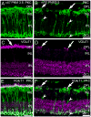
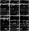
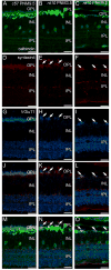
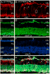
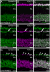
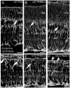
The Pde6b(rd10) (rd10) mouse has a moderate rate of photoreceptor degeneration and serves as a valuable model for human autosomal recessive retinitis pigmentosa (RP). We evaluated the progression of neuronal remodeling of second- and third-order retinal cells and their synaptic terminals in retinas from Pde6b(rd10) (rd10) mice at varying stages of degeneration ranging from postnatal day 30 (P30) to postnatal month 9.5 (PNM9.5) using immunolabeling for well-known cell- and synapse-specific markers. Following photoreceptor loss, changes occurred progressively from outer to inner retina. Horizontal cells and rod and cone bipolar cells underwent morphological remodeling that included loss of dendrites, cell body migration, and the sprouting of ectopic processes. Gliosis, characterized by translocation of Müller cell bodies to the outer retina and thickening of their processes, was evident by P30 and became more pronounced as degeneration progressed. Following rod degeneration, continued expression of VGluT1 in the outer retina was associated with survival and expression of synaptic proteins by nearby second-order neurons. Rod bipolar cell terminals showed a progressive reduction in size and ectopic bipolar cell processes extended into the inner nuclear layer and ganglion cell layer by PNM3.5. Putative ectopic conventional synapses, likely arising from amacrine cells, were present in the inner nuclear layer by PNM9.5. Despite these changes, the laminar organization of bipolar and amacrine cells and the ON-OFF organization in the inner plexiform layer was largely preserved. Surviving cone and bipolar cell terminals continued to express the appropriate cell-specific presynaptic proteins needed for synaptic function up to PNM9.5.
(c) 2010 Wiley-Liss, Inc.
Figures




Similar articles
-
Retinal organization in the retinal degeneration 10 (rd10) mutant mouse: a morphological and ERG study.J Comp Neurol. 2007 Jan 10;500(2):222-38. doi: 10.1002/cne.21144. J Comp Neurol. 2007. PMID: 17111372 Free PMC article.
-
Optimal timing for activation of sigma 1 receptor in the Pde6brd10/J (rd10) mouse model of retinitis pigmentosa.Exp Eye Res. 2021 Jan;202:108397. doi: 10.1016/j.exer.2020.108397. Epub 2020 Dec 9. Exp Eye Res. 2021. PMID: 33310057 Free PMC article.
-
Different effects of valproic acid on photoreceptor loss in Rd1 and Rd10 retinal degeneration mice.Mol Vis. 2014 Nov 4;20:1527-44. eCollection 2014. Mol Vis. 2014. PMID: 25489226 Free PMC article.
-
Neural remodeling in retinal degeneration.Prog Retin Eye Res. 2003 Sep;22(5):607-55. doi: 10.1016/s1350-9462(03)00039-9. Prog Retin Eye Res. 2003. PMID: 12892644 Review.
-
Retinal remodeling in human retinitis pigmentosa.Exp Eye Res. 2016 Sep;150:149-65. doi: 10.1016/j.exer.2016.03.018. Epub 2016 Mar 26. Exp Eye Res. 2016. PMID: 27020758 Free PMC article. Review.
Cited by
-
Regulatory effects of inhibiting the activation of glial cells on retinal synaptic plasticity.Neural Regen Res. 2014 Feb 15;9(4):385-93. doi: 10.4103/1673-5374.128240. Neural Regen Res. 2014. PMID: 25206825 Free PMC article.
-
Brg1 coordinates multiple processes during retinogenesis and is a tumor suppressor in retinoblastoma.Development. 2015 Dec 1;142(23):4092-106. doi: 10.1242/dev.124800. Development. 2015. PMID: 26628093 Free PMC article.
-
CX3CL1 (fractalkine) protein expression in normal and degenerating mouse retina: in vivo studies.PLoS One. 2014 Sep 5;9(9):e106562. doi: 10.1371/journal.pone.0106562. eCollection 2014. PLoS One. 2014. PMID: 25191897 Free PMC article.
-
Effect of degeneration stage on non-viral tissue transfection of rd10 retina ex vivo.Mol Ther Nucleic Acids. 2025 Jul 1;36(3):102616. doi: 10.1016/j.omtn.2025.102616. eCollection 2025 Sep 9. Mol Ther Nucleic Acids. 2025. PMID: 40704026 Free PMC article.
-
Presynaptic partner selection during retinal circuit reassembly varies with timing of neuronal regeneration in vivo.Nat Commun. 2016 Feb 3;7:10590. doi: 10.1038/ncomms10590. Nat Commun. 2016. PMID: 26838932 Free PMC article.
References
-
- Ahmari SE, Buchanan J, Smith SJ. Assembly of presynaptic active zones from cytoplasmic transport packets. Nat Neurosci. 2000;3:445–451. - PubMed
-
- Banin E, Cideciyan AV, Alemán TS, Petters RM, Wong F, Milam AH, Jacobson SG. Retinal rod photoreceptor-specific gene mutation perturbs cone pathway development. Neuron. 1999;23:549–557. - PubMed
-
- Barhoum R, Martínez-Navarrete G, Corrochano S, Germain F, Fernandez-Sanchez L, de la Rosa EJ, de la Villa P, Cuenca N. Functional and structural modifications during retinal degeneration in the rd10 mouse. Neuroscience. 2008;155:698–713. - PubMed
Publication types
MeSH terms
Substances
Grants and funding
LinkOut - more resources
Full Text Sources
Other Literature Sources
Miscellaneous

