. 2010 Jun 1;518(11):2090-108.
doi: 10.1002/cne.22323.

Identifying the efferent projections of leptin-responsive neurons in the dorsomedial hypothalamus using a novel conditional tracing approach

Affiliations

Identifying the efferent projections of leptin-responsive neurons in the dorsomedial hypothalamus using a novel conditional tracing approach

Laurent Gautron et al. J Comp Neurol. .

Abstract

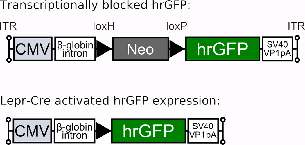
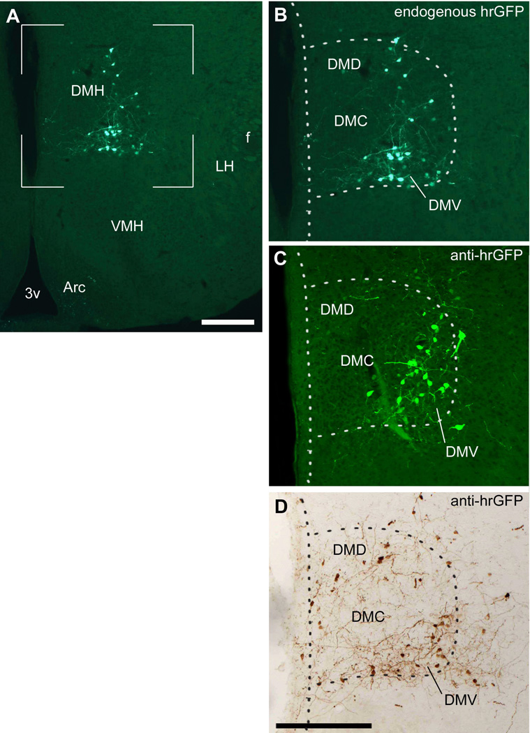
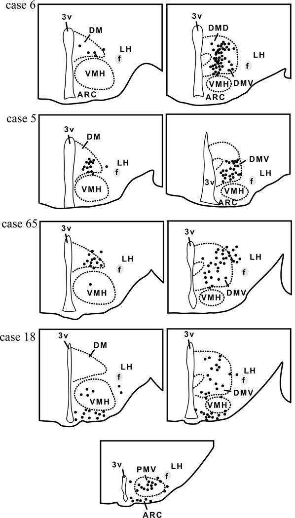
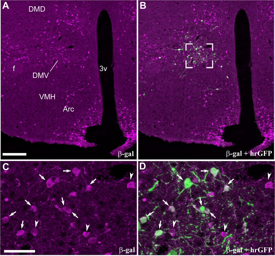
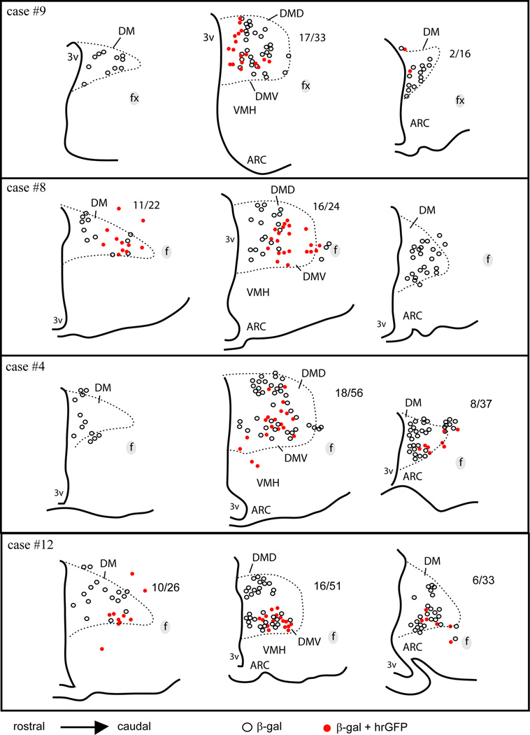
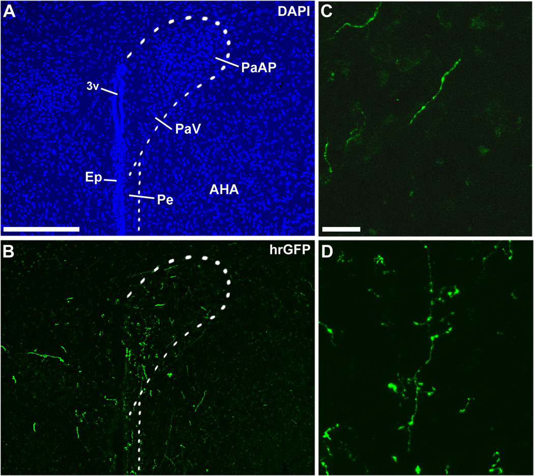
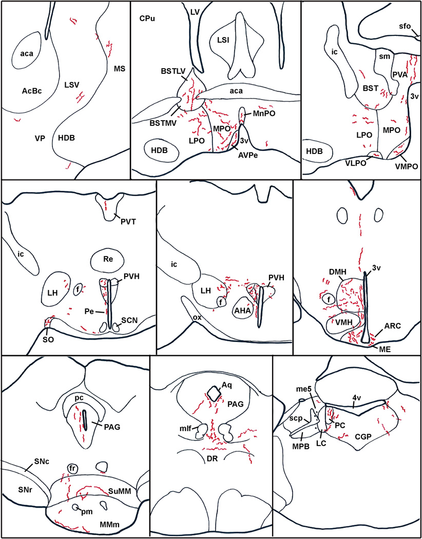
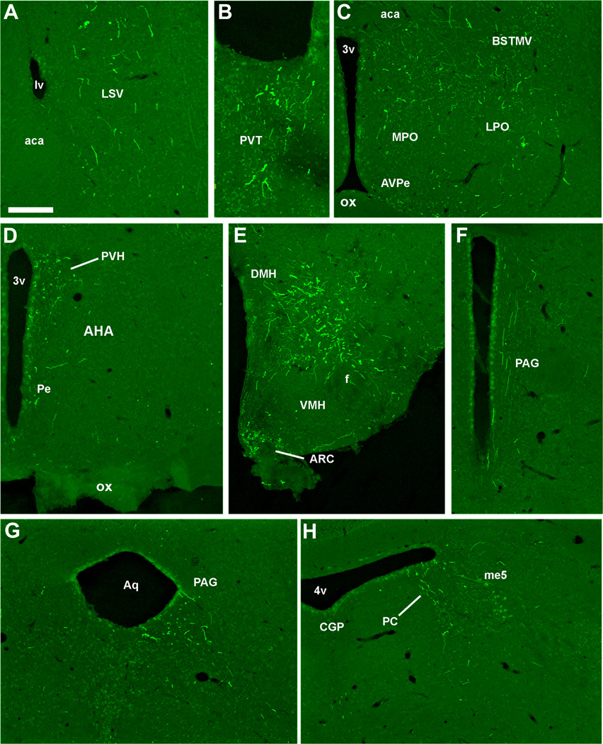
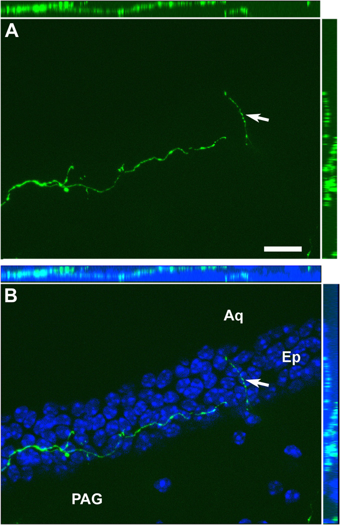
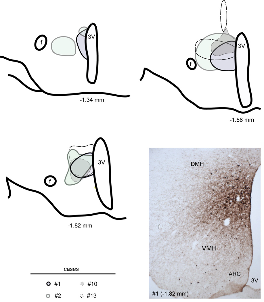
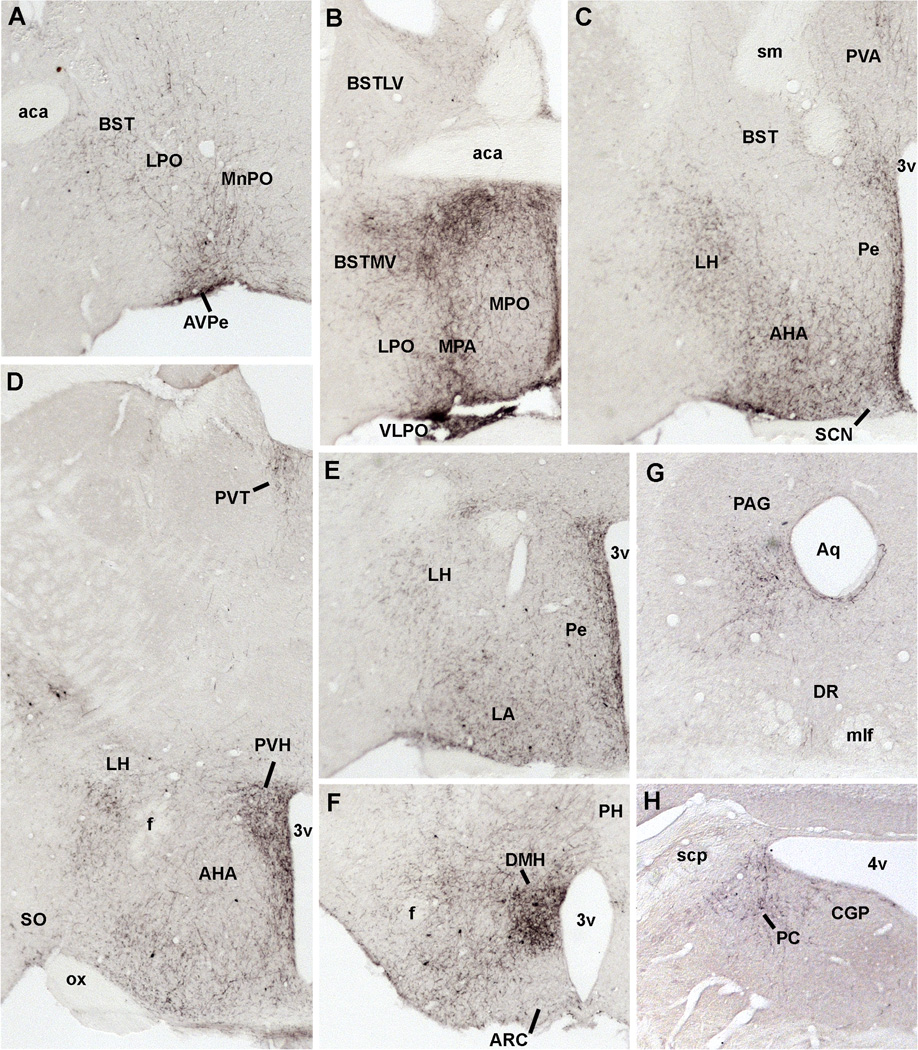
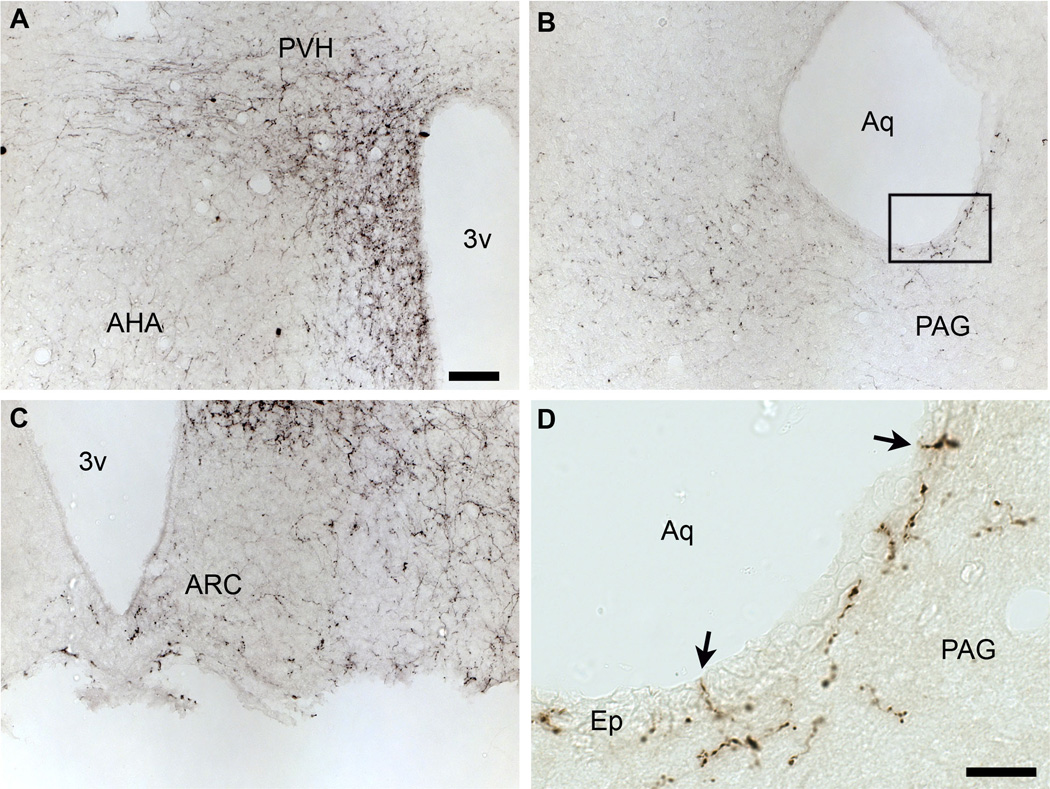
Tracing the axonal projections of selected neurons is labor intensive and inherently limited by currently available neuroanatomical methods. We developed an adeno-associated virus (AAV) that can be used for efficiently tracing identified neuronal populations. The virus encodes a humanized Renilla green fluorescent protein (hrGFP) that is transcriptionally silenced by a neo cassette flanked by LoxH/LoxP sites (AAV-lox-Stop-hrGFP). Thus, hrGFP is expressed only in neurons with Cre recombinase activity. To demonstrate the utility of this approach, the virus was injected unilaterally into the dorsomedial hypothalamus (DMH) of mice that express Cre in neurons expressing the leptin receptor. Animals with DMH injections showed robust hrGFP expression in DMH neurons, as visualized by its endogenous fluorescence or following immunolabeling. We found that hrGFP was expressed in approximately one-third to one-half of Cre-expressing neurons at the site of injection, but not in non-Cre-expressing neurons. The expression of GFP allowed us to identify the projection fields of DMH leptin-responsive neurons. Our results show hrGFP-positive axonal projections and terminals in the paraventricular nucleus of the hypothalamus, arcuate nucleus, preoptic area, bed nucleus of the stria terminalis, paraventricular thalamus, periaqueductal gray, and precoeruleus. The aforementioned pattern of projections was similar to DMH projections determined by injections of biotinylated dextran amine in the mouse DMH. Interestingly, some hrGFP-positive terminals were seen contacting the ependymal layer of the third and fourth ventricles. In summary, this approach is an effective tool for tracing axonal projections of chemically identified neurons, including leptin-responsive neurons.


Figures

Figure 1
Figure 1
Generation of a conditional hrGFP-expressing AAV vector. A loxH/loxP-flanked Neo DNA sequence was inserted to prevent transcription of the hrGFP gene. The AAV-lox-Stop-hrGFP cassette consists of a variety of elements in the following order (5’ to 3’): left inverted terminal repeat (ITR) - cytomegalovirus (CMV) immediate early promoter – human β-globin intron – loxH site – NeoR coding sequence – loxP site – hrGFP coding sequence – SV40 VP1 poly A – right ITR. The Cre-mediated removal of the Neo-Stop sequence enables the expression of hrGFP.
Figure 2
Figure 2
Expression of hrGFP in one successful injection of AAV-stop-lox-hrGFP (case #5). Endogenous fluorescence (epifluorescence illumination) is observed in many cells in the DMH but not the surrounding structures (A). Higher magnification shows that hrGFP-positive neurons are located mostly in the DMV (B). Fluorescently-labeled adjacent section (epifluorescence illumination and Apotome) (C) and DAB-labeled adjacent section (brightfield illumination) (D) revealed a comparable expression of hrGFP, with a somewhat larger number of cells and fibers. The dotted outline shows the boundary of the DMH. Abbreviations: 3v, third ventricle; Arc, arcuate nucleus of the hypothalamus; DMD, dorsal part of the dorsomedial nucleus of the hypothalamus; DMV, ventral part of the dorsomedial nucleus of the hypothalamus; DMC, compact part of the dorsomedial nucleus of the hypothalamus; f, fornix; LH, lateral hypothalamus; VMH, ventromedial nucleus of the hypothalamus. Scale bars: A=134 µm; B–D; 200 µm.
Figure 3
Figure 3
Camera lucida drawings of 2 rostral-to-caudal levels of the mouse hypothalamus illustrate the distribution of hrGFP-positive cells in 4 successful cases (LepRb-Cre mice). Each dot represents one positive neuron. Note that positive neurons are mostly restricted to the DMH in cases 5, 6 and 65, whereas hrGFP expression was observed outside of the DMH in case 18. Abbreviations: 3v, third ventricle; Arc, arcuate nucleus of the hypothalamus; DM, rostral part of the dorsomedial nucleus of the hypothalamus; DMD, dorsal part of the dorsomedial nucleus of the hypothalamus; DMV, ventral part of the dorsomedial nucleus of the hypothalamus; f, fornix; LH, lateral hypothalamus; PMV, ventral premammillary nucleus; VMH, ventromedial nucleus of the hypothalamus.
Figure 4
Figure 4
Restricted expression of hrGFP in leptin-responsive neurons as demonstrated by dual-label immunohistochemistry (case #4, LepRb-Cre-LacZ). Robust β-gal immunoreactivity (Alexa 594) is seen in leptin-responsive neurons distributed in the hypothalamus (A). By contrast, hrGFP-positive neurons (Alexa 488) are observed only in the DMH ipsilateral to the site of injection (B). High magnification photomicrographs reveal clusters of β-gal-positive neurons in the DMV (C). Dual-label labeling showing that hrGFP immunoreactivity coincided perfectly with β-gal (D). It shows that all hrGFP-positive neurons are also leptin-responsive neurons (arrows indicate colocalizations). However, some neurons positive for β-gal do not express hrGFP (arrowheads). Images have been converted to magenta-green. Scale bar in A applies to B, and scale bar in C applies to D. Abbreviations: 3v, third ventricle; Arc, arcuate nucleus of the hypothalamus; DMD, dorsal part of the dorsomedial nucleus of the hypothalamus; DMV, ventral part of the dorsomedial nucleus of the hypothalamus; f, fornix; VMH, ventromedial nucleus of the hypothalamus. Scale bars: A–B=200µm; C–D=50µm.
Figure 5
Figure 5
Camera lucida drawings of 3 rostral-to-caudal levels of the mouse hypothalamus illustrate the distribution of cells positive for β-gal alone (white dots) or β-gal and hrGFP (red dots) in 4 successful cases (LepRb-Cre-LacZ mice). Each dot represents one positive neuron. The ratio of cells positive for hrGFP over the total number of β-gal-positive neurons is indicated next to each brain section (except when no colocalization was observed). Note that the number of hrGFP-positive neurons is more important in the ventral part of the medial DMH in all cases, whereas it considerably varies in the rostral and posterior DMH. Abbreviations: 3v, third ventricle; Arc, arcuate nucleus of the hypothalamus; DM, rostral part of the dorsomedial nucleus of the hypothalamus; DMD, dorsal part of the dorsomedial nucleus of the hypothalamus; DMV, ventral part of the dorsomedial nucleus of the hypothalamus; f, fornix; VMH, ventromedial nucleus of the hypothalamus.
Figure 6
Figure 6
Immunohistochemical labeling of hrGFP-positive axonal projections in the PVH (A, B). Fibers positive for hrGFP were seen coursing toward the PVH (C), and clusters of terminals were found in the PVH itself (D). Note that fibers were more abundant on the side ipsilateral to the site of injection. The dotted outline shows the boundary of the PVH. Scale bar in A applies to B, and scale bar in C applies to D. Abbreviations: 3v, third ventricle; AHA, anterior hypothalamic area; Ep, ependyma; PaAP, anterior part of the paraventricular nucleus of the hypothalamus; PaV, ventral part of the paraventricular nucleus of the hypothalamus; Pe, periventricular hypothalamic nucleus. Scale bars: A–B=200µm; C–D=20µm.
Figure 7
Figure 7
Anatomical distribution of hrGFP-positive fibers in the brain of case #65 (LepRb-Cre). Abbreviations: 3v, third ventricle; 4v, fourth ventricle; aca, anterior commissure; AcBc, core of the accumbens nucleus; AHA, anterior hypothalamic area; Aq, aqueduct; Arc, arcuate nucleus of the hypothalamus; AVPe, anteroventral periventricular nucleus; BST, bed nucleus of the stria terminalis; BSTLV, lateroventral part of the bed nucleus of the stria terminalis; BSTMV, medioventral part of the bed nucleus of the stria terminalis; CGP; central gray of the pons; CPu, caudate putamen; DMH, dorsomedial nucleus of the hypothalamus; DR, dorsal raphé; f, fornix; fr, fasciculus retroflexus; HDB, nucleus of the horizontal limb of the diagonal band; ic, internal capsule; LH, lateral hypothalamus; LC, Locus coeruleus; LPO, lateral preoptic area; LSi, intermediate part of the lateral septal nucleus; LSV, ventral part of the lateral septal nucleus; LV, lateral ventricle; ME, median eminence; me5, mesencephalic trigeminal nucleus; MMm, median part of the medial mammillary nucleus; MnPO, Median preoptic nucleus; mlf, medial longitudinal fasciculus; MPB, medial parabrachial nucleus; MPO, medial preoptic nucleus; MS, medial septal nucleus; ox, optic chiasm; PAG, periaqueductal gray; PC, precoeruleus; Pe, periventricular hypothalamic nucleus; pm, principal mammillary tract; PVA, anterior part of the paraventricular thalamic nucleus; PVH, paraventricular nucleus of the hypothalamus; PVT; periventricular thalamus; Re, reunions thalamic nucleus; SCN, suprachiasmatic nucleus; scp, superior cerebellar peduncle; sfo, subfornical organ; sm, stria medullaris of the thalamus; SNc, compact part of the subtantia nigra; SNr, reticular part of the subtantia nigra; SO, supraoptic nucleus; suMM, median part of the supramedial mammillary nucleus; VLPO, ventrolateral preoptic area; VMH, ventromedial nucleus of the hypothalamus; VMPO, ventromedial preoptic area; VP, ventral pallidum.
Figure 8
Figure 8
Photomicrographs of hrGFP-positive fibers in representative structures from case #8 (LepRb-Cre-LacZ) including the LSV (A), PVT (B), BST (C), PVH (D), Arc and DMH (E), PAG (F, G), PC (H). Abbreviations: 3v, third ventricle; 4v, fourth ventricle; aca, anterior commissure; AHA, anterior hypothalamic area; Aq, aqueduct; Arc, arcuate nucleus of the hypothalamus; AVPe, anteroventral periventricular nucleus; BSTMV, medioventral part of the bed nucleus of the stria terminalis; CGP; central gray of the pons; DMH, dorsomedial nucleus of the hypothalamus; LPO, lateral preoptic area; LSV, ventral part of the lateral septal nucleus; LV, lateral ventricle; me5, mesencephalic trigeminal nucleus; MPO, medial preoptic nucleus; ox, optic chiasm; PAG, periaqueductal gray; PC, precoeruleus; Pe, periventricular hypothalamic nucleus; PVH, paraventricular nucleus of the hypothalamus; PVT; periventricular thalamus; VMH, ventromedial nucleus of the hypothalamus. Scale bar: A–H=200 µm.
Figure 9
Figure 9
Optical sections reconstruction (epifluorescence and Apotome) of a hrGFP-positive fiber (A) demonstrating that it is traveling inside the ependymal layer in all 3 dimensions (B). The ependymal layer of the aqueduct is revealed by DAPI staining (B). The white arrow shows an axonal branch running through the ependyma. Scale bar in A applies to B. Abbreviations: Aq, aqueduct; Ep, ependymal layer; PAG, periaqueductal gray. Scale bar: A–B= 200µm.
Figure 10
Figure 10
Camera lucida drawings of 3 rostral-to-caudal levels of the mouse hypothalamus illustrate the distribution of BDA deposits in 4 successful cases (distance from bregma is indicated below each level). Sites of injections are circled. A photomicrograph from case #1 appears in the bottom right panel. Abbreviations: 3v, third ventricle; Arc, arcuate nucleus of the hypothalamus; DMH, dorsomedial nucleus of the hypothalamus; VMH, ventromedial nucleus of the hypothalamus; f, fornix.
Figure 11
Figure 11
Photomicrographs of BDA-positive fibers in representative structures from case #1. Abbreviations: 3v, third ventricle; 4v, fourth ventricle; aca, anterior commissure; AHA, anterior hypothalamic area; Aq, aqueduct; Arc, arcuate nucleus of the hypothalamus; AVPe, anteroventral periventricular nucleus; BST, bed nucleus of the stria terminalis; BSTLV, lateroventral part of the bed nucleus of the stria terminalis; BSTMV, medioventral part of the bed nucleus of the stria terminalis; CGP; central gray of the pons; DMH, dorsomedial nucleus of the hypothalamus; DR, dorsal raphé; f, fornix; LH, lateral hypothalamus; LPO, lateral preoptic area; MnPO, Median preoptic nucleus; mlf, medial longitudinal fasciculus; MPB, medial parabrachial nucleus; MPA, medial preoptic area; MPO, medial preoptic nucleus; ox, optic chiasm; PAG, periaqueductal gray; PC, precoeruleus; Pe, periventricular hypothalamic nucleus; PH, posterior hypothalamus; PVA, anterior part of the paraventricular thalamic nucleus; PVH, paraventricular nucleus of the hypothalamus; PVT; periventricular thalamus; SCN, suprachiasmatic nucleus; scp, superior cerebellar peduncle; sm, stria medullaris of the thalamus; SO, supraoptic nucleus; VLPO, ventrolateral preoptic area; VMH, ventromedial nucleus of the hypothalamus; VMPO, ventromedial preoptic area.
Figure 12
Figure 12
Immunohistochemical labeling of BDA-positive axonal projections in the PVH (A), PAG (B, D) and Arc (C). Note the fibers traveling closely to the ventricle near the PAG (B). At high magnification, it appears that some fibers travel through the ependymal layer itself (arrows) (D). Abbreviations: 3v, third ventricle; Arc, Arcuate nucleus; AHA, anterior hypothalamic area; Ep, ependyma; PVH, paraventricular nucleus of the hypothalamus; PAG, periaqueductal gray. Scale bar: A–C= 50µm; D= 20µm.

Similar articles

Cited by

References

    1. Baver SB, Pickard GE, Sollars PJ, Pickard GE. Two types of melanopsin retinal ganglion cell differentially innervate the hypothalamic suprachiasmatic nucleus and the olivary pretectal nucleus. Eur J Neurosci. 2008;27(7):1763–1770. - PubMed
    1. Bellinger LL, Bernardis LL. The dorsomedial hypothalamic nucleus and its role in ingestive behavior and body weight regulation: lessons learned from lesioning studies. Physiol Behav. 2002;76(3):431–442. - PubMed
    1. Bjørbaek C, Elmquist JK, Frantz JD, Shoelson SE, Flier JS. Identification of SOCS-3 as a potential mediator of central leptin resistance. Mol Cell. 1998;1(4):619–625. - PubMed
    1. Bota M, Swanson LW. The neuron classification problem. Brain Res Rev. 2007;56(1):79–88. - PMC - PubMed
    1. Campbell RE, Gaidamaka G, Han SK, Herbison AE. Dendro-dendritic bundling and shared synapses between gonadotropin-releasing hormone neurons. Proc Natl Acad Sci U S A. 2009;106(26):10835–10840. - PMC - PubMed

Publication types