. 2010 May 7;6(9):1022-31.
doi: 10.1002/smll.201000028.

Near-infrared light triggers release of Paclitaxel from biodegradable microspheres: photothermal effect and enhanced antitumor activity

Affiliations

Near-infrared light triggers release of Paclitaxel from biodegradable microspheres: photothermal effect and enhanced antitumor activity

Jian You et al. Small. .

Abstract

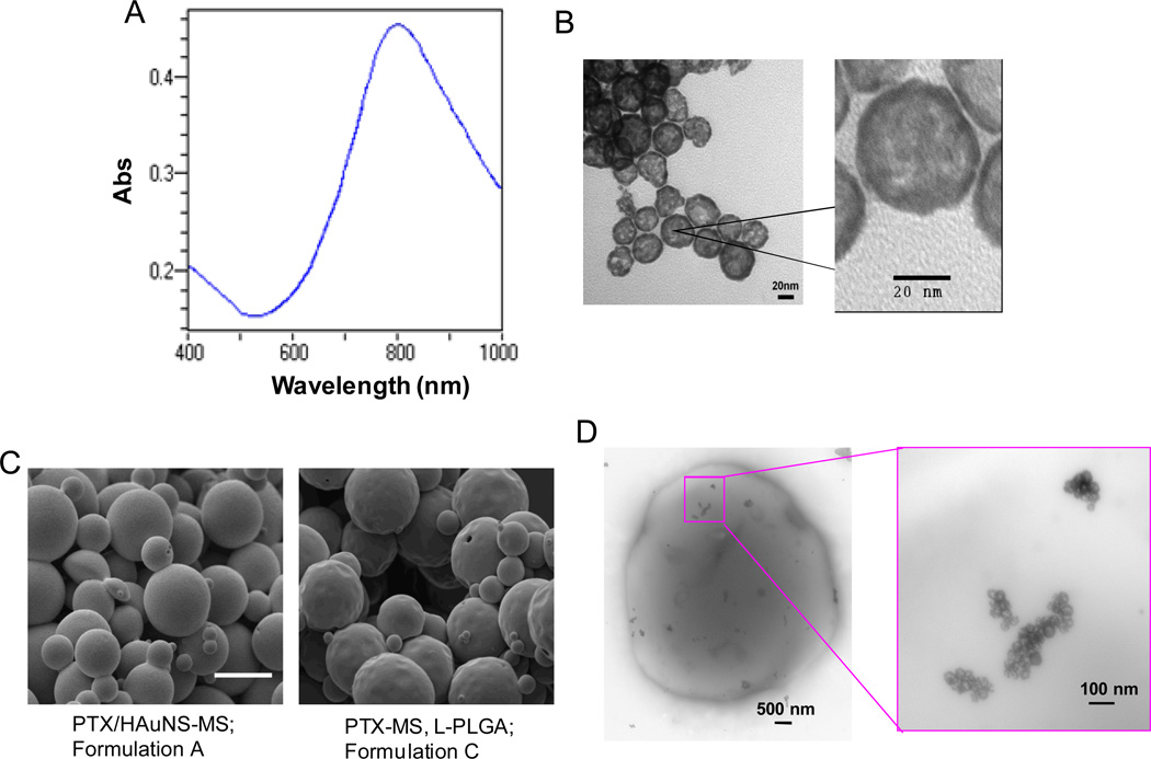
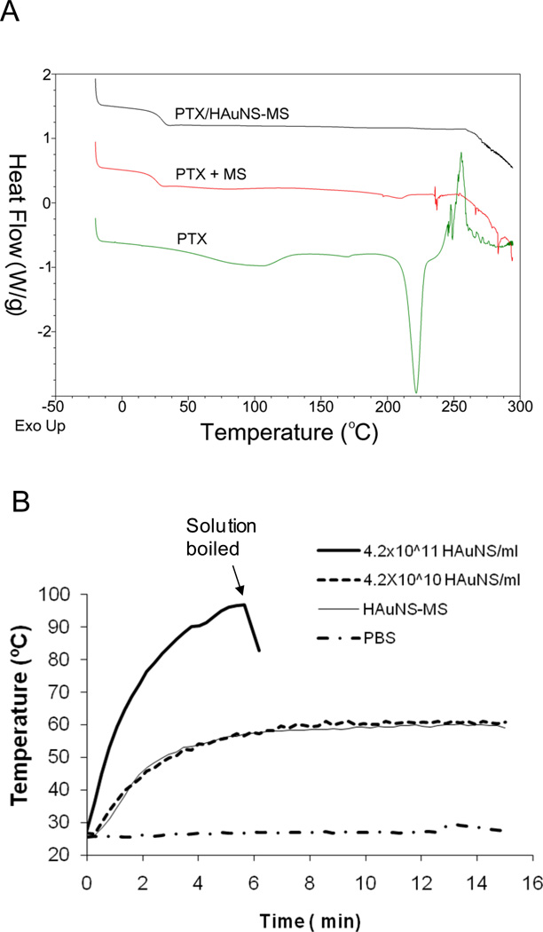
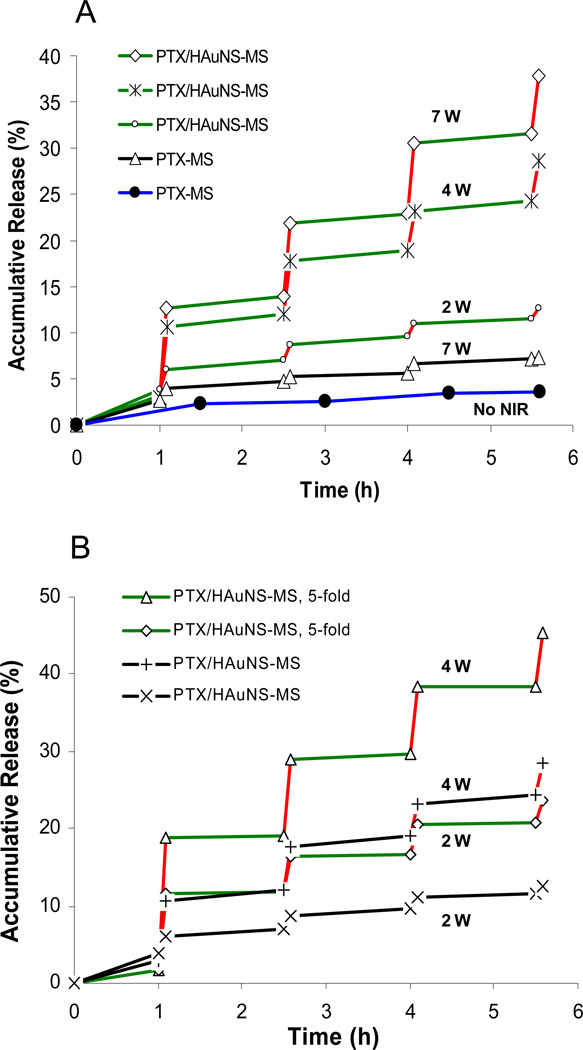
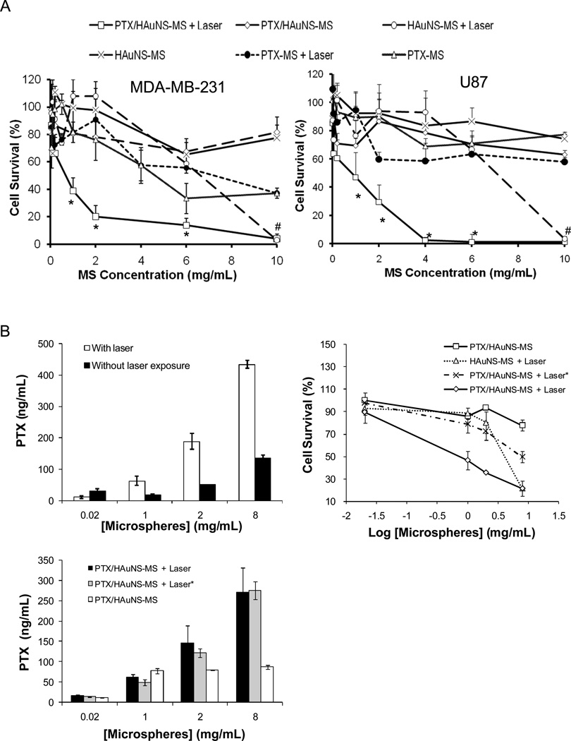
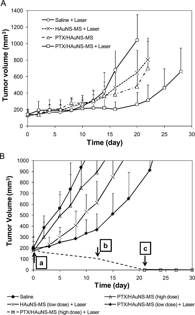
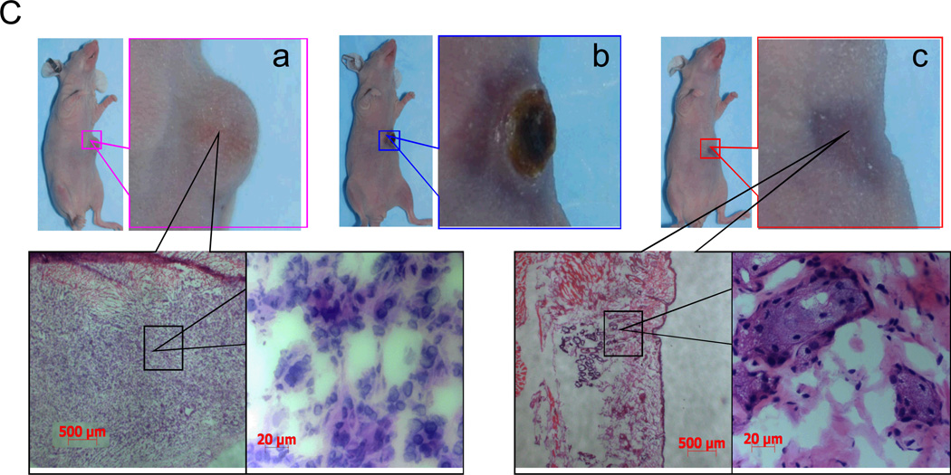
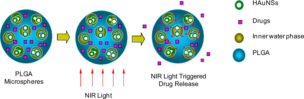
Despite advances in controlled drug delivery, reliable methods for activatable, high-resolution control of drug release are needed. The hypothesis that the photothermal effect mediated by a near-infrared (NIR) laser and hollow gold nanospheres (HAuNSs) could modulate the release of anticancer agents is tested with biodegradable and biocompatible microspheres (1-15 microm) containing the antitumor drug paclitaxel (PTX) and HAuNSs (approximately 35 nm in diameter), which display surface plasmon absorbance in the NIR region. HAuNS-containing microspheres exhibit a NIR-induced thermal effect similar to that of plain HAuNSs. Rapid, repetitive PTX release from the PTX/HAuNS-containing microspheres is observed upon irradiation with NIR light (808 nm), whereas PTX release is insignificant when the NIR light is switched off. The release of PTX from the microspheres is readily controlled by the output power of the NIR laser, duration of irradiation, treatment frequency, and concentration of HAuNSs embedded inside the microspheres. In vitro, cancer cells incubated with PTX/HAuNS-loaded microspheres and irradiated with NIR light display significantly greater cytotoxic effects than cells incubated with the microspheres alone or cells irradiated with NIR light alone, owing to NIR-light-triggered drug release. Treatment of human U87 gliomas and MDA-MB-231 mammary tumor xenografts in nude mice with intratumoral injections of PTX/HAuNS-loaded microspheres followed by NIR irradiation results in significant tumor-growth delay compared to tumors treated with HAuNS-loaded microspheres (no PTX) and NIR irradiation or with PTX/HAuNS-loaded microspheres alone. The data support the feasibility of a therapeutic approach in which NIR light is used for simultaneous modulation of drug release and induction of photothermal cell killing.


Figures

Fig. 1
Fig. 1
(A) Absorption spectrum of HAuNS showing the plasmon resonance peak tuned to the NIR region (λmax=808 nm). (B) TEM images of HAuNS revealing the morphology of the hollow nanospheres. Bar, 20 nm. (C) SEM images of PTX-loaded, HAuNS-embedded microspheres (PTX/HAuNS-MS) and microspheres containing only PTX (PTX-MS). The presence of HAuNS resulted in microspheres with a smoother surface. Bar, 10 µm. (D) TEM photographs of a PTX/HAuNS-MS showing clusters of HAuNS dispersed within the polymeric matrix.
Fig. 2
Fig. 2
(A) DSC thermograms of PTX/HAuNS-MS, pure PTX, and a physical mixture of PLGA and PTX (5% PTX, w/w). An endothermic peak at around 210°C, representing the melting point of PTX, was present in the physical mixture of PTX and MS, but not in PTX/HAuNS-MS, suggesting that PTX was dissolved in PLGA polymer at the molecular level. (B) Comparison of the temperature changes in aqueous solutions containing HAuNS and HAuNS-MS after exposure to NIR light at an output power of 4.5 W/cm2. HAuNS-loaded PLGA microspheres elevated the temperature of the water to the same extent as did HAuNS alone at a concentration of 4.2×10¹⁰ nanoparticles/mL.
Fig. 3
Fig. 3
NIR light triggered release of PTX from PTX/HAuNS-MS. Red lines represent the 5-min period during which the microspheres were irradiated with NIR laser. (A) PTX release profiles of PTX/HAuNS-MS (formulation B) at an NIR output power of 7 W (◇), 4 W (*), and 2 W (○); PTX release profiles from PTX-MS (formulation C) at an NIR output power of 7 W (△) and without NIR exposure (●). (B) PTX release from PTX/HAuNS-MS containing 4.7×10¹⁰ HAuNS/mg PLGA (formulation B) at an NIR output power of 4 W (△) and 2 W (◇); PTX release from PTX/HAuNS-MS containing 9.4×10⁹ HAuNS/mg PLGA (formulation A) at an NIR output power of 4 W (+) and 2 W (×), respectively. The spot size was 10 mm in diameter.
Fig. 4
Fig. 4
(A) Cytotoxicity in the presence and absence of NIR irradiation. MDA-MB-231 or U87 glioma cells were incubated with various microsphere formulations for 72 h. For NIR treatment, cells were irradiated with an NIR laser 4 times at an output power of 2 W for 3 min each. Cells were incubated with PTX/HAuNS-MS, PTX-MS, and HAuNS-MS. Data are presented as mean plus/minus standard deviation of triplicate measurements. *P<0.01 compared to all the other treatment groups. #p<0.01 compared to all the other treatment groups except cells treated with HAuNS-MS. (B) Effect of free PTX released into the medium on cytotoxicity against MDA-MB-231 cells. PTX concentrations in cell culture media in the presence and absence of laser irradiation (top left), and in culture media 72 h after incubation with MDA-MB-231 cells (bottom) were quantified by HPLC (n = 4). Cell survival was determined using MTT assay (n = 3) after 72h incubation (top right). Treatment with (PTX/HAuNS-MS + laser*) indicates PTX/HAuNS-MS in cell culture medium was first irradiated by NIR laser, followed by transferring the medium including microspheres into cell culture wells with MDA-MB-231 cells. The data are presented as the mean ± standard deviation.
Fig. 5
Fig. 5
(A) Antitumor effects of various treatments on U87 human gliomas grown in nude mice. The microspheres were injected intratumorally in a single dose when tumor volume reached 100 mm3. Data are presented as mean ± SD tumor volumes (n = 4–5). (B) Antitumor effects of various treatments on MDA-MB-231 human breast carcinoma inoculated into the mammary fat pads of nude mice. The microspheres were injected intratumorally in a single dose when tumor volume measured 200 mm3. Data are presented as mean ± SD tumor volumes (n = 5). Arrows indicate times at which tumors were removed for histologic analysis; (a–c) correspond to photographs a–c shown in (C). (C) Microphotographs of tumor tissues removed at different times from mice treated with PTX/HAuNS-MS at a high dose of 6.0 mg equivalent PTX/kg (2.82×10¹⁰ HAuNS particles/mouse) plus laser irradiation. Tumors were burned initially, but gradually healed and became scars. No microscopic tumor cells were found in the scar tissues.
Fig. 5
Fig. 5
(A) Antitumor effects of various treatments on U87 human gliomas grown in nude mice. The microspheres were injected intratumorally in a single dose when tumor volume reached 100 mm3. Data are presented as mean ± SD tumor volumes (n = 4–5). (B) Antitumor effects of various treatments on MDA-MB-231 human breast carcinoma inoculated into the mammary fat pads of nude mice. The microspheres were injected intratumorally in a single dose when tumor volume measured 200 mm3. Data are presented as mean ± SD tumor volumes (n = 5). Arrows indicate times at which tumors were removed for histologic analysis; (a–c) correspond to photographs a–c shown in (C). (C) Microphotographs of tumor tissues removed at different times from mice treated with PTX/HAuNS-MS at a high dose of 6.0 mg equivalent PTX/kg (2.82×10¹⁰ HAuNS particles/mouse) plus laser irradiation. Tumors were burned initially, but gradually healed and became scars. No microscopic tumor cells were found in the scar tissues.
Fig. 6
Fig. 6
Hypothetical structure of PTX/HAuNS-MS and proposed mechanism of NIR-triggered drug release from the microspheres. PTX is dispersed uniformly in the matrix of PLGA polymer, whereas HAuNS are primarily dispersed in the water phase within the microspheres.

Similar articles

Cited by

References

    1. Lee ES, Oh KT, Kim D, Youn YS, Bae YH. J. Controlled Rel. 2007;123:19. - PMC - PubMed
    1. Perelman LA, Pacholski C, Li YY, VanNieuwenhze MS, Sailor MJ. Nanomed. 2008;3:31. - PubMed
    1. Lai CY, Trewyn BG, Jeftinija DM, Jeftinija K, Xu S, Jeftinija S, Lin VSY. J. Am. Chem. Soc. 2003;125:4451. - PubMed
    1. Meers P. Adv. Drug Deliver. Rev. 2001;53:265. - PubMed
    1. Ankareddi I, Hampel ML, Sewell MK, Kim D-H. Nanotech. 2007;2:431.

Publication types

MeSH terms

LinkOut - more resources