. 2010 Oct;172(1):85-93.
doi: 10.1016/j.jsb.2010.03.021. Epub 2010 Apr 13.

High-throughput expression and purification of membrane proteins

Affiliations

High-throughput expression and purification of membrane proteins

Filippo Mancia et al. J Struct Biol. 2010 Oct.

Abstract

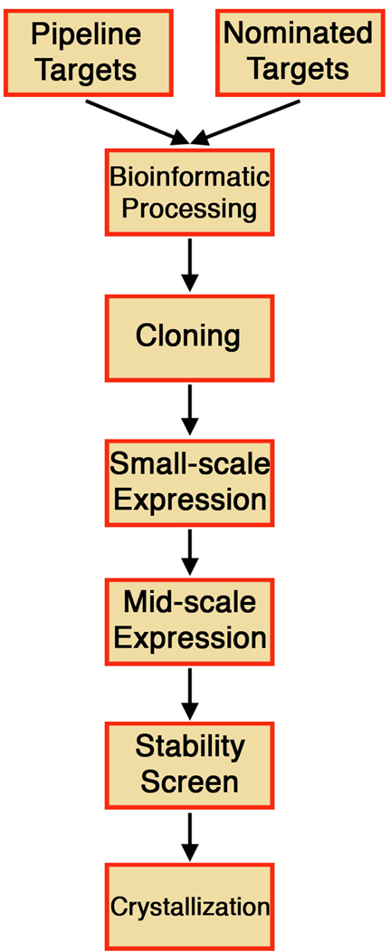
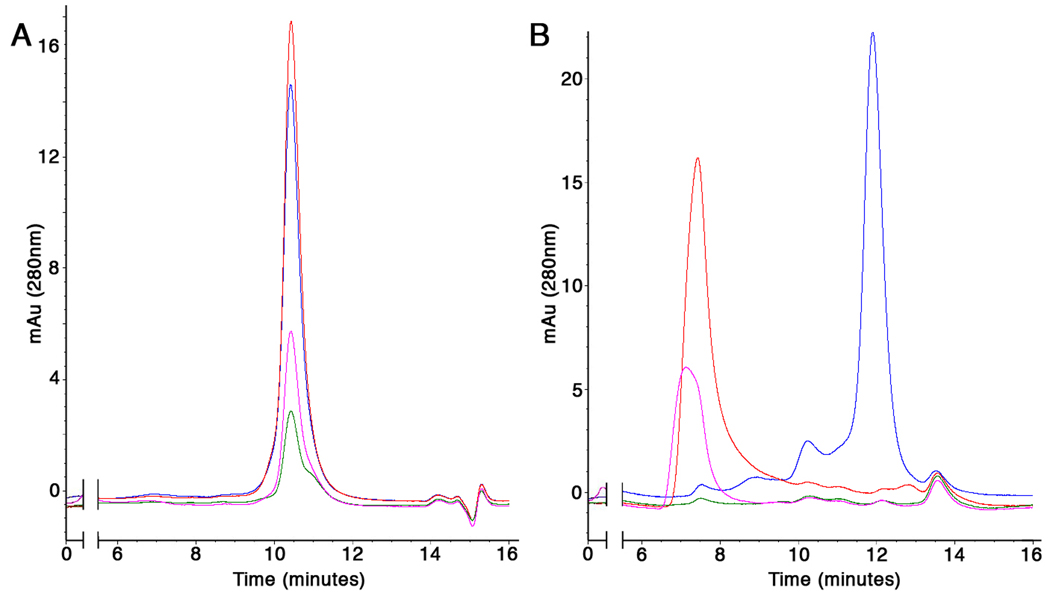
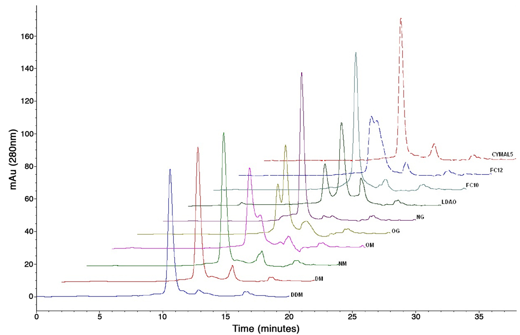
High-throughput (HT) methodologies have had a tremendous impact on structural biology of soluble proteins. High-resolution structure determination relies on the ability of the macromolecule to form ordered crystals that diffract X-rays. While crystallization remains somewhat empirical, for a given protein, success is proportional to the number of conditions screened and to the number of variants trialed. HT techniques have greatly increased the number of targets that can be trialed and the rate at which these can be produced. In terms of number of structures solved, membrane proteins appear to be lagging many years behind their soluble counterparts. Likewise, HT methodologies for production and characterization of these hydrophobic macromolecules are only now emerging. Presented here is an HT platform designed exclusively for membrane proteins that has processed over 5000 targets.


Figures

Figure 1
Figure 1
Schematic representation of the NYCOMPS HT pipeline for the identification of prokaryotic membrane proteins suitable for structural studies.
Figure 2
Figure 2
Representative results from small-scale expression tests. A 24-well SDS-PAGE gel stained with coomassie blue. Membrane proteins are purified from 0.6mL culture volumes. An omni-present contaminant is marked.
Figure 3
Figure 3
Picture of custom-made sonicator robot. Key features are indicated with arrows. Sample holder, pictured here, can be interchanged with a 96-well deep-well block or with a custom-made holder for 6, 50mL centrifuge tubes.
Figure 4
Figure 4
Representative results from mid-scale expression and purification tests Commassie-stained SDS-PAGE gel of 24 purified membrane proteins form the medium-scale pipeline. 2.5% of the total volume eluted from the metal affinity chromatography step was loaded in each lane. Two contaminant bands are marked. Interestingly, these contaminants differ from those present in the small-scale expression screens. This difference could be due to different growth conditions of the bacteria.
Figure 5
Figure 5
Stability in short-chain detergents. SEC elution profiles of 2 different proteins treated with large excess of DDM (blue), C8E4 (green), LDAO (red) and β-OG (pink). The SEC experiments are performed with DDM included in the mobile phase at twice its CMC. Stability of the protein shown in (A) is apparent when compared to the one in (B), as indicated by presents of a large percentage of aggregated material which elutes after approximately 7 minutes, in the void volume of the column.
Figure 6
Figure 6
SEC-mediated detergent exchange. The elution traces are shown for one example protein run on a SEC column equilibrated with 12 different detergents in the mobile phase. The traces are shown in a staggered array, and the matching detergent is indicated above each. This protein yielded diffracting crystals.

Similar articles

Cited by

References

    1. Wallin E, von Heijne G. Genome-wide analysis of integral membrane proteins from eubacterial, archaean, and eukaryotic organisms. Protein Sci. 1998;7:1029–1038. - PMC - PubMed
    1. Berman H, Henrick K, Nakamura H. Announcing the worldwide Protein Data Bank. Nat Struct Biol. 2003;10:980. - PubMed
    1. Henderson R, Unwin PN. Three-dimensional model of purple membrane obtained by electron microscopy. Nature. 1975;257:28–32. - PubMed
    1. Deisenhofer J, Epp O, Miki K, Huber R, Michel H. X-ray structure analysis of a membrane protein complex. Electron density map at 3 A resolution and a model of the chromophores of the photosynthetic reaction center from Rhodopseudomonas viridis. J Mol Biol. 1984;180:385–398. - PubMed
    1. Grisshammer R, Tate CG. Overexpression of integral membrane proteins for structural studies. Q Rev Biophys. 1995;28:315–422. - PubMed

Publication types

MeSH terms

LinkOut - more resources